






Abstract
Objectives
To examine the immune cell profile in the bone marrow of systemic lupus erythematosus (SLE) patients and to assess its clinical relevance.
Methods
Sixteen bone marrow samples from 14 SLE patients were compared with seven healthy control samples. The numbers of immune cells and apoptotic cells in the bone marrow were examined by immunohistochemistry. The association between immune cell subsets and clinical features was investigated.
Results
CD4+ T cells, macrophages and plasma cells were more common in the bone marrow of SLE patients than in healthy controls (p = 0.001, p = 0.004 and p < 0.001, respectively). Greater numbers of CD4+ T cells and macrophages were associated with high-grade bone marrow damage. The percentage of apoptotic cells in bone marrow of SLE patients was significantly higher than that in controls (p < 0.001) and was positively correlated with the number of plasmacytoid dendritic cells (p = 0.013). Increased number of plasma cells along with high interleukin-6 expression was correlated with anti-double stranded DNA antibody levels and the SLE disease activity index (p = 0.031 and 0.013, respectively).
Conclusion
Bone marrow from SLE patients showed a distinct immune cell profile and increased apoptosis. This, coupled with a correlation with disease activity, suggests that the bone marrow may play a critical role in the pathogenesis of SLE.
Introduction
Systemic lupus erythematosus (SLE), a chronic autoimmune disease of unknown aetiology, is characterised by the production of autoantibodies against nuclear autoantigens. Haematological abnormalities such as anaemia and thrombocytopenia are common clinical manifestations.1,2 Peripheral cellular destruction (mediated by autoantibodies), dysfunction of haematopoietic stem cells and autoimmune-mediated bone marrow damage are thought to be immunological contributors.3,4 Few studies have examined the bone marrow characteristics associated with SLE. Voulgarelis et al. examined the histological features of bone marrow specimens from 40 cytopenic SLE patients and reported prominent disruption of the normal architecture and the presence of atypical cells belonging to the erythroid and megakaryocytic lineages. 5 Fibrosis, necrosis and gelatinous transformation are also relatively common histological findings.6,7 However, no study has undertaken a detailed analysis of the immune cell profile in the bone marrow of SLE patients and its relationship with disease activity.
Several studies suggest that dysregulated apoptosis and prolonged exposure to nuclear autoantigens play a role in the pathogenesis of SLE,8,9 and others report the accumulation of apoptotic cells in various tissues in these patients.10,11 Hepburn et al. were the first to report multiple apoptotic bodies in the bone marrow of SLE patients. 12 However, the clinical relevance of these apoptotic cells and their relationship with other immune cells are unknown. Therefore, the aims of the present study were to investigate the role of the bone marrow in the pathogenesis of SLE by examining the immune cell profile and level of apoptosis in the bone marrow of SLE patients and to assess their clinical relevance.
Patients and methods
Patients and clinical information
Between 2000 and 2013, 16 bone marrow specimens were obtained from 14 SLE patients at Seoul National University Hospital. Seven bone marrow specimens of seven bone marrow transplantation donors were used as controls. All SLE patients fulfilled the revised American College of Rheumatology criteria for classification of SLE at diagnosis. 13 Information regarding specific organ involvement, disease activity (measured using the SLE disease activity index (SLEDAI)) and laboratory findings such as serum complement levels and titre of antoantibodies (anti-dsDNA, anti-Sm, anti-Ro/La and anti-cardiolipin) were obtained from medical records. SLE patients with a culture-proven infection in any organ or concurrent haematological disorders at the time of bone marrow examination were excluded. The study was approved by the institutional review board (IRB) of Seoul National University Hospital (IRB no. H-1210-040-432).
Bone marrow examination and immunohistochemistry
Haematoxylin and eosin (H&E)-stained paraffin-embedded bone marrow sections and Wright–Giemsa-stained bone marrow aspirate smears were evaluated by two independent investigators blinded to the patients’ clinical information. Bone marrow cellularity and morphological dysplasia of tri-lineage haematopoietic cells were assessed. Other histological manifestations, including oedema, fibrosis, gelatinous changes, necrosis, lymphoid follicular formation and haemophagocytosis, were also evaluated. Bone marrow necrosis was graded semiquantitatively as described by Maisel et al. 14 High-grade bone marrow damage was defined as extensive bone marrow necrosis (≥50% of the biopsy) and/or severely decreased cellularity (<30%), and low-grade bone marrow damage was defined as mild-to-moderate bone marrow necrosis (<30%). For immunohistochemistry, formalin-fixed, paraffin-embedded bone marrow sections were deparaffinised in xylene and hydrated in a graded series of ethanol solutions followed by distilled water. After heat-induced antigen retrieval in sodium citrate buffer, endogenous peroxidase activity was blocked by incubation in a peroxidase-blocking solution (Dako A/S, Glostrup, Denmark). After incubation in 5% normal goat serum for 30 min, the specimens were incubated with specific primary antibodies (see below) for 60 min at room temperature or overnight at 4 ℃, followed by horseradish peroxidase-conjugated anti-mouse or anti-rabbit Envision+ (Dako A/S) for 30 min. Staining was visualised using diaminobenzidine. Cell nuclei were counterstained with haematoxylin. For dual staining of CD4 and CD68, automated parallel staining was performed with chromoplex dual detection system (Leica Microsystems, Wetzlar, Germany) using diaminobenzidine and fast-red for chromogen.
Because cellularity of bone marrow in patients with SLE is usually decreased, the percentage of specific cellular subsets was calculated for quantification by counting positively stained cells against all the nucleated cells in a high-power field (magnification: ×400). The mean values from five or more randomly selected fields were used as a representative value for the subset. Interleukin-6 (IL-6) expression was assessed semiquantitatively as follows: 0, no staining; 1, <25% of cells stained; 2, 26–50% of cells stained; 3, 51–75% of cells stained; and 4, >75% of cells stained. All specimens were examined in a blinded manner by two independent investigators using a digital quantification system (Leica Application Suite version 3.8) connected to a light microscope (Leica DFC240, Wetzlar, Germany).
Primary antibodies
The following primary antibodies were used in the present study: mouse monoclonal anti-CD3 (Dako A/S), mouse and rabbit monoclonal anti-CD4 (Abcam, Cambridge, UK; Thermo Scientific, Rockford, IL, USA), rabbit monoclonal anti-CD8 (Abcam), rabbit monoclonal anti-CD20 (Abcam), mouse monoclonal anti-CD68 (Dako A/S), mouse monoclonal anti-CD138 (Abcam), rabbit monoclonal anti-active caspase 3 (Abcam), mouse monoclonal anti-CD303 (BDCA-2) (Merck Millipore, Darmstadt, Germany) and rabbit polyclonal anti-IL-6 (Abcam).
Statistical analysis
Continuous variables were expressed as the mean ± standard deviation and compared using the Mann–Whitney U test. Fisher’s exact test was used to compare categorical variables. Spearman’s correlation coefficient was used to examine the correlation between two continuous variables. All statistical analyses were performed using SPSS version 18.0 (SPSS Inc., Chicago, IL, USA) and p values <0.05 were considered significant.
Results
Histological features of the bone marrow
Histological features of bone marrow specimens and the clinical characteristics of the patients

WBC: white blood count; Hb: haemoglobin; SLEDAI: Systemic Lupus Erythematosus Disease Activity Index; M: male; F: female.
Immune cell profile in the bone marrow
There were no significant differences in the percentages of CD3+ and CD8+ cells in the bone marrow of SLE patients and controls. By contrast, CD4+ T cells were more common in the bone marrow of SLE patients than in healthy controls (1.82 ± 1.45% vs. 0.26 ± 0.12%, p = 0.001), as were CD68+cells (16.35 ± 7.17% vs. 8.04 ± 1.38%, p = 0.004). The cells were distributed throughout the parenchyma and showed a macrophage-like morphology, characterised by a stellate-appearance and numerous cytoplasmic processes. The percentage of CD68+ cells correlated with that of CD4+ cells (r = 0.826, p < 0.001). Since CD4 can be expressed in the cytoplasm of CD68+ macrophage, we also performed double staining of CD4 and CD68. CD4+ T cells were clearly differentiated from CD68+ cells from cellular morphology and staining pattern (Figure A1 in the appendix).
The percentage of CD20+ cells in the bone marrow was lower in SLE patients than in controls (2.04 ± 1.30% vs. 4.48 ± 0.72%, p < 0.001); however, the percentage of CD138+ cells was higher (9.72 ± 5.64% vs. 3.44 ± 0.64%, p < 0.001). CD138+ cells were more prominent in the perisinusoid area in SLE patients and were morphologically similar to plasma cells. The immune cell profiles are summarised in Figure 1.
In situ analysis of immune cells in the bone marrow (a) and their percentages in normal control (NC) and systemic lupus erythematosus (SLE) patients ((b)–(d)).
Relationship between immunohistochemical and clinicopathologic features
The percentages of CD4+ T cells and CD68+ macrophages in the bone marrow were negatively correlated with bone marrow cellularity (r = −0.518, p = 0.040 and r = −0.549, p = 0.028, respectively), which is an indirect representation of haematopoietic function. The five bone marrow specimens showing high-grade damage also showed higher percentages of CD4+ T cells and CD68+ macrophages than the other 11 bone marrow specimens of SLE patients (3.06 ± 1.50% vs. 1.26 ± 1.06% p = 0.038; 22.10 ± 7.19% vs. 13.09 ± 4.67%, p = 0.027, respectively). Also, patients with high-grade bone marrow damage showed evidence of more severe thrombocytopenia as compared with all other SLE patients (62.60 ± 68.90 × 10³/mm3 vs. 140.73 ± 77.88 × 10³/mm3, p = 0.027). These results suggest that CD4+ T cells and macrophages may play a pivotal role in bone marrow damage and subsequent thrombocytopenia (Figure 2).
Relationship between organ damage and the presence of immune cells in the bone marrow (BM) of systemic lupus erythematosus patients. Bone marrow specimens showing high-grade damage also show significantly greater infiltration by CD4+ T cells (a) and CD68+ macrophages (b).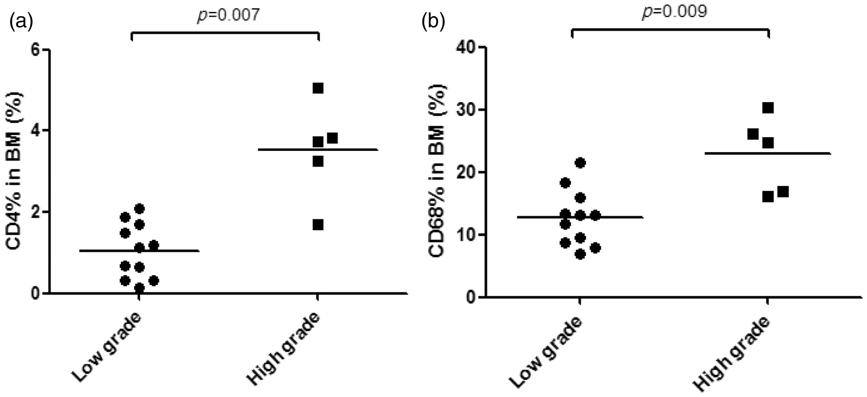
The percentage of CD138+ plasma cells in the bone marrow was significantly correlated with markers of disease activity, including anti-dsDNA antibodies and serum C4 levels (r = 0.538, i = 0.031 and r = −0.652, p = 0.006, respectively). The SLEDAI scores also correlated with the percentage of plasma cells in the bone marrow (r = 0.581, p = 0.031) (Figure 3). Interestingly, bone marrow samples from patients with active lupus nephritis at the time of bone marrow examination contained more plasma cells than those from patients without nephritis (18.28 ± 7.16% vs. 7.19 ± 2.16%, p = 0.038). The levels of other autoantibodies, including anti-Ro/La, anti-Sm and anti-cardiolipin antibodies, were not correlated with the percentage of plasma cells in the bone marrow.
Correlation between the percentage of plasma cells in the bone marrow and markers of SLE disease activity: (a) SLEDAI score; (b) anti-dsDNA antibody levels (expressed on a log scale); and (c) serum complement levels.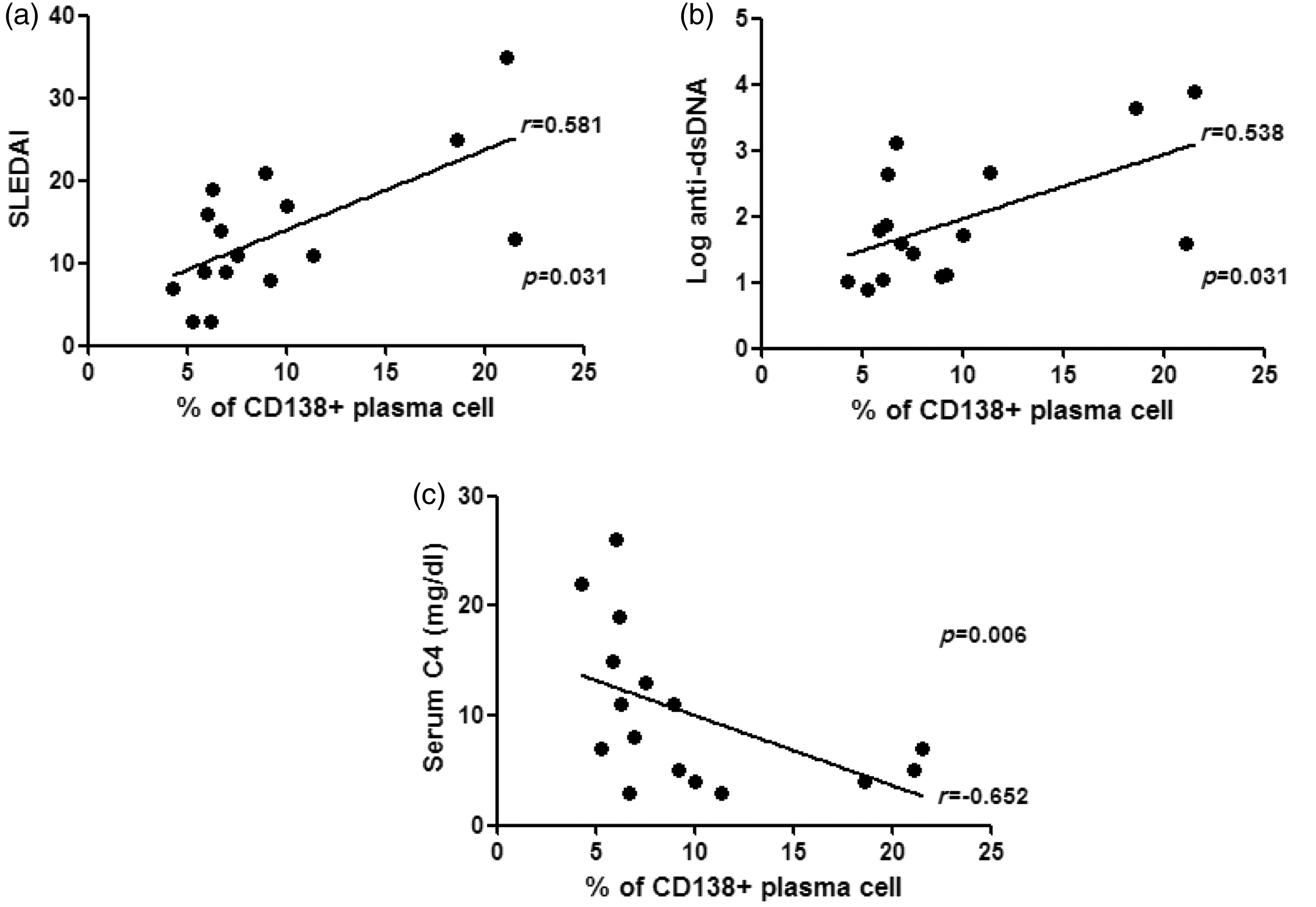
Up-regulation of IL-6 expression in the bone marrow of SLE patients
To investigate factors underlying the higher percentage of plasma cells in the bone marrow of SLE patients, we stained the bone marrow sections for IL-6. IL-6 expression was significantly higher in the SLE group; 12 (75%) SLE specimens showed grade 3 or 4 staining, whereas all the healthy control samples showed grade 0 or 1 (p = 0.001). SLE patients with high IL-6 expression in the bone marrow (≥grade 3) also had higher percentages of macrophages and plasma cells than those with low expression (18.12 ± 6.44% vs. 9.26 ± 1.99%, p = 0.008 and 11.09 ± 5.89% vs. 5.60 ± 1.16%, p = 0.020, respectively). This subgroup also showed higher anti-dsDNA levels (2.21 ± 0.96 vs. 1.13 ± 0.31, p = 0.008, expressed on a log scale) (Figure 4).
Expression of interleukin-6 (IL-6) in the bone marrow and its association with clinicopathological features. All control bone marrow specimens show low-grade IL-6 expression, whereas most systemic lupus erythematosus specimens show high-grade IL-6 expression (a). High-grade IL-6 expression in the bone marrow is significantly associated with an increase in the percentages of CD138+ plasma cells and CD68+ macrophages and with higher titres of anti-dsDNA antibodies (b).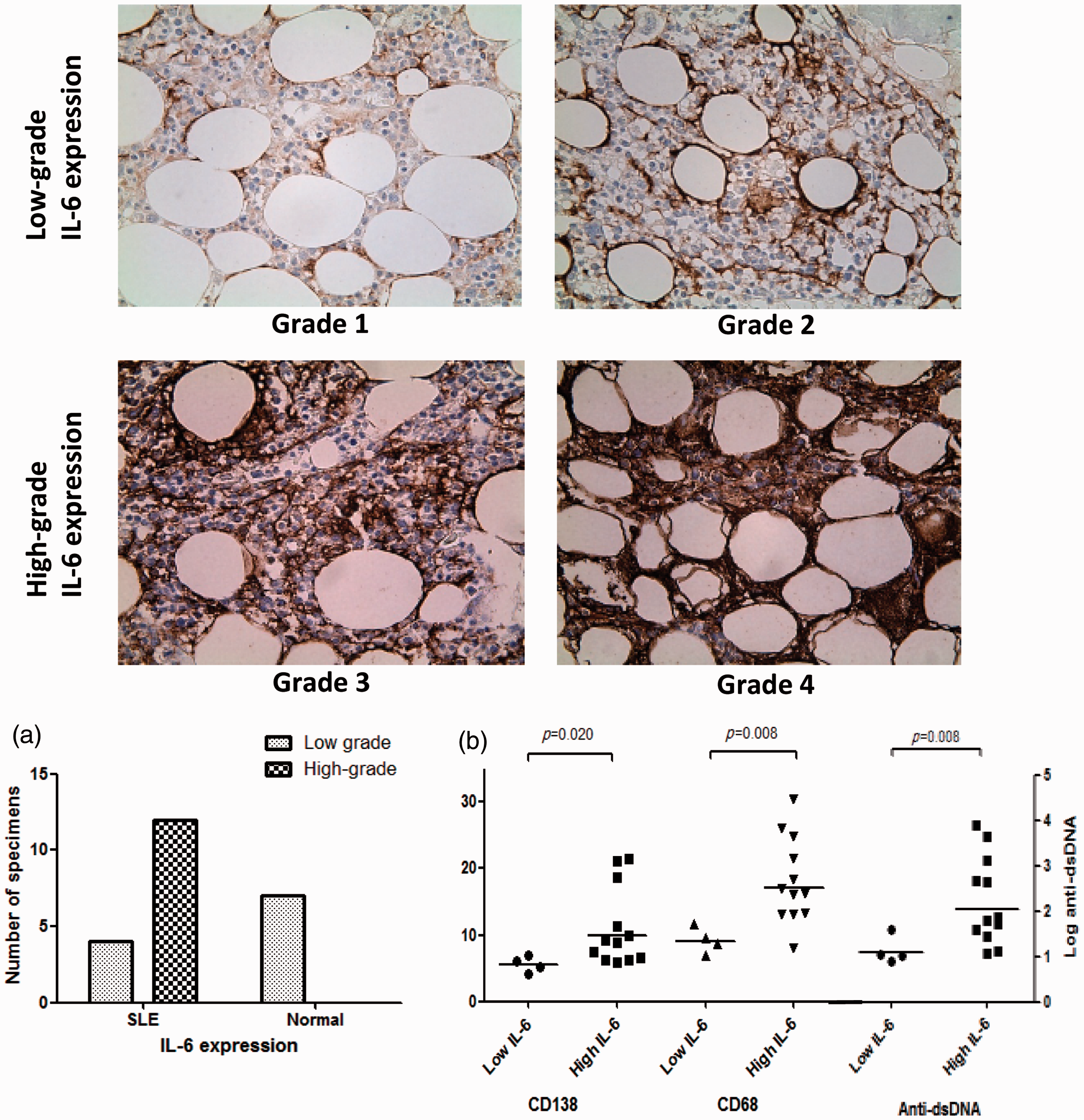
The bone marrow of SLE patients contains higher numbers of plasmacytoid dendritic cells and shows higher levels of apoptosis
Plasmacytoid dendritic cells (pDCs) are a major source of type I interferon (IFN) and are activated by immune complexes or apoptotic cells in SLE patients. Therefore, we next examined the expression of active caspase-3 and BDCA-2 (which are specific markers for apoptotic cells and pDCs, respectively)
15
in the bone marrow. The percentage of apoptotic cells in the bone marrow of SLE patients was significantly higher than that in healthy controls (2.47 ± 1.35% vs. 0.19 ± 0.22%, p < 0.001), as was the percentage of BDCA-2+ pDCs (4.60 ± 2.50% vs. 2.00 ± 1.32%, p = 0.020). There was a positive correlation between the percentage of BDCA-2+ cells in the bone marrow and that of apoptotic cells (r = 0.606, p = 0.013) (Figure 5). The percentage of apoptotic cells in the bone marrow was not correlated with that of other immune cells in bone marrow, but it was negatively correlated with serum C3 levels (r = −0.618, p = 0.011).
Expression of active caspase-3+ (apoptotic) cells and BDCA-2+ plasmacytoid dendritic cells (pDCs) in the bone marrow. The percentages of apoptotic cells and pDCs in bone marrow of systemic lupus erythematosus (SLE) patients are higher than those in the normal controls (NC) (a). There is a significant correlation between the percentages of these two cell types in the bone marrow of SLE patients (b).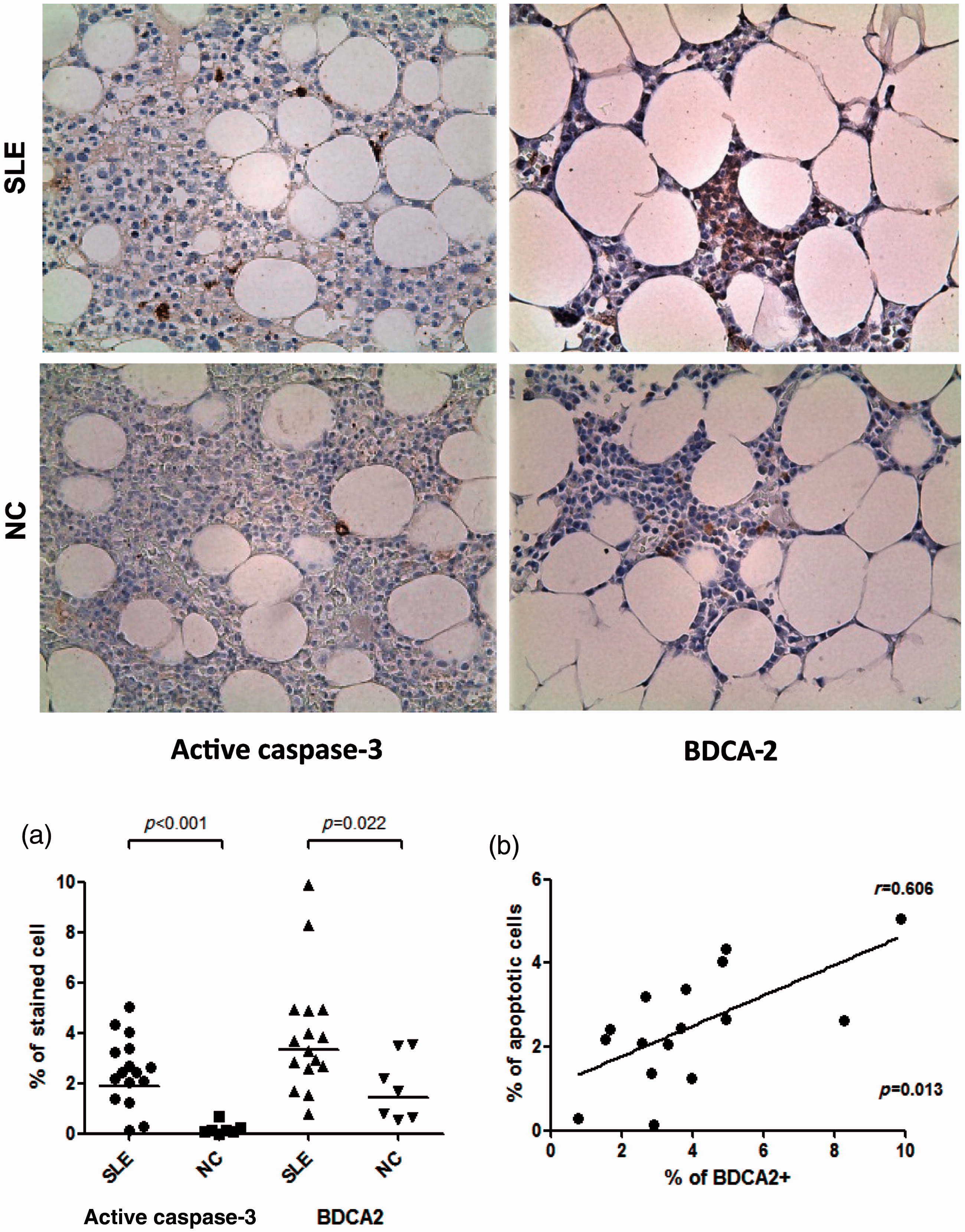
Discussion
The results of the present study showed that the immune cell profile in the bone marrow of SLE patients was significantly different from that of healthy controls, and that there was an association between the immune cell profile and the clinical features of SLE. Increased numbers of CD4+ T cells and macrophages in the bone marrow were related to more extensive bone marrow damage, and increased bone marrow plasma cells was significantly correlated with disease activity and the levels of anti-dsDNA antibodies. In addition, we found an association between an increase in the number of apoptotic cells and the presence of pDCs.
The immunohistochemical and clinicopathological features of target organs have been investigated in patients with lupus nephritis. Renal biopsy specimens from patients with lupus nephritis show infiltration of the renal interstitium by CD4+ lymphocytes, which is associated with a worsening of renal function. 16 In addition, samples from patients with this more aggressive form of nephritis show evidence of glomerular and interstitial macrophage accumulation, which is the best clinical marker of renal activity.17,18 Likewise, we found that the percentages of CD4+ lymphocytes and CD68+ macrophages in the bone marrow of SLE patients were significantly higher than those in the controls, and were closely associated with hypocellularity and high-grade organ destruction. In our experiment, the proportion of CD4+ T cells in bone marrow was small as compared with that of CD8+ T cells in both SLE patients and healthy controls. Therefore, increased CD4+ T cells did not lead to statistically significant change in the proportion of total T cells (CD3+ cells). However, CD4+ T cells rarely existed in normal bone marrow so we think that this small increase in CD4+ T cells is an important change in bone marrow of SLE patients. Taken together, these results suggest that CD4+ lymphocytes and CD68+ macrophages may be central players in bone marrow damage (as they are in damage caused to other target organs in SLE patients).
The observation that the increased percentage of plasma cells in the bone marrow of SLE patients was significantly correlated with anti-dsDNA levels is rather interesting. The majority of long-lived plasma cells reside in the bone marrow, where they produce antibodies for a long time in the absence of antigenic stimulation or T cell help.19,20 Long-lived plasma cells originate from proliferating plasmablasts in the spleen or lymph nodes, from where they migrate to the bone marrow along a chemokine gradient. 21 After arriving in the bone marrow, they must occupy a specific survival niche to mature into long-lived plasma cells. Some cytokines and adhesion molecules function as survival niches for bone marrow plasma cells. IL-6, in conjunction with B-cell-activating factor (BAFF) and C-X-C motif chemokine 12 (CXCL12) expressed by stromal cells, provides a survival signal.22,23 Because long-lived plasma cells in the bone marrow are resistant to immunosuppressive agents and B cell depletion therapy, it is assumed that they are the main cause of refractory autoimmune disease.24,25 Recently, Cheng et al. demonstrated that bone marrow plasma cells continuously secreted anti-dsDNA antibodies and caused immune complex nephritis after they were transferred from NZB/W mice to Rag1−/− mice. 26 This result is consistent with the results of the present study, which showed that the increased percentage of plasma cells in the bone marrow of SLE patients correlated with increased anti-dsDNA antibody and serum complement levels, which are significantly associated with lupus nephritis. This indicates that increased numbers of plasma cells in the bone marrow may trigger lupus by secreting anti-dsDNA antibodies; this increases the formation of anti-dsDNA-containing immune complexes, which then activate the complement system. Although we did not directly show that the plasma cells in the bone marrow secreted autoantibodies, we did identify an association between bone marrow plasma cell numbers and immune complex nephritis in SLE patients. In addition, the results indicate that increased expression of IL-6 in the bone marrow of SLE patients may be an important factor for plasma cell survival and subsequent autoantibody production. IL-6 is the most efficient survival factor for bone marrow plasma cells in vitro, and monocytes-macrophages are the main producers of the IL-6 required for plasma cell maturation in murine lymph nodes.23,27 Although little is known about the cellular source of cytokines that promote plasma cell survival in humans, we found that IL-6 expression was significantly associated with the number of macrophages in the bone marrow of SLE patients.
Dysregulated apoptosis can generate nuclear autoantigens, which act as important triggers for autoimmunity. We identified an increase in the number of apoptotic cells in the bone marrow of SLE patients, which is consistent with the results of a previous study. 12 However, we also identified an important association between the degree of apoptosis and an increase in the number of pDCs in the bone marrow of SLE patients. pDCs are a major producer of type I IFN, which plays an important role in the pathogenesis of SLE. In SLE, pDCs recognise immune complexes containing self-nucleic acids and then produce type I IFN. 28 Lovgren et al. demonstrated that late apoptotic cells release nuclear material, which induces pDCs to secrete IFN-alpha. 29 SLE patients show elevated levels of circulating apoptotic cells, which are cleared very slowly. As a result, pDCs continuously produce type I IFN. This activates conventional DCs, which in turn stimulate autoimmune T cells. 30 Patients with lupus nephritis show a marked increase in glomerular infiltration by pDCs, which is not observed in normal kidneys. Such infiltration is prominent in the kidneys of patients with active class III/IV lupus nephritis. 31 Likewise, the results reported herein suggest that increased apoptosis and increased numbers of pDCs in the bone marrow of SLE patients may promote T cell-mediated bone marrow damage via the unabated production of type I IFN.
This study has several limitations. First, the number of SLE bone marrow specimens was small, and most patients showed haematological manifestations at the time of bone marrow examination. Therefore, further study is needed to confirm the applicability of our results to SLE patients who do not show haematological manifestations. Second, the results are based on in situ analyses rather than on functional studies. However, previous studies (both in vitro studies and animal studies) support our conclusion that the bone marrow is a major target organ in SLE, which plays an important role in disease pathogenesis by acting as a source of anti-dsDNA antibodies and triggering autoimmunity by dysregulated apoptosis.
In conclusion, the results reported herein suggest that the bone marrow may play the following critical roles in the pathogenesis of SLE. First, it is a target organ that is damaged by CD4+ T cells and macrophages. Second, an increased number of apoptotic cells and subsequently pDCs may activate a pathogenic process of lupus. Third, bone marrow plasma cells, supported by IL-6, generate anti-dsDNA antibodies, which increase systemic manifestations such as nephritis.
Footnotes
Funding
This work was partially supported by 800-20120026 from the SNU college of Medicine.
Conflict of interest statement
The authors have no conflicts of interest to declare.