






Abstract
We investigated systemic lupus erythematosus (SLE) patients with epilepsy, a major and organic neurological symptom. Our aim was to test patients for the autoimmune epilepsy-associated antibodies anti-GAD, anti-NMDAR, anti-AMPAR1/2, anti-GABABR and anti-VGKC. We tested sera from ten SLE patients with current or previous episodes of epileptic seizures. In addition, sera were tested for staining on primary hippocampal neurons. The patients’ clinical and neuroimaging profile, disease activity and accumulated damage scores and therapeutic regimens administered were recorded, and correlations were evaluated. Patients were negative for all anti-neuronal autoantibodies tested, and showed no staining on primary hippocampal cells, which suggests the absence of autoantibodies against neuronal cell surface antigens. Epileptic seizures were all tonic–clonic, and all patients had high disease activity (mean SLE Damage Acticity Index score 19.3 ± 7.3). Six patients had minor or no brain magnetic resonance imaging findings, and three had major findings. 9/10 patients received immunosuppression for 5 ± 4 months, while anti-convulsive treatment was administered to all patients (4.2 ± 3 years). Our results suggest that the majority of SLE-related epileptic seizures cannot be attributed to the action of a single antibody against neuronal antigens. Studies with larger neuropsychiatric SLE populations and stricter inclusion criteria are necessary to verify these findings.
Introduction
Systemic lupus erythematosus (SLE) is a systemic autoimmune disease, affecting mainly women of reproductive age. The central nervous system (CNS) is often affected in SLE patients, hence the need to form definitions, correlations, and exclusions for neuropsychiatrics lupus erythematosus (NPSLE), that include 11 separate central nervous and eight peripheral nervous system manifestations. 1 The calculated incidence and prevalence of neurological involvement in SLE varies between studies, owing to different study design, diagnostic approach, and patient baseline characteristics.2,3
The neurological repertoire of SLE includes major and organic symptoms, such as epileptic seizures, in up to 10% of NPSLE patients. Epilepsy can also manifest as a symptom of autoimmune encephalopathy (AE), and may be associated with neuronal autoantibodies. Autoimmune encephalopathies invariably present with rapid cognitive decline over days to weeks, irritability, and memory loss, while almost 50% of the patients also present with epileptic seizures and psychiatric disturbances. The most noteworthy laboratory finding in AE-epilepsy is the association with antibodies, measured by cell-based assays, 4 which are directed against neuronal cell surface antigens, including the voltage-gated potassium channel complex (VGKC), N-methyl-D-aspartate receptor (NMDAR, NR1 subunit), α-amino-3-hydroxy-5-methyl-4-isoxazolepropionic acid receptors 1 and 2 (anti-AMPAR1/2), and gamma-aminobutyric acid B receptor (GABABR). 5
In contrast to AE, most SLE-associated autoantibodies are directed against intracellular antigens, including antibodies associated with neurological phenotypes–for example, anti-ribosomal P. 6 However, a small number of recent studies have described the existence of antibodies directed against the NMDA and GABAB receptors in NPSLE, mostly with methods that recognize linear antigenic epitopes7,8 and, in a recent study, also by a cell based assay. 9 The aim of the present study was to determine whether a major NPSLE symptoms, such as epilepsy, could be attributed to the presence of neuronal antibodies against extracellular antigens, like those described in the AE syndromes.
Patients and methods
Patients
We retrospectively evaluated an SLE cohort for epileptic seizures. We singled out 39 out of 459 SLE patients (actively followed in our department for 3–20 years), who presented with epileptic seizures during the course of their disease. Archived sera were available from ten SLE patients (33.9 ± 15.2 years at blood sampling, 90% females). All patients fulfilled the 1997 American College of Rheumatology (ACR) SLE diagnostic criteria. Neurological assessment was based on the 1999 ACR definitions, exclusions, and correlations for NPSLE 1 and the 2010 EULAR recommendations for the management of SLE with neuropsychiatric manifestations. 10 The SLE Disease Activity Index (SLEDAI)—Safety of Estrogens in Lupus Erythematosus National Assessment (SELENA) Modification, was used to evaluate disease activity, whereas accumulated damage due to the disease was assessed by the Systemic Lupus International Collaborating Clinics (SLICC)/ACR Damage Index (SDI). Blood samples from five patients were drawn during acute neurological involvement, whereas blood from the remaining five patients was drawn during an inactive phase, following the onset of neurological manifestations by a mean time period of 2.6 ± 2.1 years. The University of Athens Faculty of Medicine Ethics committee approved the study.
Detection of neuronal autoantibodies
Anti-NMDAR (NR1 subunit), anti-AMPAR1/2, anti-GABABR, and anti-VGKC (CASPR2 and LGI1 antigens) autoantibody testing was performed by an indirect immunofluorescence test (Euroimmun AG), which employs BIOCHIP slides coated with human embryonic kidney (HEK) cells transfected with the corresponding antigens. This method of antibody detection primarily detects antibodies directed against extracellular epitopes. Anti-glutamic acid decarboxylase (GAD) antibodies were detected using a commercial ELISA kit (Euroimmun AG).
To identify antibodies against other, non-characterized, neuronal cell surface antigens, we stained primary hippocampal neurons, which constitute an established substrate for screening for novel specificities. Primary hippocampal neurons were isolated from E16 mice embryos, as previously described, 11 and were cultured for seven days in vitro before use. All patient sera were diluted 1/75 in 1% normal donkey serum (NDS) in phosphate-buffered saline (PBS), and incubated with neurons for 2 h at 37℃. Neurons were then fixed with 4% paraformaldehyde in PBS for five minutes and permeabilized with 0.3% Triton in PBS for another five min utes. An anti-human fluorochrome-conjugated secondary antibody was applied at 1/750 dilution (Alexa Fluor 488, Invitrogen) for 1 h, and cells were mounted with a fluorescent mounting medium (Dako, Agilent Technologies) and imaged using a fluorescent microscope (Zeiss Axiophot).
Statistical analysis
Continuous variables are presented as mean ± SD. Categorical variables are presented as frequencies. Normality was tested using the one sample Kolmogorov Smirnov Test. The chi-square test (categorical variables) and the T-test (quantitative variables) were used to evaluate associations between subgroups of patients with CNS involvement. Spearman’s rank correlation coefficient was applied to assess statistical dependence between patients’ clinical characteristics. The level of statistical significance was set at P ≤ 0.05. SPSS statistical software was used for all analyses (SPSS Hellas Inc., Athens, Greece).
Results
Clinical and imaging findings
Epileptic seizures were all generalized tonic–clonic, and in three patients they evolved to status epilepticus, requiring intubation and ICU hospitalization. Electroencephalogram (EEG) was performed in five cases and was normal in two of them, whereas typical epileptiform discharges presented in the three remaining cases. Routine CSF analysis was normal in all patients. Magnetic resonance imaging (MRI) revealed microinfarcts and non-specific white matter lesions in four patients, while it was normal in two. In the remaining patients, three presented with an image reminiscent of Posterior Reversible Leukoencephalopathy Syndrome (PRESS, two evolved to status epilepticus), and one had an image of cerebral vasculitis, which also led to status epilepticus and intubation.
Patient clinical characteristics at CNS involvement
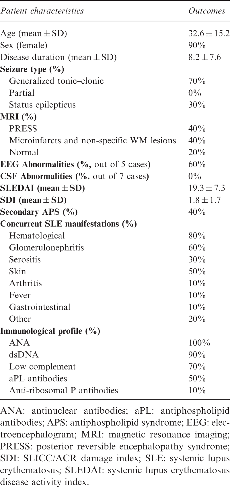
ANA: antinuclear antibodies; aPL: antiphospholipid antibodies; APS: antiphospholipid syndrome; EEG: electroencephalogram; MRI: magnetic resonance imaging; PRESS: posterior reversible encephalopathy syndrome; SDI: SLICC/ACR damage index; SLE: systemic lupus erythematosus; SLEDAI: systemic lupus erythematosus disease activity index.
Autoantibody profile
All patients with epilepsy were negative for all anti-neuronal autoantibodies tested. Furthermore, we did not observe any staining on primary cultured hippocampal cells, which suggests the complete absence of antibodies against neuronal cell surface antigens. All patients were ANA positive, with anti-dsDNA antibody positivity and low complement levels in nine and seven cases, respectively. Finally, five patients were positive for antiphospholipid antibodies, and one patient was positive for anti-ribosomal P antibodies.
Treatments and outcome
Nine patients received induction immunosuppressants during the acute phase of CNS involvement, for a mean period of 5 ± 4 months (Figure 1). Immunosuppressive regimens were, in descending order, intravenous pulses of steroids plus cyclophosphamide (eight patients), IVIg (four patients), high doses of oral steroids (three patients), and rituximab (three patients), while one patient also underwent plasmapheresis. Four patients were given more than one regimen to achieve symptom remission. Anticoagulation or anti-platelet agents were administered in seven patients, including all those positive for antiphospholipid antibodies. Finally, anti-convulsive treatment was administered to all patients (mean treatment duration 4.2 ± 3 years).
Therapeutic regimes in SLE patients with epilepsy. All combinations of immunosuppressive regimens that were administered to SLE patients during active neurological involvement are presented.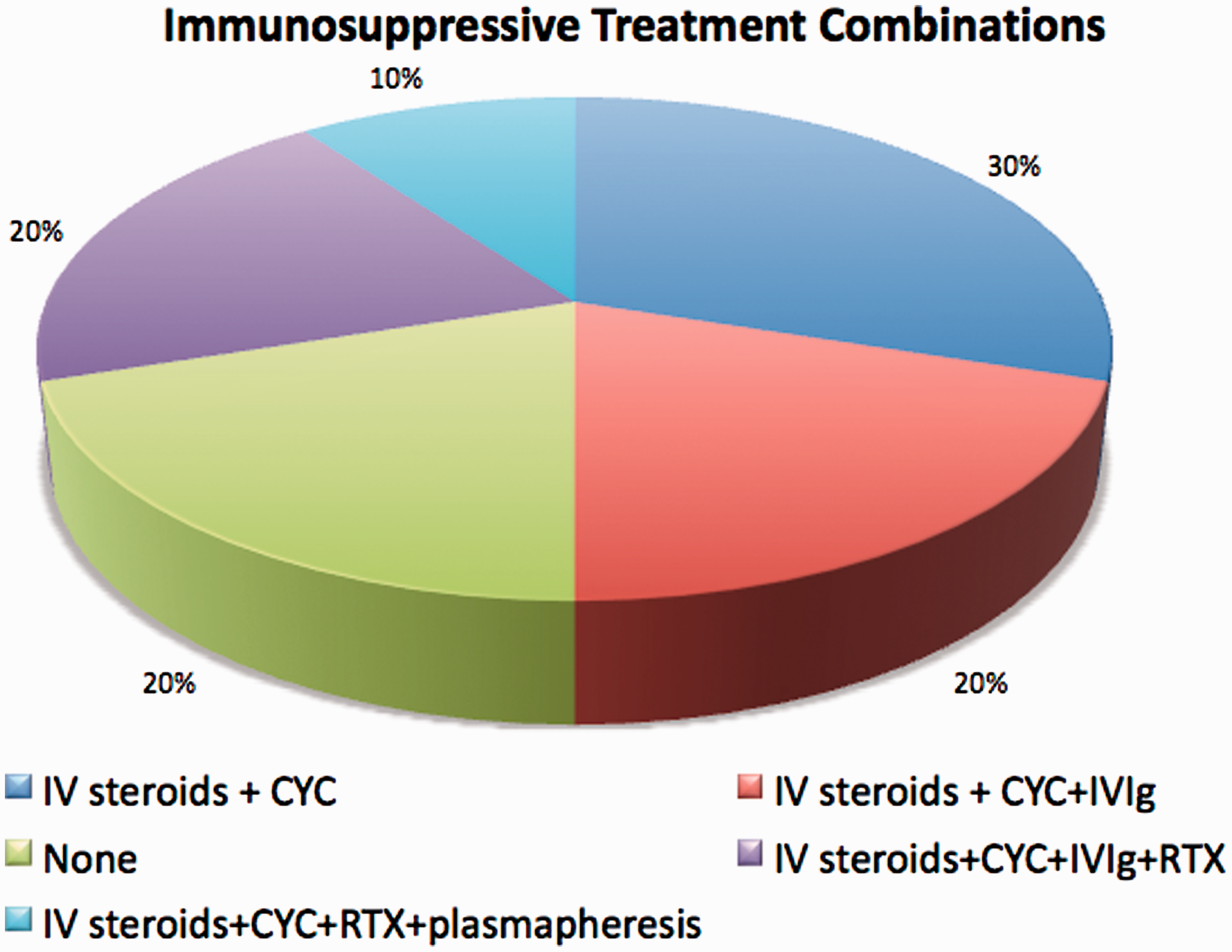
Three patients had a relapsing course, since they had previous exacerbations of their symptoms. However, no new relapses were observed from blood sampling to the conduction of the study. Finally, cumulative damage due to the disease at the end of follow up was 2.4 ± 1.5, an SDI score that was not significantly different from the mean SDI during CNS involvement (P > 0.05).
Clinical and laboratory correlations
Patients with major MRI lesions (n = 4, with PRESS or vasculitis) had higher disease activity during CNS involvement, and they required symptomatic treatment for a longer period of time (all P < 0.05). SLEDAI scores at CNS involvement did not correlate with worse cumulative damage at the end of follow up, or with longer duration of treatment. On the contrary, coexistence of secondary APS or antiphospholipid antibodies was related to a trend towards worse SDI values at the end of follow up (P < 0.01).
Discussion
We present a retrospective case series of patients with epileptic seizures in the context of NPSLE. NPSLE is a heterogeneous entity, which includes various major and minor neurological manifestations, attributed to diverse pathogenetic mechanisms. Our hypothesis was that epilepsy (a major neurological NPSLE manifestation) might be due to the presence of specific anti-neuronal autoantibodies. This has been previously shown for myelitis, another major NSPLE manifestation, which in many cases is due to antibodies against AQP4. 3
Epileptic seizures, either as an isolated manifestation or in the context of autoimmune encephalopathy, have been associated with antibodies against neuronal cell surface antigens. Epileptic seizures are a major feature in VGKC, NMDAR, and GABABR associated syndromes (found in about 70%–80% of patients), and a minor feature in AMPAR and GAD syndromes. A recent animal passive transfer model has confirmed these clinical associations. 12 These antigens, apart from GAD, are extracellular receptors or synaptic protein complexes that mediate synaptic transmission. Antibodies to GAD65, the cytoplasmic enzyme that catalyzes the conversion of L-glutamate to GABA in inhibitory synapses, are more often associated with stiff-person syndrome, but there are an increasing number of reports describing refractory epilepsy cases. 13
Brain MRI in VGKC, AMPAR, and GABABR encephalopathy cases usually shows increased FLAIR/T2 signal involving one or both temporal lobes, without contrast enhancement. In other encephalopathy syndromes, MRI is often normal, or with mild transient cortical–subcortical changes. Overall, in these autoantibody-associated epilepsy and encephalopathy cases, response to immunotherapy is good, while immunosuppressive treatments have poor results.
NPSLE pathogenesis is arguably complex and ambiguous. It includes mechanisms such as microangiopathy, intrathecal cytokine synthesis, and the action of several autoantibodies, including antiphospholipid and anti-ribosomal P antibodies. 14 In other systemic disorders, such as primary antiphospholipid syndrome, the occurrence of seizures was associated with smoking and stroke. 15 Regarding NPSLE epileptic seizures, information is scarce, since no specific factor is known to directly contribute to their emergence. Known risk factors include antiphospholipid antibody-associated microangiopathy and generalized inflammation, in the context of high systemic disease activity. 16 Moreover, the LUMINA cohort study has proposed that the reduction of the seizure threshold is due to the activation of the hypothalamic–pituitary–adrenal axis from pro-inflammatory cytokines. 17
An interesting finding is the association of anti-dsDNA antibodies that cross-react to the NMDA receptor in SLE patients with diffuse neuropsychiatric manifestations, such as cognitive dysfunction and emotional disturbances. 18 These antibodies are primarily determined with ELISA, and bind the NR2A and NR2B subunits of the NMDA receptor. Even though patients with focal NPSLE manifestations, such as epilepsy, also harbor these cross-reactive antibodies, no specific correlation with epileptic seizures has been unequivocally demonstrated.19–21 Further, no other studies have associated NPSLE with any known antibodies against neuronal cell surface antigens. The exception is a study that describes a positive correlation between neurological involvement and GABABR antibodies in an SLE case series of SLE patients; 8 nevertheless, this study has the weakness that the antibodies were determined with only a single method.
Our study demonstrates no positivity for anti-NMDAR or anti-GABABR antibodies. We can assume that even the SLE-related neurological manifestations, which resemble autoimmune encephalitis features, are most likely attributed to more complex mechanisms than to the action of a single antibody against neuronal cell membrane antigens. This conclusion is supported by the different neuro-imaging and laboratory findings in SLE epilepsy patients compared to AE/epilepsy patients. All patients with epileptic seizures in our study had normal CSF findings, whereas the majority of patients with AE/epilepsy have mild pleocytosis and increased CSF protein. 5 Furthermore, no MRI findings reminiscent of AE were seen in any of our patients; 60% of NPSLE patients in our study had minor, non-specific findings or normal MRI imaging.
SLE-epilepsy patients in our study showed a satisfactory therapeutic response following immunosuppressive treatment for a mean time period of 5 ± 4 months and complementary symptomatic therapy for 4.2 ± 3 years. AE patients also respond to immunotherapy, even though the approach is different. 22 The first line of treatment for AE/epilepsy patients with specific antibodies is often plasmapheresis, followed by IVIg, in contrast to the administration of steroids and cyclophosphamide in NPSLE cases.
Our study has the limitation of a small sample size and the heterogeneous timing of presentation of the neuropsychiatric events. In spite of these limitations, our study suggests that SLE-related epileptic seizures cannot be attributed to the action of a single antibody against neuronal antigens. Studies with larger NPSLE populations and with stricter inclusion criteria, possibly after exclusion of other potential causes, such as antiphospholipid antibodies and vasculopathy, are necessary to verify our findings.
Footnotes
Acknowledgements
Dr E Kampylafka collected clinical data, performed experiments, analyzed data and drafted the manuscript. Dr H Alexopoulos analyzed data, drafted and critically revised the manuscript. Ms P Fouka performed experiments. Professor HM Moutsopoulos critically revised the manuscript. Professor M Dalakas critically revised the manuscript. Professor AG Tzioufas supervised the study and critically revised the manuscript.
Declaration of Conflict of Interest
The author(s) declare no potential conflict of interest with respect to the research, authorship and/or publication of this article.
Funding
The author(s) disclosed receipt of the following financial support for the research, authorship, and/or publication of this article: This work was supported by the Special Research Account, University of Athens, and the Institute of Autoimmune Systemic and Neurological Diseases, Athens, Greece.