






Abstract
B cells play a pivotal role in the pathogenesis of autoimmune diseases. In patients with systemic lupus erythematosus (SLE), the percentages of plasmablasts and IgD−CD27− double-negative memory B cells in peripheral blood are significantly increased, while IgD+CD27+ IgM memory B cells are significantly decreased compared to healthy donors. The phenotypic change is significantly associated with disease activity and concentration of autoantibodies. Treatment of B-cell depletion using rituximab results in the reconstitution of peripheral B cells in SLE patients with subsequent improvement in disease activity. Numerous studies have described abnormalities in B-cell receptor (BCR)-mediated signaling in B cells of SLE patients. Since differences in BCR signaling are considered to dictate the survival or death of naïve and memory B cells, aberrant BCR signal can lead to abnormality of B-cell subsets in SLE patients. Although Syk and Btk function as key molecules in BCR signaling, their pathological role in SLE remains unclear. We found that Syk and Btk do not only transduce activation signal through BCR, but also mediate crosstalk between BCR and Toll-like receptor (TLR) as well as BCR and JAK-STAT pathways in human B cells in vitro. In addition, pronounced Syk and Btk phosphorylation was observed in B cells of patients with active SLE compared to those of healthy individuals. The results suggest the involvement of Syk and Btk activation in abnormalities of BCR-mediated signaling and B-cell phenotypes during the pathological process of SLE and that Syk, Btk and JAK are potential therapeutic targets in SLE.
Introduction
B cells play a pivotal role in the initiation and perpetuation of systemic lupus erythematosus (SLE) by producing autoantibodies. Autoantibodies are usually present for many years before the diagnosis of SLE. 1 Over time, autoantibodies evolve to have higher affinity, undergo isotype switching and spread to recognize new epitopes during the development of SLE. 1 Autoantibodies are not passive bystanders but critical effectors responsible for many clinical and pathological manifestations, including cytopenias, thromboembolic disease, neuronal damage, skin rashes, arthritis, and renal disease. B cells also influence autoimmune disease through multiple antibody-independent mechanisms.2,3 For example, through antigen presentation and production of cytokines and costimulatory factors, B cells regulate T-cell activation and polarization. Moreover, B cells induce lymphoid neogenesis through tumor necrosis factor-beta (TNF-β), which recruits and activates follicular dendritic cells, thus generating lymphoid follicles.
The relative importance of autoantibody-dependent vs. -independent effects of B cells is usually difficult to assess. Chan et al.4–6 examined this issue in a series of studies using a mouse model in which B cells expressed only membrane immunoglobulin-M (mIgM). Mice with only mIgM have mature B cells, but these cells do not secrete Ig. They compared the development of lupus in MRL/lpr mice with mIgM-only B cells and MRL/lpr mice with normal B cells. In the group with normal B cells, the survival rate was 50% at 32 weeks, whereas it was 50% at 56 weeks in mice with mIgM-only B cells (p < 0.0007), and 90% at 56 weeks in mice with no B cells (p < 0.0001). Thus, even the lack of Ig-secreting B cells significantly influenced the course of lupus in this particular animal model. These results suggest that B cells play a critical role in SLE pathology not only by autoantibody production but also cytokine production and antigen presentation.
Abnormality of B-cell subsets in SLE patients
The abnormalities of certain peripheral B-cell subsets in SLE compared with healthy individuals were reviewed recently. 7 A central finding in phenotyping peripheral blood B cells in SLE is the substantial increase in antigen experienced IgD−CD27+ class-switched memory B cells, 8 which are less susceptible to immunosuppressive therapy.7,8 Importantly, the peripheral memory B-cell receptor (BCR) repertoire in SLE is shaped by abnormal selection, exaggerated somatic hypermutation, and increased receptor editing.9,10 A large pool of memory B cells poses a risk for autoimmunity since these cells have lower activation thresholds; regulation by FcγRIIb receptors may be reduced, 11 and these cells can be rapidly activated in a non-antigen-specific manner by the combination of Toll-like receptor (TLR) agonists and a proliferation-inducing ligand (APRIL) or B-cell activating factor (BAFF) as well as by the combination of cytokines, such as interleukin (IL)-21. 12
Another memory B-cell subset particularly enhanced in patients with SLE has been identified in which memory B cells do not express CD27.13–15 While IgD−CD27− B cells expressing mutated BCRs have been identified both in the tonsils and in very small numbers in peripheral blood in healthy donors,16,17 large numbers of IgD−CD27− memory B cells were found in the peripheral blood of patients with SLE and a great increase was associated with increased disease activity and active renal disease. 14
Only a few reports examined the function of IgD+CD27+ IgM memory B cells in SLE patients. Rodríguez-Bayona et al. 18 reported CD95, CD80, CD86, CXCR3 and CXCR4 overexpression in IgD+CD27+ IgM memory B cells of SLE patients. The actual developmental origin of IgM memory cells and their specific role in functional immune responses remain to be elucidated. In this regard, IgM memory cells, which may develop through germinal center (GC)-independent pathways, were originally proposed to represent the human functional equivalents of B1 and marginal zone (MZ) B cells and to represent the critical cellular compartment for protection against infections with encapsulated organisms.19–23 Under the latter condition, IgM memory B cells are capable of differentiating into IgG-secreting plasma cells. 24 Interestingly, the proportion of normal human IgM memory B cells with auto-reactive BCR is much lower than that observed in naïve B cells, suggesting the existence of an additional checkpoint to prevent self-reactivity at this level. 11
One study from our laboratory found significantly higher percentages of plasmablasts and IgD−CD27− memory B cells and significantly lower percentage of IgD+CD27+ IgM memory B cells in peripheral blood of SLE patients compared to control participants. Other groups also reported similar results.2,18,25 Interestingly, the percentage of IgD+CD27+ IgM memory B cells was also significantly lower in active SLE patients compared to patients with inactive SLE. In addition, significantly high levels of antinuclear antibodies (ANA), anti-double-stranded DNA (anti-dsDNA) and anti-extractable nuclear antigen (anti-ENA) antibodies (Ab) were associated with low numbers of IgD+CD27+ IgM memory B cells. 18 In addition, the percentage of IgD−CD27− memory B cells in peripheral blood of SLE patients correlated significantly with disease activity and disease-specific autoantibodies, such as anti-dsDNA, anti-Sm, anti-ribonucleoprotein (anti-RNP), and 9G4.13,14 These results stress the potential involvement of B-cell subset abnormalities in the pathology of SLE, and suggest that therapies designed to target B cells, such as rituximab, an anti-CD20 antibody, could be effective against lupus.
Phenotypic changes in B cells following rituximab treatment in SLE patients
Next, we investigated the effects of treatment on B-cell abnormalities and determined changes in B-cell subsets during relapse in order to clarify the association between abnormality of B-cell subsets and SLE pathology. Rituximab is a human-mouse chimeric monoclonal antibody that targets the CD20 antigen, a B-cell-specific antigen, and causes depletion of CD20-expressing pre-B and mature B cells. Randomized, placebo-controlled trials of rituximab failed to demonstrate added value over conventional therapy both in non-renal lupus (Exploratory Phase II/III SLE Evaluation of Rituximab (EXPLORER)) and lupus nephritis (Lupus Nephritis Assessment with Rituximab (LUNAR)).26,27 However, rituximab has recently been reported to show rapid effects and prolonged efficacy in some patients with refractory SLE.28–34 We analyzed the longitudinal changes in B-cell differentiation patterns in long-term responders to rituximab therapy. 35 Rituximab treatment resulted in rapid (within 28 days) disappearance of naïve B cells, IgD−CD27− memory B cells and IgD−CD27+ B memory cells from the peripheral blood of SLE patients. On the other hand, plasmablasts were persistently present in the peripheral blood of these patients until Day 28, although they also disappeared completely from the peripheral blood by six months after rituximab treatment. Naïve B cells showed recovery within three to nine months after treatment, whereas depletion of memory B cells and plasma cells was still observed for more than six years.
On the other hand, SLE patients with relapse after prolonged remission by rituximab therapy showed a quite different B-cell phenotypic pattern. For example, a 16-year-old female with lupus nephritis (World Health Organization (WHO) type IV) with high disease activity achieved SLE remission after rituximab treatment, with rapid disappearance of double-negative memory B cells and class-switched memory B cells by Day 28. However, the disease relapsed at 1.5 years after rituximab treatment, with simultaneous reappearance of the butterfly rash, increase in anti-dsDNA antibody titer, and recurrence of proteinuria. Interestingly, the percentages of IgD−CD27− memory B cells, IgD−CD27+ memory B cells and plasmablasts increased just before relapse.35,36
BCR activation plays an important role in SLE
Clinical and experimental evidence indicate the close relationship between IgD−CD27− and IgD−CD27+ memory B cells and SLE pathology.2,13,14,18,25 However, the origin of these cells remains to be elucidated. One possible mechanism for the increase in these memory B cells is differences in BCR signaling, which is reported to dictate the survival or death of naïve and memory B cells.37,38 Thus, BCR signaling abnormalities could be responsible for the large number of pathological memory B cells in SLE patients.
The BCR does not only convey a specific antigen-binding capability to each B cell, but also plays a critical role in B-cell development, activation and survival, thus regulating the ultimate fate of B cells.39,40 B cells of SLE patients display augmented signaling responses, including increased tyrosine phosphorylation and calcium flux both spontaneously 41 and in response to BCR ligation,42–44 compared to healthy control B cells. Broad kinome array analysis highlighted changes in the activation of several key kinases in SLE B cells, such as increased phosphorylation of phosphoinositide 3-kinase and Akt-1 (PKB), as well as abnormal phosphatase activity, indicated by decreased SH2-containing inositol phosphatase (SHIP) phosphorylation. 45 In spite of an overall reduced level of Lyn, the lipid rafts of B cells of SLE patients are large and intensely stained, and contain high levels of phosphorylated Lyn, suggestive of an enhanced activation state 46 or possibly enhanced “tonic” (ligand-independent) signaling. Finally, both SHP-1 and CD45 are down-regulated in B cells of some patients, 47 possibly indicating that compensatory pathways are operative in SLE. Low activity of phosphatase and tensin homolog (PTEN), a phosphatase that serves to downregulate BCR downstream signals, has been described in SLE B cells. 44 Overall, the data strongly suggest that BCR-related pathways are aberrantly upregulated in SLE B cells. This is in line with the results of genome-wide association studies that linked B-cell signaling genes, such as protein tyrosine phosphatase, non-receptor type 22 (lymphoid) (PTPN22), B-cell scaffold protein with ankyrin repeats 1 (BANK1) and BLK proto-oncogene, Src family tyrosine kinase (BLK), with SLE.48–50
Roles of Syk and Btk in SLE B cells
Work over the past 25 years has established that protein phosphorylation is a fundamentally important mode of intracellular signal transduction. 51 Following the completion of the human genome project, the identity of all players has been clarified: There are 518 kinases in the human kinome, which can be divided into eight families. Recent evidence suggests that a combination of three stimuli, BCR triggering (first signal), cognate T-cell help, such as CD40 (second signal) and TLR stimulation by endogenous nucleic acids and immunocomplexes or cytokines, such as IL-4 and IL-21, produced by follicular helper T cells (third signal), induces the most robust B-cell proliferation and differentiation, thus recapitulating the pathogenesis of SLE.52–55 In this regard, spleen tyrosine kinase (Syk), Btk and Janus kinase (JAK), which are tyrosine kinases downstream of BCR, TLR and cytokine receptor, are potentially attractive therapeutic targets for B cell-mediated pathological process in SLE.
Syk is a 72 kDa non-receptor type protein tyrosine kinase (PTK) 56 that is activated via multi-chain immune receptors, such as BCR and Fc receptor (FcR), and is widely expressed in immunocompetent cells, such as mast cells, macrophages, neutrophils and B cells.57,58 BCR ligation by antigens prompts activation of various protein kinases, including Lyn, a Src-family kinase, and Syk. 51 Activation of Syk is a key event for further propagation of downstream signaling molecules in B cells, 59 and Syk blockade prevents the development of skin and kidney lesions in lupus mice.60,61 Our current understanding of BCR-mediated Syk activation, however, is extrapolated mainly from rodent studies.
We investigated the role of Syk in human B-cell activation in vitro. BCR-crosslinking, in conjunction with stimulation by soluble CD40L (sCD40L) and CpG, induced efficient B-cell proliferation, cytokine production (IL-6, IL-10 and tumor necrosis factor-alpha (TNF-α)) and differentiation, especially in CD27+ memory B cells, and these effects were significantly abrogated by Syk inhibitors. Our results showed that Syk-mediated BCR signaling is prerequisite for optimal induction of TLR9 and tumor necrosis factor receptor-associated factor (TRAF)-6, thereby allowing efficient propagation of CD40- and TLR9-signaling critical for various human B cell functions, especially in memory B cells. 62 We also used flow cytometry to examine the expression and phosphorylation of Syk in peripheral naïve (CD19+CD27−) and memory (CD19+CD27+) B cells of SLE patients. The results showed significantly high expression levels of Syk and p-Syk in B cells from active SLE patients compared to controls, but the level was not different between naïve and memory B cells. Other studies showed co-expression of CD86 with p-Syk in B cells. 63 Adding support to our findings, other studies reported a high proportion of a particular CD27− B-cell subset with intracellular Syk over-expression in SLE. 64
Bruton’s tyrosine kinase (Btk) is another key intracellular kinase known to regulate B-cell function. Btk mutations in humans cause X-linked agammaglobulinemia (XLA), a severe inherited disease associated with deficiency of B cells in peripheral blood and severely low Ig production. 65 On the other hand, Kil et al. 66 demonstrated that transgenic mice that overexpress Btk exhibit spontaneous GCs and large number of plasma B cells, with ANA production and lupus-like autoimmune diseases. Others showed that Btk deficiency was associated with arrest of B-cell development at the pre-B-cell stage. 67 The growing interest in Btk stems from its role as part of the BCR and pre-BCR signal transduction pathway. 68 In general, the initial step in engagement of BCR results in two interrelated interactions, between Btk-PH domain with PIP3 and Btk-SH2 domain with B-cell linker (BLNK), which in turn recruit Btk to the plasma membrane with activation of phospholipase-Cγ (PLCγ), leading to transduction of downstream signaling pathways, including Ca2+ influx, antibody generation and B-cell development. 69 Importantly, the therapeutic use of Btk inhibitors is currently being tested in clinical trials in patients with B-cell lymphoma 70 and in a series of in vivo/vitro experiments of autoimmune diseases. 71
We examined the role of Btk in human B cell activation in vitro. The results showed that the combination of sCD40L, an anti-BCR, and IL-21 caused robust gene expression of activation-induced cytidine deaminase (AICDA), B-cell CLL/lymphoma 6 (BCL6) and X-box binding protein 1 (XBP-1), and IgG production, which were significantly abrogated by ONO-A, a Btk inhibitor. Although p-STAT1 and p-STAT3 were induced by IL-21 with anti-BCR, sCD40L, p-STAT1 in the nucleus, but not cytoplasm, was exclusively impaired by a Btk inhibitor. 72 In addition, the level of Btk phosphorylation in B cells was also significantly higher in SLE patients than in controls. 36
Roles of JAK in human B-cell differentiation
The JAK family is essential for the signaling pathways of various cytokines, growth factors and hormones and is implicated in the pathogenesis of autoimmune diseases. 73 JAK is a tyrosine kinase that constitutively binds to the cytoplasmic region of the cytokine receptors. The JAK family is composed of four members, JAK1, JAK2, JAK3 and Tyk2 and is activated instantly after cytokines bind to their receptor expressed on the cell surface. 74 Subsequently, JAKs activate signal transducers and activators of transcription (STAT) proteins that directly translocate from the cell membrane into the nucleus to regulate the transcription of the target genes, comprising the signaling pathway, the so-called JAK-STAT pathway.73–76 However, whether its action involves B-cell activation remains unclear.
We investigated the effects of tofacitinib on the gene regulatory network that controls B-cell class-switching and differentiation. 77 Our results showed CD19+ B-cell stimulation with anti-BCR, sCD40L and IL-4, with or without tofacitinib, a JAK inhibitor. Stimulation with anti-BCR, sCD40L and IL-4 induced robust expression of AICDA, BCL6 and XBP-1 in B cells, and these effects were abrogated by tofacitinib in a dose-dependent manner. Interestingly, BACH2 was repressed by addition of IL-4, but this effect was suppressed by tofacitinib in a dose-dependent manner. In addition, the strong induction of IL-6 gene expression and protein production by BCR, sCD40L and IL-4 was significantly abrogated by tofacitinib in a dose-dependent manner. Meanwhile, tofacitinib, even when used at the highest dose, did not affect IL-10, which is produced by regulatory B cells. These results suggest that tofacitinib suppresses B cell activation, differentiation and class switching, while maintaining B-cell regulatory function. 77 In support of our conclusion, Onda et al. 78 reported that tofacitinib reduced the numbers of CD127+ pro-B cells and impaired the formation of GCs in the mouse, without affecting Ig levels.
Summary
B-cell subset abnormalities are strongly involved in the pathology of SLE. B-cell depletion therapy with rituximab results in reconstitution of peripheral B cells with subsequent improvement in disease activity. Several studies have demonstrated the presence of abnormalities in BCR-mediated signaling in B cells of SLE patients. Since differences in BCR signaling determine the survival or death of naïve and memory B cells, aberrant BCR signals could lead to abnormalities in B-cell subset functions in SLE patients. We found that Syk and Btk not only play a fundamental role in the regulation of BCR signaling, but also mediate crosstalk with TLR and JAK-STAT pathways in human B cells, resulting in B-cell activation in SLE patients (Figure 1). The results suggest that tyrosine kinases, such as Syk, Btk and JAK, could be potentially suitable in new therapies targeting B-cell-mediated pathological processes in autoimmune diseases such as SLE.
Pathways involved in B-cell activation and differentiation in autoimmune disease.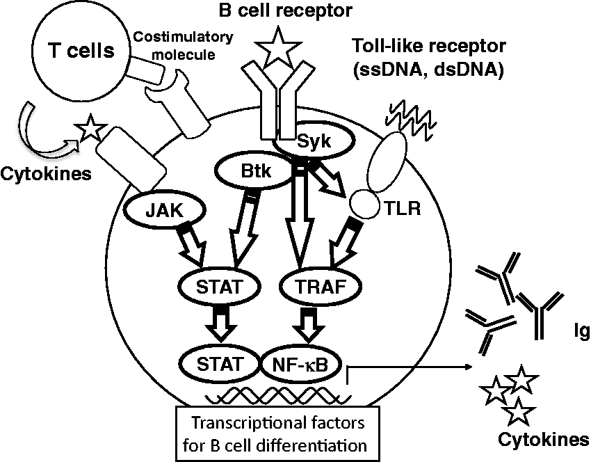
Footnotes
Declaration of conflicting interests
The author(s) declared the following potential conflicts of interest with respect to the research, authorship, and/or publication of this article: Y. Tanaka has received consulting fees, lecture fees, and/or honoraria from Mitsubishi-Tanabe, Eisai, Chugai, Abbott Japan, Astellas, Daiichi-Sankyo, Abbvie, Janssen, Pfizer, Takeda, Astra-Zeneca, Eli Lilly Japan, GlaxoSmithKline, Quintiles, MSD, and Asahi-Kasei, and has received research grants from Bristol-Myers, Mitsubishi-Tanabe, Abbvie, MSD, Chugai, Astellas, and Daiichi-Sankyo. S. Iwata declares no conflicts of interest.
Funding
The author(s) disclosed receipt of the following financial support for the research, authorship, and/or publication of this article: this work was supported in part by Research on Rare and Intractable Diseases and Research Grants-In-Aid for Scientific Research by the Ministry of Health, Labor and Welfare of Japan, the Ministry of Education, Culture, Sports, Science and Technology of Japan, and the University of Occupational and Environmental Health (UOEH), Japan, and a UOEH Grant for Advanced Research.