
Abstract
Objective
Several studies have evaluated the prevalence of rheumatoid factor (RF) and anti-citrullinated proteins antibodies (ACPA) in systemic lupus erythematosus (SLE) patients but no data are available on the anti-carbamylated proteins (anti-CarP), a new biomarker for rheumatoid arthritis (RA). We evaluated the anti-CarP prevalence in SLE patients with joint involvement and the associations with different phenotypes.
Methods
Seventy-eight SLE patients with joint involvement were enrolled (F/M 73/5; mean ± SD age 47.6 ± 11.2 years; mean ± SD disease duration 214.3 ± 115.6 months). As control groups, we evaluated SLE patients without joint manifestations (N = 15), RA (N = 78) and healthy individuals (HS, N = 98). Anti-CarP were assessed by home-made ELISA in all patients and controls, RF and ACPA in SLE patients with joint involvement (commercial ELISA kit).
Results
The prevalence of anti-CarP in SLE patients with joint involvement was similar to RA (p = NS) and significantly higher compared with SLE without joint involvement and HS (p < 0.0001, p < 0.0001, respectively). Four patients were positive for all three antibodies: seventy-five percent of these showed Jaccoud arthropathy. Fourty-five percent of ACPA-ve/RF-ve patients were anti-CarP + ve.
Conclusions
The evaluation of anti-CarP in SLE joint involvement demonstrated a prevalence of almost 50%, similar to RA and significantly higher than SLE without joint involvement and HS.
Introduction
Systemic lupus erythematosus (SLE) is a chronic autoimmune disease characterized by the production of several autoantibodies.1–3
Joint involvement is one of the most common clinical manifestations occurring in up to 90% of SLE patients at any time during the disease course and with different degrees of severity. From a clinical point of view, SLE patients may experience arthralgia, arthritis, and deforming non-erosive arthritis (Jaccoud arthropathy, JA). 4
A number of studies have investigated the presence of rheumatoid arthritis (RA)-related autoantibodies in SLE patients with joint involvement, rheumatoid factor (RF) having a prevalence ranging from 17% to 45.4% and anti-citrullinated proteins antibodies (ACPA) showing a frequency between 4.4% and 27.3%.5–13
More recently, the study conducted by Goddard et al. identified the presence of ACPA in about 7% of SLE patients regardless of joint involvement. 14
Among the several post-translational protein modifications, citrullination is catalyzed by peptidyl arginine deiminase (PAD) inducing the production of citrullinated peptides, whereas carbamylation is a non-enzymatic process consisting of the addition of a cyanate group of self-proteins. 15 Once carbamylated, self-proteins lose their native tertiary structure; this process can generate new epitopes, resulting in the production of anti-carbamylated proteins antibodies (anti-CarP). 15 Anti-CarP antibodies have been found in the sera of RA patients before the onset of symptoms and, interestingly, they can be detected in about 16% of seronegative RA patients. 16 The prognostic value of these autoantibodies was also demonstrated: patients with inflammatory arthralgia and positive for anti-CarP antibodies seem to be at higher risk of developing arthritis than the negative ones. 17 For this reason, anti-CarP antibodies have been proposed as a new biomarker for RA useful for diagnostic and prognostic purposes.
Very few data are available concerning the prevalence of these autoantibodies in SLE patients with joint involvement. In 2016, Pecani et al. identified the presence of anti-CarP in 13.3% of SLE patients, regardless of the presence of articular manifestations and without providing data concerning the association with disease phenotypes. 18 In the same year, Ziegelasch and colleagues analyzed 441 well-characterized SLE patients within two European cohorts, identifying anti-CarP in 9.8% of participants. Moreover, a subgroup of patients was evaluated by imaging methods in order to study erosive damage, showing an association between radiographically detected erosions and anti-CarP antibodies. 19
Moving from this premise, the primary aim of the present study was to evaluate the prevalence of anti-CarP and other RA-related autoantibodies in a cohort of SLE patients with joint involvement. Moreover, we aimed at analyzing the association between anti-CarP antibodies and any laboratory and clinical feature.
Methods
Consecutive SLE patients with a clinical history of joint involvement (arthralgia or arthritis), attending the Lupus Clinic of the Rheumatology Unit, Sapienza University of Rome (“Sapienza Lupus Cohort”), were evaluated. SLE diagnosis was performed according to the revised 1997 American College of Rheumatology (ACR) criteria. 20 Arthralgia was defined as the presence of recurrent (minimum three episodes) or persistent (minimum six weeks) pain or stiffness (lasting at least 30 minutes) of at least one joint during patient’s clinical history. Arthritis was defined as the occurrence of at least one episode of clinical synovitis (swelling, effusion or tenderness) and at least 30 minutes of morning stiffness of at least one joint, eventually confirmed by magnetic resonance imaging or ultrasound. Both arthralgia and arthritis should not have been better explained by any “other disease.”
The study was performed according to the protocol and good clinical practice principles and Declaration of Helsinki statements and was approved by the Ethics committee of the Sapienza University of Rome, Policlinico Umberto I, Rome, Italy. All the patients gave signed consents. The clinical and laboratory data were collected in a standardized computerized electronically filled form, including demographics, past medical history with the date of diagnosis, comorbidities and previous and concomitant treatments.
Patients were divided according to the presence of arthralgia or arthritis. Moreover, patients with arthritis were further divided according to the presence of non-deforming arthritis (NDA) and JA.
Disease activity was assessed by using the SLE Disease Activity Index 2000 (SLEDAI-2K). 21
As control groups, we enrolled 15 SLE patients without joint manifestations, 78 patients affected by RA, according to the ACR 2010 criteria 22 and 98 healthy subjects (HS) age and sex matched.
All SLE patients with joint involvement and RA controls underwent blood draw to detect RF, ACPA, and anti-CarP antibodies. All autoantibody assays were carried out in duplicate, commercial enzyme-linked immunosorbent assay (ELISA) kits were used and the results were evaluated according to the manufacturers’ instructions. ACPA titers were obtained using commercial ELISA kits (Delta Biologicals, Rome, Italy). Values above 25 U/ml were considered positive. A solid-phase ELISA kit was performed in order to determine immunoglobulin (Ig)G, IgA, and IgM isotypes of RF (Diamedix, Miami, FL, USA). Values above 10 U/ml were considered positive.
Anti-CarP antibodies were detected in all participants (cases and controls) by a home-made ELISA using carbamylated fetal calf serum (Ca-FCS) and non-modified FCS as antigens. Ca-FCS was obtained using the method described by Shi et al. 23
In brief, Nunc Maxisorp polystyrene plates (Thermo Scientific, Waltham, MA, USA) were coated overnight at 4℃ with non-modified FCS and Ca-FCS (12 µg/ml in 0.05 M Na2CO3/NaHCO3buffer, pH 9.6). After washing, plates were blocked with phosphate-buffered saline (PBS) containing 1% bovine serum albumin (BSA) (Sigma, St Louis, MO, USA) for six hours at 4℃. Subsequently, the wells were incubated with patient serum diluted 1:50 in PBS/0.05% Tween20 (PBS-T)/BSA 1% overnight at 4℃. Alkaline phosphatase-conjugated anti-human IgG antibodies (Sigma, St Louis, MO, USA) were diluted in PBS-T/BSA 1% (1:1000) and incubated for two hours at room temperature. Paranitrophenyl-phosphate was used as a substrate and the optical density (OD) was measured at a 405 nm wavelength. All assays were performed in duplicate and the absorbance of control wells (non-modified FCS) was subtracted to account for non-specific binding. A titration curve of two positive reference sera with medium to high ELISA immunoreactivity for Ca-FCS was performed to show the performance of the tests and to transform the absorbance of Ca-FCS to arbitrary units per milliliter (aU/ml). The cut-off was established as the mean OD + 3 SD of 56 age- and sex-matched healthy individuals (blood donors) and then the obtained value was converted into aU/ml (corresponding to 340 aU/ml).
Ultrasonographic (US) assessment
A subgroup of SLE patients underwent US assessment. The examination was performed by using a MyLab70 XVision Gold (Esaote, Genova, Italy) machine equipped with a multifrequency linear array transducer (6–18 MHz) with application of Power Doppler (PD, PFR 750 Hz, Doppler frequency 11.1 MHz, gain 50% and low filters). According to the international US guidelines, a systematic bilateral multiplanar grayscale and PD examination was performed at metacarpophalangeal and proximal interphalangeal joints level in order to identify inflammatory features. Specifically, we evaluated the prevalence of patients with US signs of synovitis (synovial effusion and/or hypertrophy and/or PD) in at least one joint.
Moreover, the presence of erosions was assessed and we evaluated the percentage of patients with erosive damage in at least one joint. All abnormalities were defined according to the Outcome Measures in Rheumatology (OMERACT) definitions.24,25
Statistical analysis
We used version 5.0 of the GraphPad statistical package. Normally distributed variables were summarized using the mean ± SD, and non-normally distributed variables by the median and range. Frequencies were expressed by percentage. Wilcoxon’s matched pairs test and paired t-test were performed accordingly. Univariate comparisons between nominal variables were calculated using chi-square test or Fisher’s exact-test where appropriate. Spearman’s test was used to assess the correlations. Two-tailed p values were reported, and p values less than 0.05 were considered significant.
Results
Demographic, clinical and laboratory features of the enrolled SLE patients with joint involvement
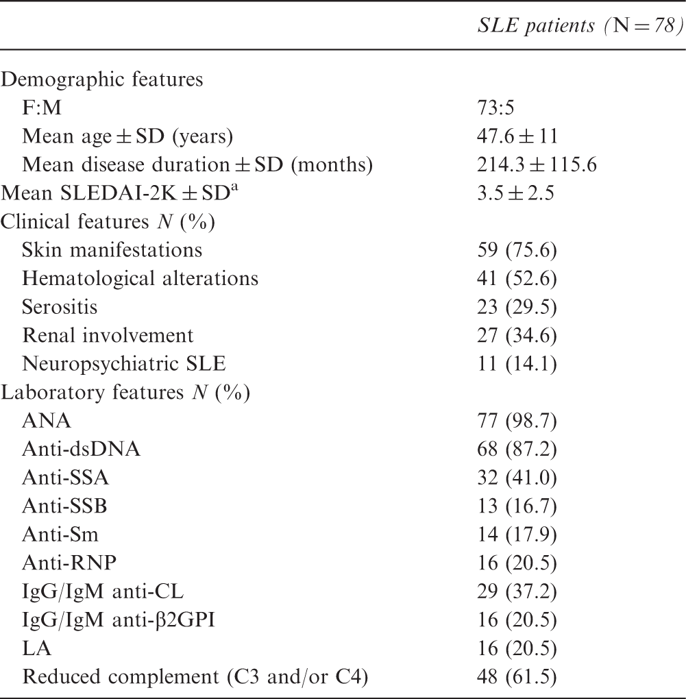
SLE: systemic lupus erythematosus; F: female; M: male; SD: standard deviation; ANA: antinuclear antibodies, CL: cardiolipin; β2GPI: β2 glycoprotein I; LA: lupus anticoagulant; Anti-dsDNA: anti-double-stranded DNA; Ig: immunoglobulin.
At the time of anti-carbamylated protein (anti-CarP) testing.
Seventeen SLE patients (21.8%) experienced arthralgia during their disease history, the remaining 61 arthritis: specifically, non-deforming arthritis (NDA) in 43 patients (70.5%) and JA in 18 (23.1%).
As controls, we enrolled:
– Fifteen SLE patients without joint manifestations (3M/12F), mean age ± SD43.3 ± 11.6 years; mean disease duration ± SD 180.0 ± 84.3 months) – Seventy-eight RA patients (12 M/62 F, mean age ± SD 59.3 ± 12.1 years, mean disease duration ± SD 157.9 ± 108.7 months; treatment: glucocorticoids 33 patients (42.3%), at least one disease-modifying antirheumatic drug (DMARD) 48 (61.5%), tumor necrosis factor (TNF) antagonist drugs 26 (33.3%), biological drugs other than TNF antagonists 17 (21.8%)) – 98 HS (24 M/74 F, mean age ± SD 55.2 ± 11.0 years).
We tested anti-CarP antibodies in all participants (cases and controls). SLE patients with joint involvement showed a prevalence of anti-CarP antibodies similar to RA patients (46.1% versus 39.7%, p = NS) and significantly higher compared with SLE patients without joint involvement (13.3%, p < 0.0001) and HS (2.4%, p < 0.0001). Focusing on SLE patients with joint involvement, 48 (61.5%) patients were positive for RF and 15 (19.2%) for ACPA. In SLE patients, the prevalence of ACPA was significantly lower than RF (p < 0.000001) and anti-CarP antibodies (p = 0.00007). No correlation between SLEDAI-2K values and anti-CarP titers was identified.
In RA controls, we identified the presence of ACPA and RF in 51 (65.4%) and 63 (80.8%) patients, respectively. This prevalence was significantly higher in comparison with SLE patients with joint involvement (p = 0.005 for RF, p < 0.0001 for ACPA).
Moreover, Figure 1 reports the number and the percentage of single-positive, double-positive and triple-positive SLE patients. Eleven patients (14.1%) were negative for all autoantibodies.
Distribution of single-positive, double-positive and triple-positive systemic lupus erythematosus patients.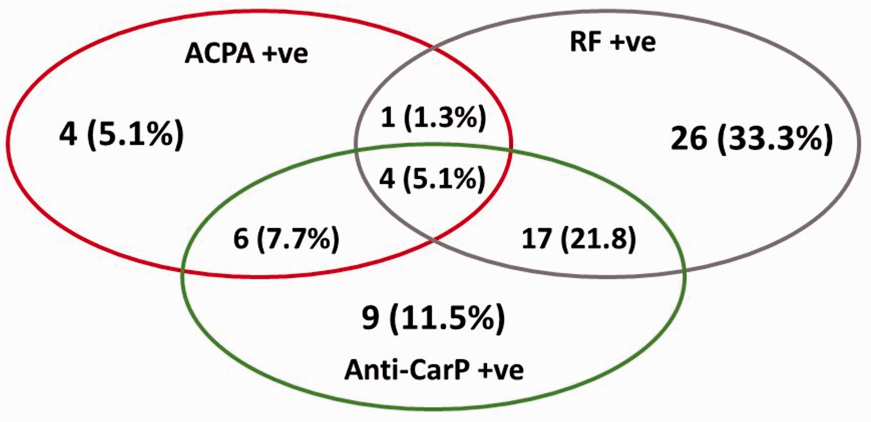
Notably, anti-CarP antibodies were identified in 45% of double-negative (ACPA-ve/RF-ve) patients, with a mean ± SD titer of 963.3 ± 1348.9 AU/ml. Interestingly, when considering the four patients who were triple positive, three of these showed JA. Moreover, the same patients satisfied both classification criteria for SLE and RA, allowing a re-classification as rhupus.17,18
Figure 2 reports data on the distribution of autoantibodies according to the phenotype of joint involvement.
Prevalence of autoantibodies positivity in patients with different joint involvement phenotypes: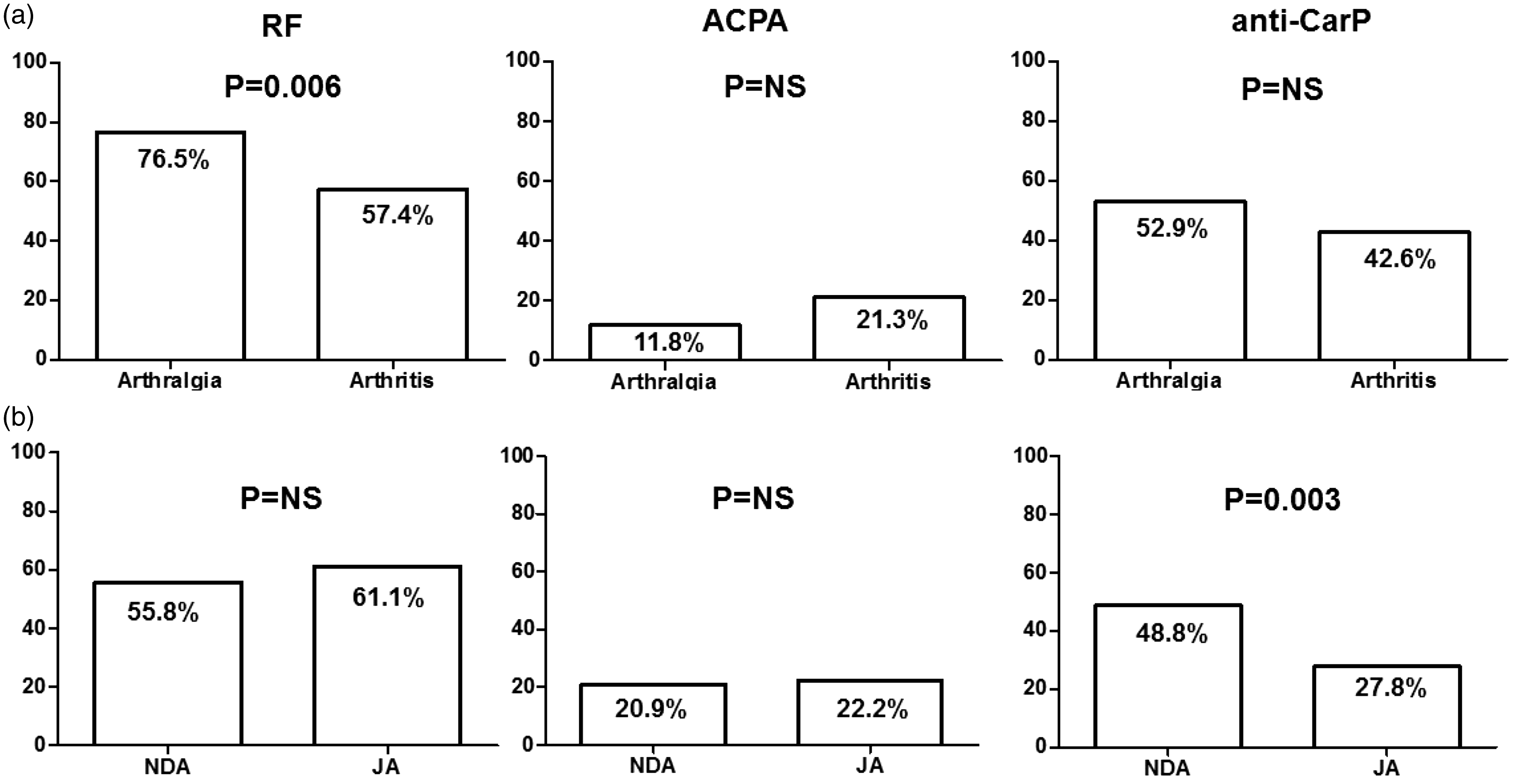
Comparing patients with arthralgia and those with arthritis, RF was significantly more frequent in the first group (76.5% versus 57.4%, p = 0.006; Figure 2(a)), while there were no differences in ACPA or anti-CarP antibodies. Regarding patients with arthritis, anti-CarP antibodies were significantly more frequent in patients with NDA than JA (48.8% versus 27.8%, p = 0.003; Figure 2(b)).
None of the autoantibodies tested (RF, ACPA or anti-CarP) showed any other clinical or laboratory feature than joint involvement.
The US evaluation of a subgroup of 36 SLE patients (M/F 3/33, mean age ± SD 50.7 ± 11.0 years, mean disease duration ± SD 243.3 ± 126.1 months) identified the presence of synovitis in at least one joint in 66.7% of cases. On the other hand, among the patients evaluated by US, 50% of them (18/36 individuals) showed at least one swollen joint, with presence of synovitis in 55.5%.
Moreover, we identified the presence of erosive damage in at least one joint in 15/36 patients (41.7%). The presence of anti-CarP was significantly associated with erosive damage: a total of 63.1% of anti-CarP positive patients showed US-detected erosive damage, compared with 41.2% of anti-Carp negative (p = 0.002). Three out of four triple-positive patients showed erosive damage.
Discussion
In the present study, we evaluated the prevalence of anti-CarP antibodies in SLE patients with joint involvement: almost 50% of the patients resulted positive for anti-CarP antibodies. This prevalence was similar to that identified in RA patients and significantly higher than in SLE patients without joint involvement and HS.
Interestingly, with regard to SLE patients with joint involvement, 45% of double-negative (ACPA-ve/RF-ve) patients were anti-CarP antibody positive.
Joint involvement is a frequent manifestation in SLE patients, leading to disability and affecting quality of life. The majority of patients experience arthralgia or non-erosive arthritis, without structural damage. However, a subset of patients may develop more severe joint involvement, with the appearance of deformities and erosive damage. 4 To date, no specific biomarker has proved useful to identify SLE patients at risk of developing articular phenotypes that are more aggressive. Moreover, within SLE-associated autoantibodies, only anti-La/SSB has been linked with the development of joint involvement, as demonstrated by data from the large cohort published by Cervera and colleagues in 1993. 26 Since several similarities exist between the musculoskeletal manifestations of SLE and RA patients, recent research has aimed at analyzing the role of RA-related autoantibodies in SLE patients. RF, despite a prevalence up to 45% of SLE patients, does not seem associated with joint involvement in SLE.5–13 Interestingly, in the present cohort, a high frequency of RF was identified in patients with arthralgia, significantly higher compared with other RA-related autoantibodies.
Conversely, ACPA have gained great interest since these antibodies can be found in SLE patients with joint involvement with a prevalence ranging from 4.4% to 27.3%.5–13 The identification of higher titers and prevalence, ranging from 20% to 60%, in the subset of SLE patients with radiographically detected erosive arthritis, suggested their possible role as a biomarker of a more aggressive phenotype.5–13 The meta-analysis conducted by Budhram and colleagues in 2014, evaluating SLE patients with erosive arthritis, identified a good specificity (91.8%) but a low sensitivity (47.8%) of ACPA. 27 It is possible to speculate that SLE patients with erosive arthritis could be distinguished in two subpopulations: those ACPA + ve sharing RA-like pathogenic pathways, and the ACPA-ve ones, in which other pathogenic mechanisms may be suspected.
In this view, anti-CarP antibodies may play a role in this latter condition. It is noteworthy to observe that 30%–40% of ACPA-ve RA patients present with erosive damage. Anti-CarP antibodies have been detected in both ACPA-negative and ACPA-positive RA patients; however, a higher prevalence was observed in ACPA-ve. In our cohort, 45% of SLE patients with joint involvement, who were ACPA-ve/RF-ve, were anti-CarP positive. This percentage is higher compared with seronegative RA patients, in whom anti-CarP antibodies have been found in about 16% of cases. 16 This result could suggest the possible role of these antibodies as biomarkers in ACPA-ve/RF-ve SLE patients.
Carbamylation is a novel molecular pathway that determines the immunological tolerance break with the generation of new epitopes, inducing the production of autoantibodies. 15 This non-enzymatic post-translational modification consists of the addition of a cyanate group on self-proteins, determining the change of polymerization ability, sensitivity to proteinases, and avidity of antibody-antigen binding. 28
Anti-CarP antibodies do not seem to be associated with any specific genetic (i.e. human leukocyte antigen (HLA) shared epitope) or environmental factor (i.e. smoking) that, instead, seems detrimental in the development of ACPA.29,30 This may suggest that the development of anti-CarP antibodies could follow distinct pathogenic pathways. 21
Shi et al. studied a population of healthy individuals with arthralgia in whom the presence of anti-CarP antibodies seemed associated with a higher risk of developing RA, independently of ACPA positivity. 23 The evaluation of healthy first-degree relatives of RA patients identified a prevalence of anti-CarP ranging from 9.2% to 18.3%.31,32
Furthermore, anti-CarP antibodies have also been detected in other than-RA inflammatory conditions. Chimenti and colleagues found higher levels and prevalence of anti-CarP antibodies in psoriatic arthritis patients compared with healthy individuals. 33 More recently, the study conducted by Bergum et al. on 78 patients affected by Sjögren’s syndrome demonstrated a frequency of 27% of anti-CarP, which was associated with higher focus scores of salivary gland biopsy. 34 No studies have been performed on anti-CarP antibodies in SLE so far. Scinocca et al. evaluated anti-homocitrullinated fibrinogen antibodies, which are generated through an alternative carbamylation pathway. 35 Indeed, homocitrulline is generated by carbamylation reaction of cyanate with the primary aminic group of lysine residues. 24 Of 37 SLE patients, two patients (5%) were weakly positive for these autoantibodies, with a prevalence significantly lower compared with RA and similar to healthy individuals. 29 This result may suggest that homocitrullination is not a key pathway in SLE.
More recently, two studies provided data about the prevalence of anti-CarP in SLE patients showing a lower frequency of these antibodies in comparison with our results.18,19 In particular, we identified a prevalence higher than 40% in comparison with 13.3% and 9.8% observed in the studies conducted by Pecani and Ziegelasch and colleagues in 2016, respectively.18,19 We hypothesize that this discrepancy could be due to the differences in the studied cohorts. In the previous studies, the authors evaluated the anti-CarP prevalence irrespective of the presence of joint involvement; conversely, in our study we selected only patients with this specific manifestation.
Furthermore, in the analysis conducted by Ziegelasch et al., the presence of anti-CarP was associated with radiographically detected erosions, but not with clinically detected arthritis. 19
Our results may suggest that anti-CarP antibodies may play a role in the development of specific clinical features of SLE. The mechanism underlying the development of anti-CarP antibodies in SLE patients has not been identified. As known, carbamylation develops at the tissue level during an inflammatory status when myeloperoxidase (MPO) is released from neutrophils. 36 These cells play an important role in SLE pathogenesis: Their specific kind of death, called NETosis, is a potential source of autoantigens, with the release of several molecules, such as MPO. 37 To summarize, we could speculate that MPO, deriving from NETosis, could locally increase carbamylation, leading to the break of immune tolerance and to the production of antibodies directed against carbamylated proteins.
Another interesting result from our study is that anti-CarP autoantibodies may be able to discriminate patients with NDA from JA patients. Indeed, a higher number of SLE patients with NDA present with anti-CarP antibodies. This could indicate distinct pathogenic mechanisms associated with the development of different phenotypes.
Finally, by the US assessment of a subgroup of SLE patients, we identified a significant association between anti-CarP and erosive damage. Undoubtedly, this result should be confirmed in larger SLE populations.
In conclusion, to the best of our knowledge, this is the first study focusing on the prevalence of anti-CarP in a cohort of patients affected by SLE with joint involvement, identifying a prevalence higher than 40%.
Certainly, a possible limitation of our analysis is the small size of the control group constituted by SLE patients without joint involvement. However, the prevalence of this specific manifestation is so high, making the selection of this subgroup difficult.
Notably, these autoantibodies seem to be associated with NDA. Moreover, anti-CarP antibodies were present in almost half of ACPA-ve/RF-ve patients, even higher than in seronegative RA. Further investigations on larger cohorts of patients are necessary to clarify the possible role of anti-CarP antibodies as biomarkers of SLE joint involvement.
Footnotes
Declaration of conflicting interests
The authors declared no potential conflicts of interest with respect to the research, authorship, and/or publication of this article.
Funding
The authors received no financial support for the research, authorship, and/or publication of this article.