
Abstract
Platelet activation and decrease in platelet count characterize the development of the most feared form of antiphospholipid syndrome (APS), i.e. catastrophic APS (CAPS). We aimed to assess if immuno-affinity purified anti-β2-glycoprotein I (aβ2GPI) antibodies enhance platelet activation inducing a significant flow obstruction in a platelet function analyzer (PFA). Affinity purified aβ2GPI antibodies were obtained from 13 triple positive patients with a strong lupus anticoagulant (LA) and high titers of IgG anticardiolipin antibodies (aCL) and IgG aβ2GPI. Platelet activation stimulated by adenosine diphosphate (ADP) in the presence or absence of aβ2GPI was measured by the expression of P-selectin on platelet surface using flow cytometry. P-selectin expression remained close to baseline when normal whole blood was incubated with aβ2GPI alone. When stimulated using aβ2GPI combined with ADP, P-selectin expression (28.42 ± 5.15% vs. 20.98 ± 3.94%, p = 0.0076) was significantly higher than ADP alone. Closure time of normal whole blood passed through the PFA was significantly shorter using affinity purified aβ2GPI than control IgG both in Col/ADP (160.1 ± 62.1 s vs. 218.6 ± 43.8 s; p = 0.021) and Col/EPI cartridges (149.5 ± 26.7 s vs. 186.9 ± 45.5 s; p = 0.030). Thus, platelet activation is enhanced by aβ2GPI antibodies with a consequent premature closure in a PFA, possibly resembling that in microcirculation in patients with CAPS.
Keywords
Introduction
Antiphospholipid syndrome (APS) is a systemic autoimmune disease characterized by the presence of at least one clinical event among vascular thrombosis and/or pregnancy morbidity and the presence of persistently elevated levels of circulating antiphospholipid antibodies (aPL), such as anticardiolipin antibodies (aCL), anti-β2-glycoprotein I (aβ2GPI), and the lupus anticoagulant (LA). 1 These antibodies and platelets are considered the key players in the pathogenesis of arterial thrombosis. 2 Anti-β2-GPI auto-antibodies enhance platelet activation, as evidenced by some authors monitoring calcium mobilization and platelet thrombus formation. 3 It has been shown in vivo that the aβ2GPI antibody/β2GPI complex is targeted at the platelet thrombus. 3 During an infection disease platelet can be activated and an infection disease frequently precedes the development of the most feared form of APS, i.e. catastrophic APS (CAPS).4,5
Our group has shown that aβ2GPI antibodies isolated during the catastrophic phase induce higher platelet activation than those isolated during the quiescent phase of APS. 6 During the acute phase of the disease patients develops thrombocytopenia indicating a direct involvement of platelets in the thrombotic microangiopathy. 7
The aim of this study was twofold: first to evaluate platelet activation by means of surface P-selectin expression after in vitro stimulation with adenosine diphosphate (ADP) in the presence of immuno-affinity purified aβ2GPI antibodies, and to assess if activated platelets induce a significant flow obstruction measured by a platelet function analyzer (PFA).
Materials and methods
Blood samples
Whole blood samples from 13 healthy volunteers were collected with minimal stasis using a 21 gauge needle into 129 mM sodium citrate polypropylene tubes, with the first 3–4 mL of blood drawn being discarded. 8 None of volunteers had known platelet disorders nor took aspirin or other antiplatelet medications in the previous 10 days. Blood was maintained at room temperature and analyzed within 3 h. 9 Patients were regularly followed up with the determination of aPL. They gave their written consent for the use of their plasma in this study.
Patients’ aβ2GPI affinity purification
aβ2GPI antibodies were purified from 13 triple positive (LA+, aCL+, aβ2GPI+) APS patients by affinity chromatography. 10 Briefly, plasma was loaded onto a prepacked polypropylene column (HiTrap NHS-activated, GE Healthcare, Stockholm, Sweden) that had been coupled with purified human β2GPI, according to the manufacturer’s instructions. After extensive washing with phosphate-buffered saline (PBS), aβ2GPI antibodies were eluted from the column using 0.1 mol/L glycine, 0.5 mol/L NaCl, pH 2.8, and immediately buffered with 1 mol/L Tris, pH 8.4. The purity of the resulting aβ2GPI antibody preparations was checked by sodium dodecyl sulfate–polyacrylamide gel electrophoresis, and the protein concentration was measured by optical density at 280 nm. The antigenic specificity of eluted antibodies was determined using ELISA. 11
Platelet activation evaluated by flow cytometry
Platelet activation stimulated by ADP in the presence or absence of aβ2GPI antibodies was measured by the expression of P-selectin on platelet surface through flow cytometry. Briefly, within 2 h after blood sampling, whole blood from seven healthy donors was incubated with aβ2GPI antibodies from 10 APS patients (final concentration 18 µg/mL) or control IgG from two healthy donors for 15 min at room temperature, and then stimulated with ADP (final concentration 20 µM) for another 15 min. A setup experiment was made with a FS-log/SS-log plot and two fluorochrome combinations (PE/APC). FS and SS were set to log-scale for detection of small particles. Settings were checked with proper fluorochrome and isotype matched controls. P-selectin positivity was calculated as percentage of both CD41a(PE) and CD62p(APC) population. Samples were acquired by CyFlow Space Partec flow cytometry and analyzed by Partec FloMax™ software; 20,000 events in platelet gate were acquired with a slow flow rate.
Five microliters of mixture were diluted in 45 µL HBS buffer (0.01 M Hepes, pH 7.4, 0.14 M NaCl, and 2.5 mM CaCl2 solution), and incubated with titered antibodies CD41a (PE) and CD62p (APC) for 15 min in the dark. One milliliter of HBS buffer was added before acquisition.
Platelet function analyzer
A PFA (PFA-200, Siemens AG, Munich, Germany) was used to test platelet function in the environmental milieu of whole blood. A small volume of blood was aspirated at high shear (5000–6000 s−1) through an aperture (150 µm in diameter) in a membrane coated with collagen and ADP (Col/ADP) or collagen and epinephrine (Col/EPI). The time required to obtain full occlusion of the aperture is reported as “closure time” (CT). 12
We analyzed platelet activation in 13 normal whole blood incubated for 15 min at room temperature with affinity purified aβ2GPI from 13 triple positive patients at 18 µg/mL final concentration. As a control, normal IgG at the same concentration from 13 normal subjects was used.
Statistical analysis
All results were expressed as mean ± SD and were analyzed using computer software (GraphPad, Prism 7, La Jolla, CA, USA). Data distribution was evaluated by D’Agostino–Pearson test, and Mann–Whitney test was used for non-parametric statistics. A p-value ≤ 0.05 was considered statistically significant.
Results
Clinical and laboratory characteristics of triple positive APS patients
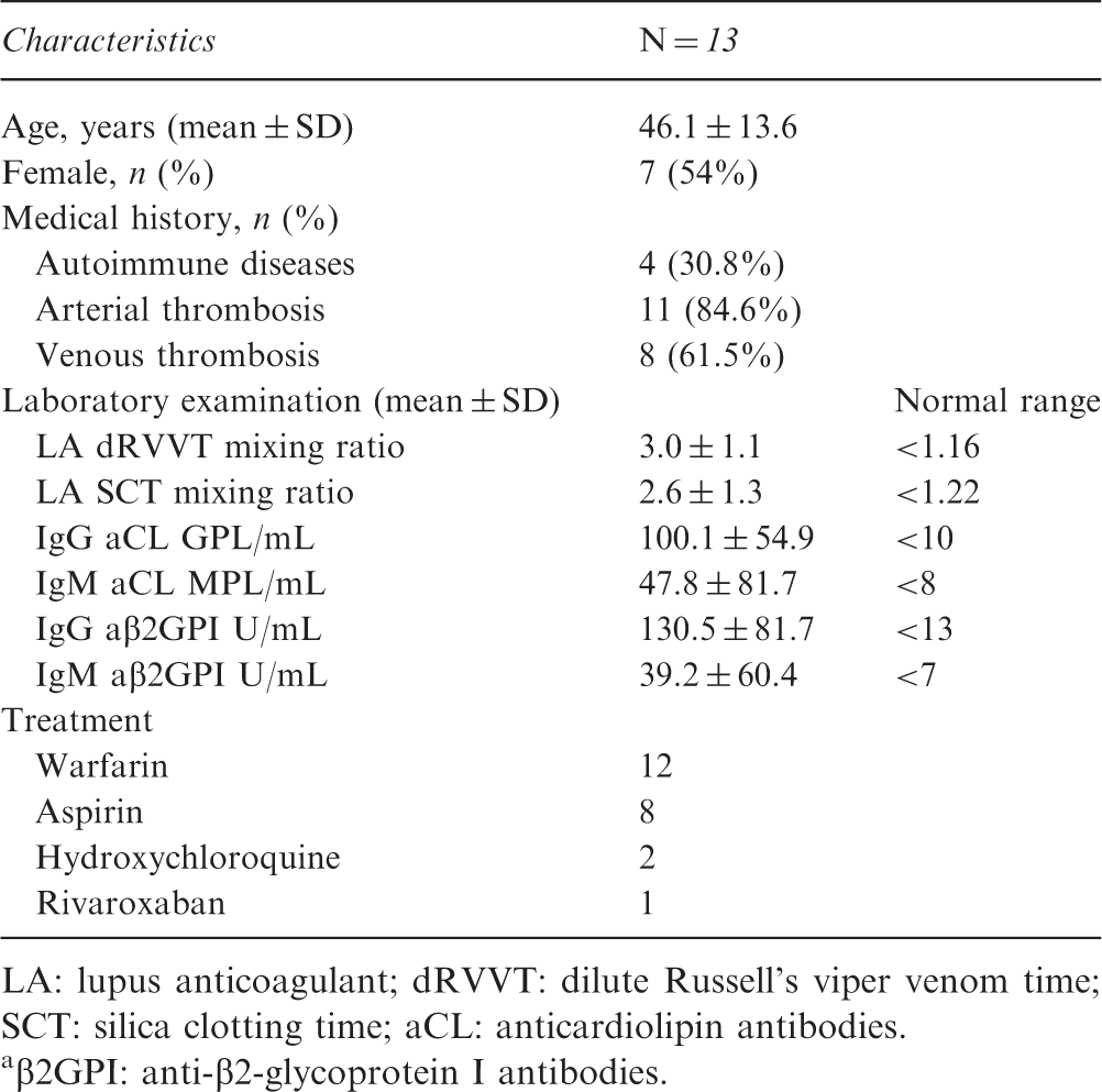
LA: lupus anticoagulant; dRVVT: dilute Russell’s viper venom time; SCT: silica clotting time; aCL: anticardiolipin antibodies.
β2GPI: anti-β2-glycoprotein I antibodies.
Characteristics of affinity purified materials
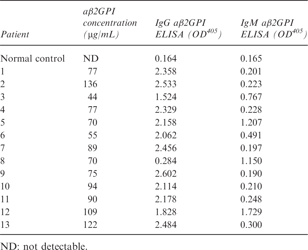
ND: not detectable.
Platelet activation by aβ2GPI antibodies
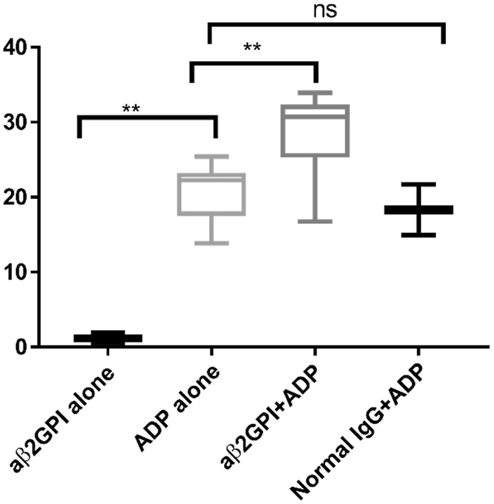
In Figure 1(a) and (b), a representative experiment showing platelet detection and identification by anti CD41a is reported. CD41a and CD62 (P-selectin) positive platelets upon stimulation with aβ2GPI antibodies, ADP, and ADP plus aβ2GPI antibodies are shown in Figure 1(c) to (e). P-selectin expression was set at around 1% at baseline for resting platelets. As shown in Figure 2, after incubation with affinity purified aβ2GPI from triple positive patients, P-selectin expression remained close to baseline (1.13 ± 0.92% vs. 1.19 ± 0.74%, p = 0.66). After stimulation of normal whole blood with ADP, P-selectin expression increased to 20.98 ± 3.94% (p < 0.0001). When whole blood was stimulated with combined ADP and aβ2GPI, P-selectin expression (28.42 ± 5.15%, p = 0.0076) was significantly higher than ADP alone, while normal IgG with ADP (18.32 ± 4.75%, p > 0.05) showed no statistical difference compared to only ADP.
P-selectin expression on platelets surface. Typical experiment using set of flow cytometry. (a) FS-log/SS-log plot set to distinguish the platelet cloud; (b) gate from (a), fluorochrome range to determine the CD41a-PE (FL2) positive platelet population; (c) gate from (b), both CD41a-PE and CD62p-APC (FL3) plot, platelets pre-incubated only with aβ2GPI; (d) gate from (b), both CD41a-PE and CD62p-APC plot, platelets pre-incubated only with ADP (20 µM); (e) gate from (b), both CD41a-PE and CD62p-APC plot, platelets pre-incubated with aβ2GPI and ADP. Comparison of P-selectin expression after pre-incubation with aβ2GPI and/or ADP (20 µM).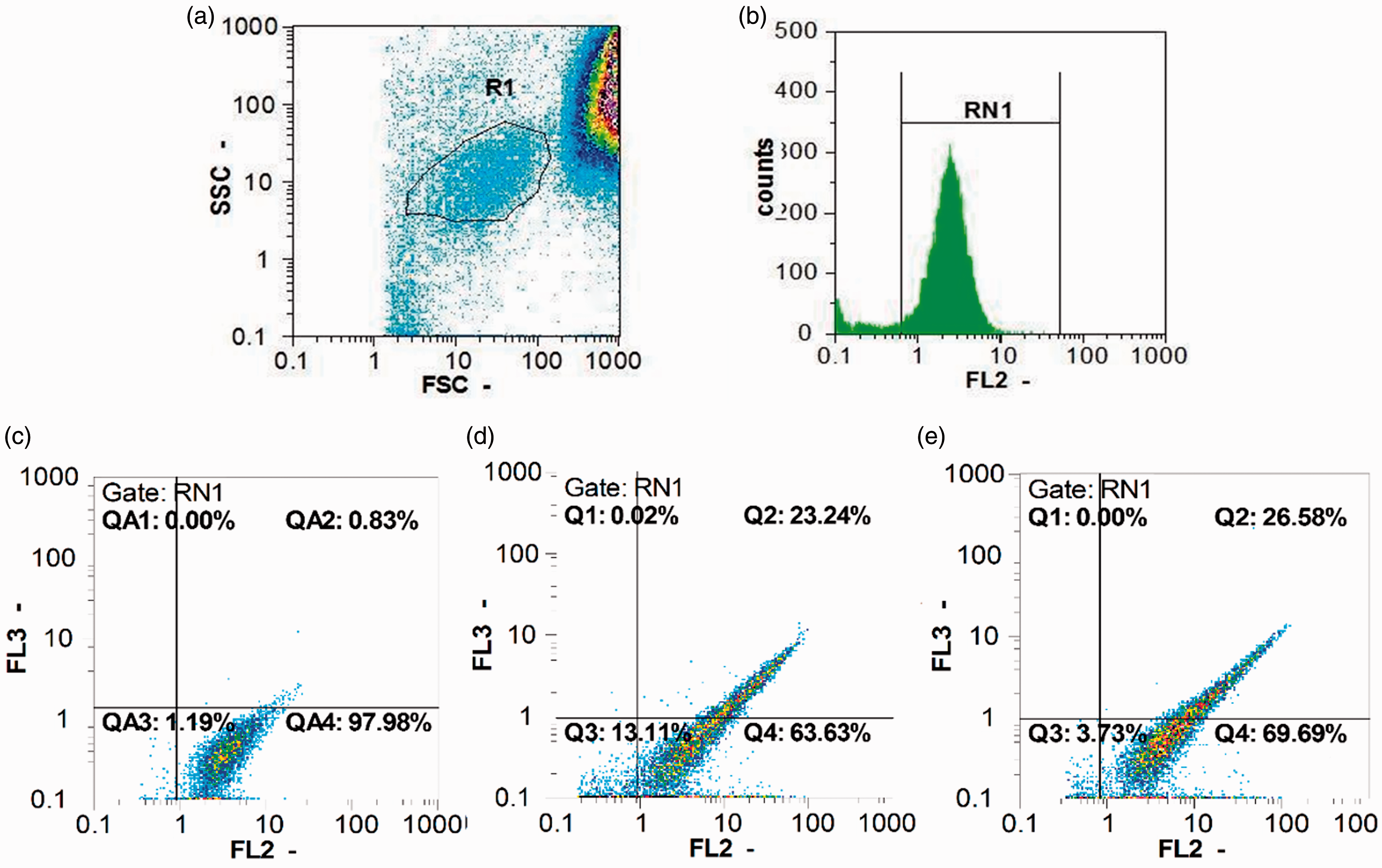
Platelet function analyzer
Eleven of 13 affinity purified aβ2GPI preparations determined a shortened CT using the Col/ADP cartridges while in the remaining two preparations the CT was similar to that of control IgG. Four of 13 preparations determined a premature occlusion of COL/EPI cartridges, seven reduced the CT, while in the remaining two the CT was close to that of normal control. Overall, the 13 affinity purified aβ2GPI from triple positive APS patients compared to control IgG from normal subjects significantly reduced the CT. This reduction was significantly both using Col/ADP cartridge (149.5 ± 26.7 s vs. 186.9 ± 45.5 s, respectively; p = 0.021) and Col/EPI cartridges (160.1 ± 62.1 s vs 218.6 ± 43.8 s, respectively; p = 0.030) (Figure 3).
PFA200 detected closure time using normal whole blood pre-incubation with control IgG (100 µg/mL) or affinity purified aβ2GPI antibodies.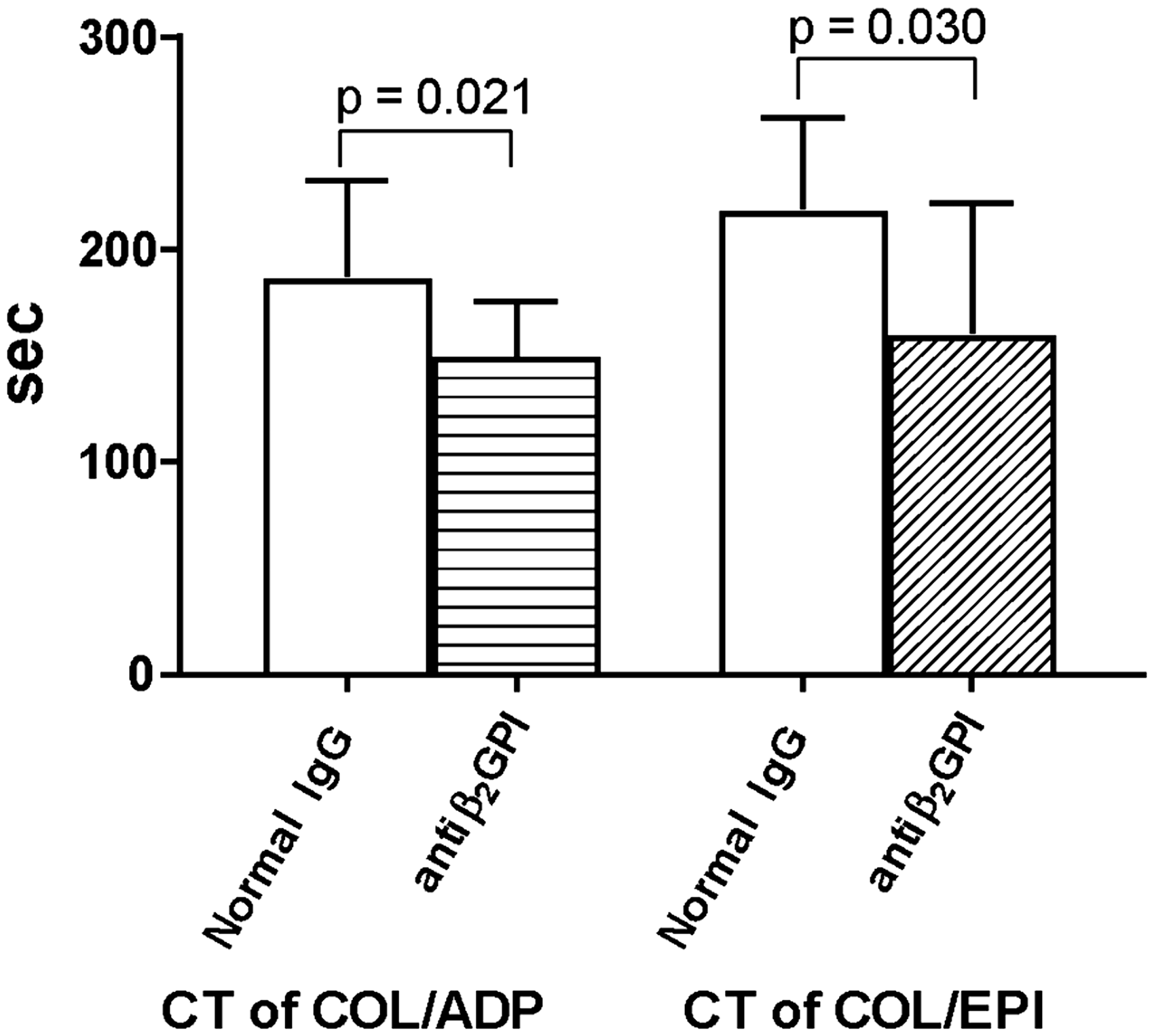
Discussion
Platelet activation in primary APS was previously reported by showing increased urinary excretion of thromboxane metabolites,13,14 accelerated spontaneous platelet aggregation, 15 higher levels of platelet CD62p, 16 increased circulating platelet-leukocyte complexes and platelet activation, 6 and an increased number of platelet microparticles. 17 In the first part of our study, we have shown that platelets incubated with affinity purified aβ2GPI antibodies do not promote platelet activation. Conversely, exposure of P-selectin significantly increased when platelets were stimulated with ADP in the presence of affinity purified antibodies. Thus, as previously suggested, antibodies can bind to phospholipids in platelet membranes but perturbation of the membrane must first occur. 18 The mechanism of antibody binding upon platelet activation may reside on the generation of PF4 tetramers that bind two β2GPI molecules simultaneously allowing their dimerization and recognition by aβ2GPI antibodies. 19 As a confirmation of our experiments, Espinola et al. proposed that the prothrombotic properties of aPL may be explained in part by their ability to enhance the activation of platelets pretreated with low doses of ADP or thrombin receptor agonist peptide. 20 In vivo platelet perturbation induced by infection may determine a massive platelet perturbation that might precede the catastrophic phase of the disease. However, higher platelet P-selectin expression can also be induced by IgG anti β2GP1 themselves when purified during the catastrophic as compared to those purified during quiescent phase. 21 Therefore, different circulating aβ2GPI antibodies might be present in different phases of the disease. One explanation could be a trigger inducing a reshuffle of β2GPI epitopes during catastrophic phase of APS and a consequent change in antibody specificity.
In the second part of experiments, we tested the ability of platelet plus aβ2GPI antibodies to adhere and aggregate in condition of shear stress. We presumed that the PFA might resemble the blood flow of human small arterial vessels. In fact, citrated whole blood is pushed at a high shear stress rate through a capillary that has at its end a collagen-coated membrane, in which a defined microscopic aperture (147 µm) is present, filled with either ADP or EPI. Arterioles as vessels on the arterial side of the microcirculation are 10–100 µm in diameter and the shear rates experienced by platelets in small arteries (approx. 2000 s−1) may sharply increase in diseased arteries.22,23 aPL promote leukocyte–endothelial cell adhesion and endothelium perturbation antagonizing eNOS via β2GPI and its receptor apoER2, 24 and this my increase the shear rate of a normal vessel. The PFA employed in this study aspirates blood samples at a shear around 5000–6000 s−1, in the presence of Col/ADP and Col/EPI thus possibly mimicking an in vivo situation. The shortened closure time in PFA 200 caused by co-incubation of aβ2GPI auto-antibodies with normal whole blood is in line with the hypothesis that a similar pathogenic mechanism could occur in vivo ending into the catastrophic phase of the disease. A massive platelet consumption/deposition in the microcirculation with consequent organ failure determines a sharp drop of platelet count. 7 Moreover, a decreased β2GPI concentration observed during the catastrophic phase of the disease indicates that platelets, β2GPI. and aβ2GPI antibodies are major players in this thrombotic microangiopathy. 25 Although this was a conditional recommendation due to very low certainty of evidence, the recently published practice guideline on diagnosis and management of the CAPS acknowledges the role of platelets and suggests using antiplatelet agents as add-on therapy of first-line treatment. 26 In conclusion, this study shows that in vitro pre-incubation of normal whole blood with aβ2GPI antibodies from APS patients significantly increase platelet activation by ADP. Moreover, platelets activation in condition of shear stress in the presence of aβ2GPI antibodies realized a premature occlusion of the aperture in a PFA, possibly reproducing the small arteries occlusion in CAPS patients.
Footnotes
Acknowledgments
Authors would like to thank all patients and blood donors participating in this work.
Declaration of conflicting interests
The authors declared no potential conflicts of interest with respect to the research, authorship, and/or publication of this article.
Funding
The authors received no financial support for the research, authorship, and/or publication of this article.