
Abstract
Objective
To describe the prevalence, clinical manifestations, histopathological features, and antibody profile associated to skin involvement in patients with catastrophic antiphospholipid syndrome (CAPS).
Methods
We performed a cross-sectional study of the patients included in the “CAPS Registry,” a registry developed by the European Forum on Antiphospholipid Antibodies (aPL)1,2 This database contains data from patients with CAPS collected from April 1992 to December 2024. Demographic clinical manifestations, laboratory features (including aPL antibodies), pathological findings in involved skin, and outcome were retrieved.
Results
Cutaneous involvement was described in 377 episodes (47%) from 361 patients. A female sex prevalence of 49.4% was observed. The average age was 39.4 (SD ± 17.9) years. The more frequent skin manifestations observed were livedo reticularis (17.6%), skin necrosis (13%), ischemic ulcers (10.4%), skin ischemic (9.7%), skin purpura (7.1%), gangrene (5%), splinter haemorrhages (2.6%), and Raynaud’s phenomenon (1.5%). The most frequently affected organs in CAPS episodes with skin involvement were kidneys (72%), lungs (61%), central nervous system (54.5%), and heart (54.4%). Thrombocytopenia and hemolysis features were more common in patients with skin involvement (p < 0.05 for all). Thrombotic microangiopathy was the predominant pathological finding, present in 82 (68.3%) episodes. No difference regarding mortality were found in episodes with or without skin involvement (29.3% vs 32.1%, p = 0.42).
Conclusion
The presence of skin involvement in patients with CAPS is frequent. The most frequent manifestations were livedo reticularis and skin necrosis. Skin involvement does not seem to be associated to mortality.
Introduction
Catastrophic antiphospholipid syndrome (CAPS) is a severe form of APS, characterized by widespread thrombosis and multiorgan failure that develops within a week and affects 1% of APS patients,1,2 yet it carries a high mortality rate if not promptly diagnosed and treated. 3
Among the various organs affected in CAPS, the skin is frequently involved. The prevalence of cutaneous involvement in CAPS Registry was 43.1%. 4 These manifestations can be key for an early diagnosis, as skin lesions may provide the first clinical signs of the underlying systemic disease.5,6 Skin involvement in CAPS includes livedo reticularis, purpura, skin necrosis, and gangrene, as well as more specific findings such as ischemic ulcers and Raynaud’s phenomenon.7,8 Although skin involvement is often easily recognized, the underlying histopathological mechanisms remain incompletely understood. 4
A retrospective analysis of French APS/Systemic Lupus Erythematosus (SLE) registry showed that half of CAPS patients exhibited cutaneous manifestations, with a broad spectrum of clinical presentations. 8
The aim of this study is to assess the prevalence, clinical manifestations, histopathological features, and antibody profiles related to skin involvement in patients with CAPS.
Methods
We performed a cross-sectional study of the patients included in the “CAPS Registry,” a registry developed by the European Forum on Antiphospholipid Antibodies (aPL).1,2 This database contains data from patients with CAPS collected from April 1992 to December 2024.
Patients in the “CAPS Registry” were reported to the registry coordinators or published in the medical literature. Data standardization was achieved through the utilization of anonymous forms completed and submitted by the attending physicians. Published case reports are identified through periodic systematic reviews of PubMed. All patients met the current classification criteria of CAPS. The “CAPS Registry” project obtained approval from the ethical committee for clinical research at Hospital Clinic, Barcelona.
Patients with clinical, or pathological evidence of skin involvement were compared with patients without skin involvement. Demographic variables such as gender and age, underlying SLE were recorded. Additionally, laboratory features, lupus anticoagulant (LA), IgG or IgM of anticardiolipin antibodies (aCL) and IgG or IgM anti-ß2-glycoprotein I antibodies (aß2GPI) presence were also registered. Stratification according to single, double, and triple positivity was performed only in patients with complete aPL data.
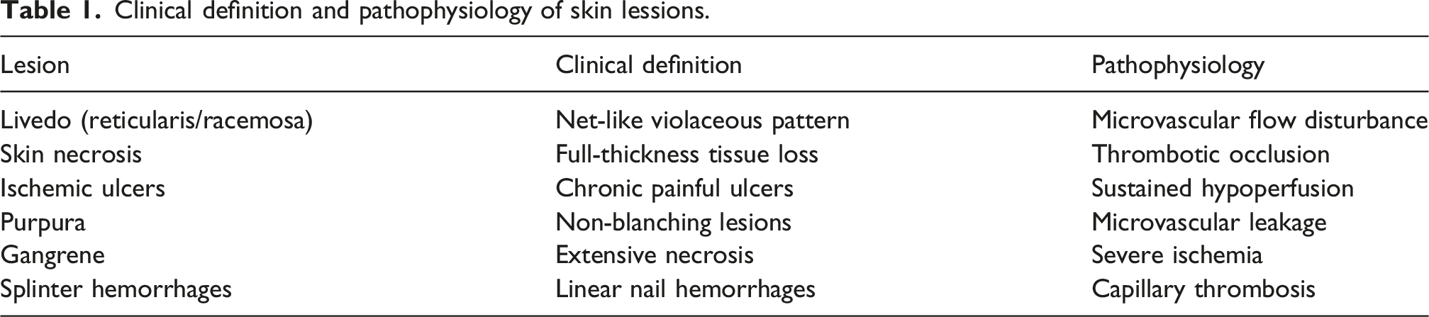
Clinical definition and pathophysiology of skin lessions.
Continuous variables were expressed as mean ± standard deviation (SD) and 95% confidence intervals (95% CI), while categorical variables were presented as frequencies and percentages. Differences between groups for continuous variables were assessed using the Student’s t-test. The chi-square test was used to compare categorical variables, and Fisher’s exact test was applied when appropriate. Variables that showed statistical significance in the univariate analysis were subsequently included in a multivariate analysis.
A p value <0.05 was considered statistically significant. A stepwise logistic regression was employed to ascertain the variables retained in the final model. Variables included in the multivariate model were selected based on clinical relevance and a p-value <0.10 in univariate analysis. Collinearity between variables was assessed using correlation analysis. In cases of high correlation, only one variable was retained based on clinical interpretability to avoid redundancy in the model. All statistical tests were two-tailed; only associations with p < 0.05 were considered statistically significant. The statistical analysis was performed with IBM-SPSS v.22 statistical software.
Results
General characteristics
Up to December 2024, the CAPS Registry included 854 patients with 875 episodes of CAPS. However, 90 episodes presented missing data in at least one relevant variable, reflecting incomplete documentation inherent to retrospective data collection. These episodes were excluded from the statistical analyses.
Finally, 756 patients with 785 episodes of CAPS were included.
Demographic characteristics, organ involvement, and laboratory manifestations in patients with catastrophic antiphospholipid syndrome with and without skin involvement.
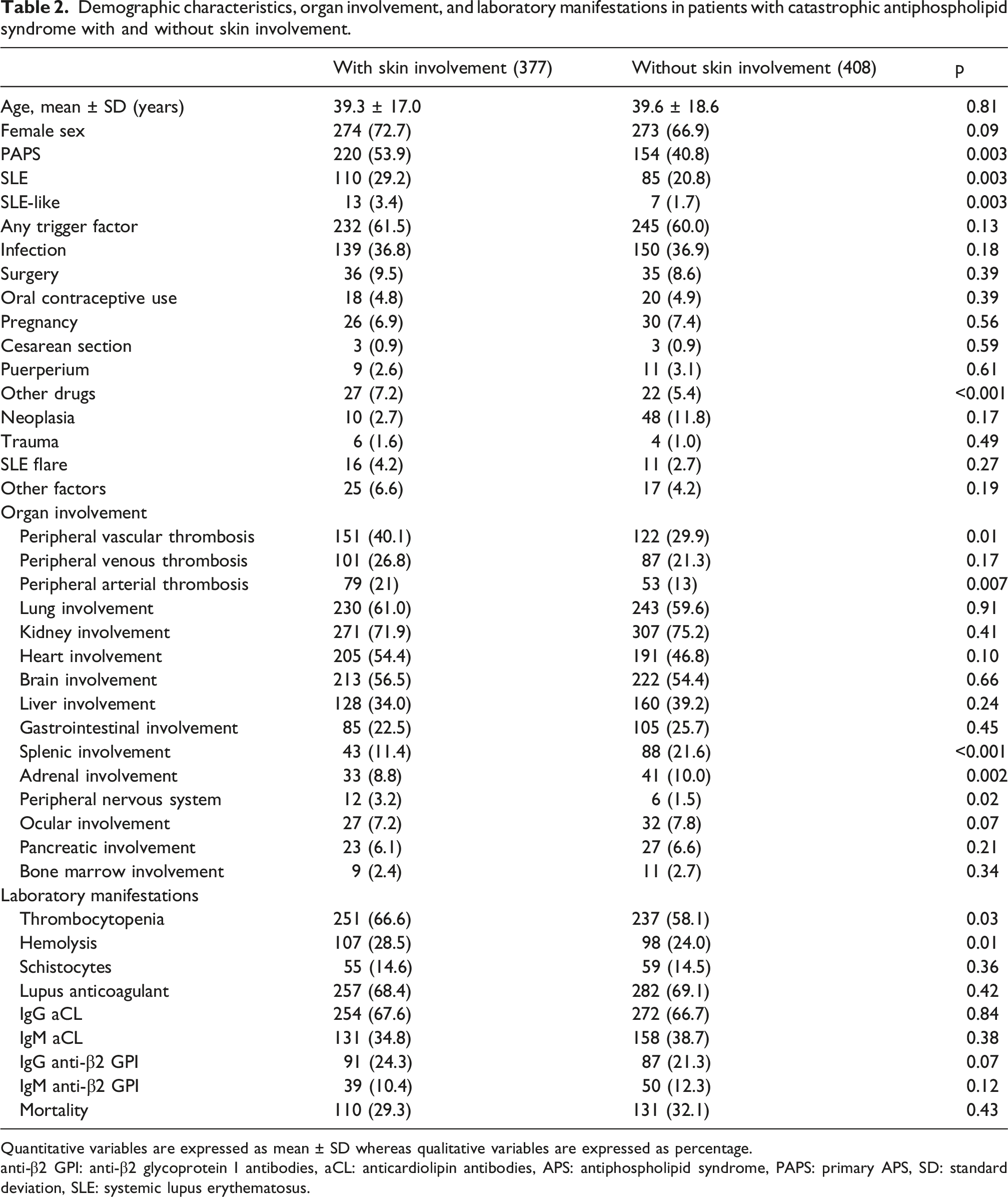
Quantitative variables are expressed as mean ± SD whereas qualitative variables are expressed as percentage.
anti-β2 GPI: anti-β2 glycoprotein I antibodies, aCL: anticardiolipin antibodies, APS: antiphospholipid syndrome, PAPS: primary APS, SD: standard deviation, SLE: systemic lupus erythematosus.
The average age was 39.3 (SD ± 17.0) years. One hundred and 10 patients (29.2%) had associated SLE, and 154 (40.8%) had primary APS (PAPS).
The most frequently affected organs in CAPS episodes with skin involvement were kidneys (71.9%), followed by lungs (61%), central nervous system (54.5%), and heart (54.4%) (Table 2).
Clinical manifestations
The more frequent skin manifestation was livedo (reticularis or racemosa) present in 17.6% of episodes of CAPS with skin involvement, followed by skin necrosis in 13% (Image 1), ischemic ulcers in 10.4%, skin ischemic in 9.7%, skin purpura in 7.1%, gangrene in 5%, and splinter haemorrhages in 2.6%. In 34.6% of episodes with skin involvement, the specific type of lesion was not detailed. Extensive cutaneous necrosis associated with thrombotic microangiopathy in catastrophic antiphospholipid syndrome. Large, sharply demarcated violaceous-to-black necrotic plaque with a surrounding erythematous rim on the lateral thigh. The lesions demonstrate full-thickness skin necrosis, reflecting severe ischemic injury secondary to microvascular thrombosis. This pattern represents an advanced stage of cutaneous involvement.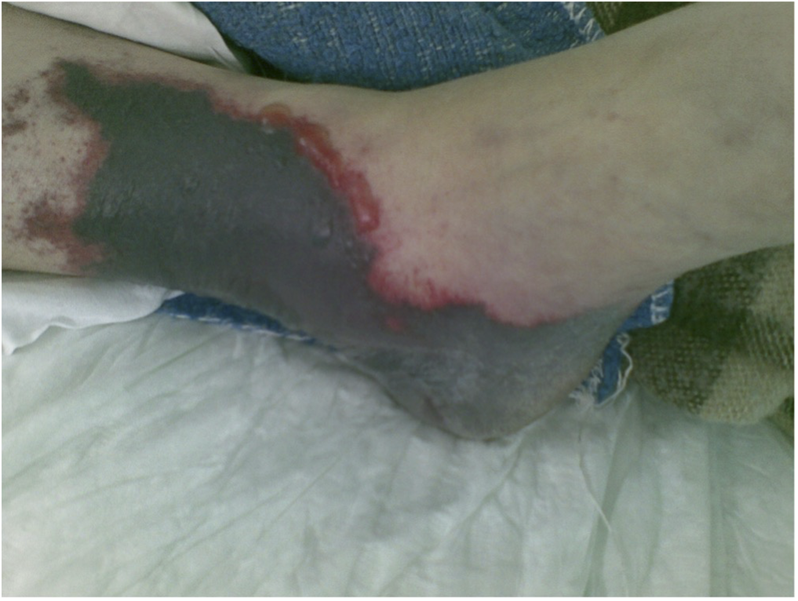
Laboratory findings and antibody profile
Thrombocytopenia and hemolytic features were significantly more frequent in patients with skin involvement (p = 0.03 and p = 0.01, respectively) (Table 2).
No significant differences were observed in the frequency of antiphospholipid antibodies between CAPS episodes with and without cutaneous involvement (Table 2). Stratification of antiphospholipid antibody profiles was available in 438 episodes with complete serological data. The remaining patients had incomplete antibody profiles or were reported as having antiphospholipid syndrome without detailed characterization of the antibody profile. Overall, 126 episodes (15.8%) were classified as triple positive, 102 (12.8%) as double positive, and 210 (26.3%) as single positive. Episodes with triple-positive antiphospholipid antibodies showed statistically significant associations with skin necrosis (17.5% vs 13.6%), Raynaud’s phenomenon (3.2% vs 1.3%), and digital ischemia (15.1% vs 9.8%) (p < 0.05 for all comparisons). Double positivity was also significantly associated with a higher frequency of livedo reticularis (27.5% vs 17.9%) and ischemic ulcers (16.7% vs 10.6%) (p = 0.04 and p = 0.027, respectively). In contrast, single antibody positivity was not consistently associated with specific cutaneous manifestations.
Histopathological findings
Anatomopathological data were available for 193 episodes, obtained from skin biopsy (n = 120) or autopsy (n = 73). The predominant pathological finding in skin samples was thrombotic microangiopathy (TMA), identified in 82 episodes (68.3%), followed by vasculitis in 7 (0.8%), cutaneous necrosis in 4 (0.5%), combined necrosis and thrombosis in 3 (0.3%), fibrosis in 3 (0.3%), capillaritis in one (0.1%), and combined necrosis and vasculitis in one (0.1%). In 29.6% pathological findings were not specified.
Mortality
Mortality did not differ significantly between CAPS episodes with and without cutaneous involvement (29.3% [110/377] vs 32.1% [131/408], p = 0.42).
Multivariate analysis
A multivariate logistic regression analysis was performed to identify independent variables associated with cutaneous involvement. The model included clinical variables such as pulmonary, cerebral, and cardiac manifestations, thrombocytopenia, haemolysis, presence of schistocytes, IgG isotype against cardiolipin and IgM β2-glycoprotein I. The logistic regression model did not show statistical significance (Chi-square = 6.05; p = 0.7). The Hosmer–Lemeshow goodness-of-fit test did not indicate a good fit (p = 0.136). The analysis showed no significant findings.
Discussion
Skin involvement was identified in 43.1% of episodes. The most frequent cutaneous manifestations were livedo (17.6%) and skin necrosis (13%), and the predominant pathological finding was TMA (68.3%).
Livedo (reticularis or racemosa) is a characteristic cutaneous finding featuring a net-like reddish-blue discoloration, reflecting impaired blood flow in the dermis. When this pattern becomes persistent is frequently associated with small-vessel thrombosis.5,7,8 A systematic distinction between livedo subtypes—namely livedo reticularis and livedo racemosa—was not consistently feasible due to the retrospective nature of the registry, and both patterns were therefore analyzed together under the term “livedo.” According to established classification criteria, these entities should be differentiated when possible. 9 Recent data suggest that microangiopathy is more specifically associated with the racemosa subtype.1 10
Skin necrosis was observed in 13% of patients with cutaneous involvement. This manifestation is often severe and typically affects distal areas such as the fingers, toes, ears and nose, where vascular supply is naturally limited. 3 Ischemic ulcers, were identified in 10.4% of patients with skin involvement. These lesions are typically painful and their presence reflects prolonged vascular compromise and they may become chronic, often requiring long-term wound management.5,7,8
Purpura was present in 7.1% of patients with skin involvement in CAPS. These lesions arise from the rupture of small dermal vessels, occurring in the context of thrombosis and microvascular leakage.4,5,7,8
Gangrene represents the most severe form of cutaneous ischemia, it was observed in 5% of patients. When tissue loss is extensive, amputation of the affected areas, may be required.2,5
In clinical practice, obtaining histopathological confirmation in CAPS may be challenging due to the critical condition of patients and the frequent use of anticoagulation. Skin lesions frequently appear in the early stages of the disease and may serve as an important diagnostic clue.77,8 Sampling from the peripheral erythematous area rather than the central necrotic zone may increase the diagnostic yield for detecting thrombotic microangiopathy. 11
The skin is particularly vulnerable to the effects of TMA, which is characterized by endothelial injury, platelet aggregation, and fibrin deposition, leading to occlusion of small dermal vessels. This ischemic process manifests clinically as livedo reticularis, purpura, necrosis, and, in the most severe cases, gangrene.12,13
The diagnostic value of skin biopsy should be emphasized. Sampling from the peripheral erythematous or violaceous area of the lesion is generally recommended to increase diagnostic yield, as viable tissue is more likely to demonstrate active vascular changes. When feasible, deep incisional or excisional biopsies are preferred to adequately assess small- and medium-sized vessels.
Regarding laboratory abnormalities, only microangiopathic features such as thrombocytopenia and hemolysis were more frequent in CAPS episodes with cutaneous involvement.
In our cohort, triple-positive patients appeared to show a higher frequency of ischemic and necrotic lesions. This observation is biologically plausible, as a higher aPL burden has been associated with increased thrombotic risk and more severe clinical phenotypes.
Our findings align with those reported of a recent multicenter cohort study, 7 in which cutaneous involvement was observed in approximately half of the patients, these manifestations were interpreted as the clinical expression of widespread microvascular thrombosis affecting the dermal circulation.
This study has several limitations that warrant consideration. First, its retrospective design inherently restricts the ability to establish causality and introduces potential sources of bias, including misclassification and incomplete clinical documentation. Second, despite representing one of the largest cohorts assessing cutaneous involvement in CAPS, the sample size remains limited due to the rarity of the syndrome. This may reduce the statistical power, particularly in multivariate analyses, and increase the likelihood of type II errors. Third, stratification of aPL profiles was only feasible in a subset of patients with complete serological data. Therefore, the apparent relationship between aPL profile and specific skin lesions should be considered exploratory. Furthermore, the potential role of non-criteria aPL such as anti-phosphatidylserine/prothrombin antibodies, markers of complement activation, or genetic thrombophilic factors, was not assessed. Finally, considerable heterogeneity in treatment regimens may act as a confounder when interpretating clinical outcomes, including mortality. We did not stratify patients according to the timing or intensity of immunosuppressive or anticoagulant therapy.
Despite its limitations, this study has notable strengths. It represents one of the largest cohorts focused on cutaneous involvement in CAPS, integrating clinical, immunological, and histopathological data. Inclusion of skin biopsy findings provides insight into microvascular pathology, often lacking in previous studies.14,15 Additionally, systematic comparison between patients with and without cutaneous involvement allows a more refined characterization of the CAPS phenotype associated with skin manifestations.
Conclusion
Cutaneous involvement in CAPS is common, with manifestations ranging from livedo reticularis and purpura to necrosis and gangrene, reflecting the severity of microangiopathic injury. Further research is needed to elucidate mechanisms driving cutaneous involvement in CAPS and refine therapeutic strategies aimed at preventing thrombotic events in the skin and vital organs.
Supplemental material
Supplemental material - Skin involvement in the catastrophic antiphospholipid syndrome: A review from CAPS registry
Supplemental material for Skin involvement in the catastrophic antiphospholipid syndrome: A review from CAPS registry by A. Ponce, I. Rodríguez-Pintó, I. Pons, A. Jerez, P. Toledano, T. Rossinol, B. Vilaplana, G. Espinosa, R. Cervera, on behalf the CAPS Registry Project Group/European Forum on Antiphospholipid Antibodies in Lupus.
Footnotes
Funding
The authors received no financial support for the research, authorship, and/or publication of this article.
Declaration of conflicting interests
The authors declared no potential conflicts of interest with respect to the research, authorship, and/or publication of this article.
Supplemental material
Supplemental material for this article is available online.
References
Supplementary Material
Please find the following supplemental material available below.
For Open Access articles published under a Creative Commons License, all supplemental material carries the same license as the article it is associated with.
For non-Open Access articles published, all supplemental material carries a non-exclusive license, and permission requests for re-use of supplemental material or any part of supplemental material shall be sent directly to the copyright owner as specified in the copyright notice associated with the article.