






Abstract
Epilepsy is a neurological condition defined by time; it is characterized by a lifelong tendency for recurrent, unpredictable, and unprovoked seizures, during which people lose control over parts of body-mind function. Diagnosing seizures involves using electroencephalograms to represent and classify brain waves in relation to clock time. Drawing upon ethnographic fieldwork in a North American teaching hospital, this paper shows that as neurologists learn to diagnose seizures, they internalize clock time norms for normal and abnormal brain waves. The paper demonstrates how these temporal norms work to assign a set of aesthetics to brain waves: patterns that conform to clock-time norms are beautiful, whereas hard-to-classify patterns are ugly. These aesthetic judgments follow diagnostically complex patients in future hospital visits, who become known, for instance, as “the patient with the ugly EEG.” The paper critiques this ascription of labels to patients and situates the role of the electroencephalogram's clock time in this predicament. It concludes with a speculative design project that reorients the relationship between temporality and embodiment by using the heartbeat as a situated and co-produced alternative to the standardized and invariant clock. Ultimately, the paper argues that the aesthetics of medical technology are fundamental to clinical care, thereby opening up new directions for research at the intersection of critical time studies and disability studies.
Keywords
Introduction
Epilepsy is an invisible chronic illness and disability characterized by recurrent and unpredictable seizures. Seizures are transient events during which people lose control over parts of body-mind function. This can mean the rhythmic twisting of a person’s wrist, sudden inexplicable feelings of joy, or involuntary spasms of the whole body. Since antiquity, epilepsy has occupied the minds of healers, philosophers, physicians, and scientists, who have believed it to arise from divinity, possession, depravity, genius, contagion, humoral tendency, pathophysiology, and psychopathology at different times and places (Temkin, 1971).
As recently as the late nineteenth century, epilepsy remained a contested object between neurology and psychiatry and it was only the development of the electroencephalograph that resolved the debate, placing epilepsy firmly within neurology (Jacoby and Baker, 2010: 616). An electroencephalogram (EEG) shows brain waves: electrical activity from multiple parts of the brain in the form of waves displayed over a horizontal axis of clock time. The EEG “radically changed our understanding of epilepsy” (Hacking, 1998: 32) and soon became routine “especially in epilepsy diagnosis” (Jefferys, 2010: 638). Indeed, it continues to serve as a fundamental tool of present-day clinical neurology (Millett, 2001a: 522).
I conducted ethnographic fieldwork at a large teaching hospital in the Midwestern region of the United States, observing epilepsy specialists as they learnt to diagnose seizures. That is, I observed clinicians transition from novices who see only “squiggly lines” into experts who see seizures in the EEG. This paper shows that, in the process of acquiring this “professional vision” (Goodwin, 1994), clinicians internalize universal clock time norms for brain activity. Further, these temporal norms, when combined with the rhythms of EEG interpretation and the larger medical system, work to assign a set of aesthetics to brain activity that is reminiscent of the spectacular and ugly connotations historically associated with visible disabilities in public spaces. This aesthetics is produced during clinical diagnosis; i.e. in the physical absence of the patient, at the second-by-second level, and by careful, highly skilled, and caring doctors, in a manner that remains disconnected from, and renders less visible, the patient’s lived experience of seizures. Importantly, in diagnostically complex patients, the ugliness of an EEG continues to remain associated with the patient in future hospital visits.
I critique this ascription of labels to patients and examine the role of the EEG’s clock time in objectifying and distancing doctors from the patient’s suffering. I conclude by discussing how the aperture of clinical diagnosis could be broadened to incorporate lived experience. In line with recent calls for a recuperative imaginary of clock time (Bastian, 2018; Pschetz et al., 2016), I propose the figure of the heartbeat as a situated and co-produced alternative to the standardized and invariant clock. Ultimately, by critically investigating the power of the EEG as a temporal regime, I argue that the aesthetics of medical technology are fundamental to clinical care and open up new directions for research at the intersection of critical time studies and disability studies.
Related work
This paper is in conversation with scholarship in the sociology of health and illness, disability studies, and critical time studies. Sociologists of health and illness have long been interested in the study of chronic illness and its implications for those diagnosed. Scholarship has variously used qualitative and quantitative sociological approaches to examine the effects of stigma, uncertainty, variations in health, increased dependence on others, repeated interactions with the medical complex, and institutionalization on people’s notions of identity and selfhood; self-presentation; familial, social, and work relationships; mechanisms of coping and disclosure; adherence to treatment; and lived experience. Foundational work by Michael Bury and Kathy Charmaz has found that chronic illness is experienced as a biographical disruption (Bury, 1982) or interruption (Charmaz, 1991) that makes people rethink commonly held assumptions about ability, social participation, and self-reliance. Chronic illness exposes people to the (un)certainty of medical knowledge and life. Charmaz (1991) further reports that people, particularly those with intrusive illnesses, respond by adjusting their schedules and expectations to accommodate the illness and its demands. This work has been influential in making visible narratives that were seldom acknowledged, highlighting the impact of socioeconomic variables on illness experience, making the case for affordable health services, and informing the design of patient-centered health-tracking devices (Barbarin et al., 2015; Barbarin et al., 2016; Charmaz, 2007; Chen, 2010, 2011; Pierret, 2003). Overall, however, sociological literature tends to align with the medical model in that chronic illness is viewed as a primarily negative influence on individuals and their social relations, with narratives of suffering and overcoming chronic illness running through the literature (Charmaz and Rosenfeld, 2009). This is perhaps understandable given that the aim is to speak to and be taken seriously by the medical complex and health policy makers, but it has given birth to what Charmaz (2007) calls a “sociology of suffering”.
Arising out of the disability rights movement, the academic field of disability studies has sought to counter these dominant understandings of disability and chronic illness. Drawing upon a mix of methodological traditions in the humanities and social sciences, scholars have used literary studies, cultural theory, historical analysis, qualitative sociology, auto-ethnography, medical and sociocultural anthropology, art practice, architecture, and action research to highlight the social construction of normalcy, deviance, and disability (Clare, 1999; Davis, 1995, 2006; Garland-Thomson, 1996, 1997; Kuppers, 2004; Linton, 1998; Shakespeare, 1998b). Additionally, as in queer feminist theory, scholars have argued that disability in fact allows for creative ways of being and living by disrupting taken-for-granted normative models of agency and self-reliance in favor of communities built around interdependence and care (Clare, 2017; Kafer, 2013; Kuppers, 2014). Early work centered around pushing for a social model of disability, which argued that society is disabling by not making all buildings accessible or by imposing strict time schedules, for example. Disability, therefore, needs to be addressed at a societal level, not through medical treatment of and intervention on individuals (Clare, 2017; Kafer, 2013; Shakespeare, 1998a). More recently, scholars have recognized that, particularly in the case of chronic illness, the social model of disability overlooks the role of suffering and the usefulness of palliative care (Clare, 2017; Shakespeare, 2010; Wendell, 2001). Present-day disability scholarship shares a complicated relationship with notions of cure, arguing for societal accommodations and palliative care while remaining in opposition to the individual-based and curative medical model.
Critiquing reductionist analyses of time that ignore the complexity of relations involved in its production and maintenance, critical time studies scholarship has called for social theoretic accounts that move past “oversimplified, single-stranded descriptions or typifications” and take seriously “the various meaningful connectivities among persons, objects, and space continually being made in and through the everyday world” (Munn, 1992: 92–116). Contemporary disability studies scholarship responds to this call through the notion of crip time as a site of contestation between various political stances on time and futurity. Alison Kafer (2013), for example, uses an intersectional feminist critique of capitalist notions of productivity to advocate for crip time as a flexible reorientation to time. Crip time is also the experiential time of prognosis; that is, of coping with the paradox of statistics: an average of 1.16 out of 1000 people with epilepsy experience sudden death, but am I, an individual with epilepsy, amongst the 1.16 or the 998.84 (Jain, 2007)? The medical imaginary that Kafer names curative time and futurity is a time and future in which disability has been cured through medical intervention. Crip time must buy into it while resisting it: people with chronic illness must often depend on medication to manage symptoms, thereby supporting the medico-scientific complex that continues to involuntarily confine, experiment on, and sterilize fellow disabled people (Kafer, 2013: 25–27).
Importantly, crip time represents an outright rejection of the clock and clock time. Despite its rhetorical power, therefore, the theoretical construct of crip time turns out to be difficult to translate from the realm of theoretical construct to the realm of design. Noting that this rejection of clock time occurs across philosophy, art, literature, and cultural theory, Michelle Bastian asks us instead to consider the redemptive potential of clocks. She proposes a “critical horology” that underscores the deeply political nature of clocks while insisting on their potential for redesign (Bastian, 2018). A temporal design framework, Bastian and colleagues argue, would instead focus on challenging dominant narratives of time and highlighting alternative networks of temporalities that “reveal actors, practices and forces that determine social coordination within specific contexts” (Pschetz et al., 2016). This paper responds to these calls, using an ethnographic study of epileptic seizure diagnosis to show that the use of clock time in combination with a multiplicity of contingent temporalities, actors, and forces, works to assign problematic aesthetics to brain activity. I conclude with a speculative attempt to design alternative temporalities in service of a disability-informed seizure diagnosis.
Research method and context
This paper draws upon ongoing ethnographic research in the pediatric neurology facility of a large teaching hospital in the Midwestern region of the United States. 1 This facility offers specialized diagnostic, therapeutic, and surgical care for people with epilepsy, seeing over 3000 patients each year. In regular intervals starting March 2018, I observed four epilepsy fellows and two clinical neurologists engaged in the diagnosis and treatment of epilepsy at this facility. Epilepsy fellows are post-residency doctors undergoing a year-long intensive training in epilepsy care and spend a majority of their time learning to diagnose seizures using EEG. These six practitioners (three women and three men) were purposively sampled to prioritize excellence in clinical practice and gender balance to generate high-quality data on the clinical perspective of epilepsy (Creswell, 2007).
This paper specifically draws upon 100 hours of participant observation in the facility’s EEG reading room and 20 informal open-ended interviews with these practitioners. Epilepsy fellows spend a majority of their working hours in the EEG reading room while on fortnight-long EEG reading shifts. The EEG reading room is an approximately 10 feet long by 12 feet wide room in which, from 8 a.m. until at least 5 p.m. every day, epilepsy fellows read EEGs—typically alone, except when presenting their interpretations to supervising neurologists. The room has five computers, each installed with a commercial EEG software suite and connected to the hospital’s intranet, internet, and medical records system.
I spent between one and four hours in the EEG reading room during each observation session, varying the day of the week and time of day to gain a realistic portrait of epilepsy fellows’ everyday work practices. I paid particular attention to instructional interactions between fellows and supervising neurologists to capture their explicit articulation of diagnostic work. Informal interviews were conducted as needed, to ask context-specific questions during observation and for post-facto clarification. I audio-recorded interviews and wrote field notes after each observation session. Field data were analyzed using an iterative and inductive process consisting of regular analytic memoing and thematic analysis. I use triangulation, respondent validation, long-term involvement, and mentor feedback during monthly data sessions to ensure the validity of my findings (Creswell, 2007; Emerson et al., 2011). I use pseudonyms to preserve the anonymity of my interlocutors. Quotes are lightly edited to preserve readability, and use square brackets to indicate changes in emotion, intonation, and pace. In the field, I am conscious of the privileges of my outsider status, critical distance, and academic freedom. I am deeply implicated in this work both as the instrument of data collection and as a person with epilepsy who benefits from medical intervention.
This paper is also informed by a close reading of the second edition of the Handbook of EEG Interpretation by William O. Tatum (Tatum, 2014), a popular clinical text intended as a quick and practical reference to EEG interpretation; and of present-day neurological research disseminated by international organizations such as the International League Against Epilepsy. This helps ensure that my arguments, grounded in practices and conventions specific to my interlocutors and field site, are in conversation with the broader clinical discourse on epilepsy.
Seizure diagnosis is deeply concerned with classification and representation
A person qualifies for a diagnosis of epilepsy if, in the absence of provocative factors, they have at least two epileptic seizures separated by more than 24 hours or have one seizure and an over 60% risk of seizure recurrence (Fisher et al., 2014). While seizures are particularly emblematic of epilepsy, conditions such as fever, alcohol withdrawal, and severe hyponatremia (low blood sodium) can also provoke seizures in people who do not have epilepsy. Further, not all seizure-like events are epileptic: physiological conditions such as syncope (fainting) and hypocapnia (reduced carbon dioxide in the blood); and psychological conditions such as anxiety and post-traumatic stress can produce convulsions and other episodic phenomena that can be mistaken for epileptic seizures. To complicate matters, these “common imitators” can co-occur with epilepsy (Brodtkorb, 2013).
Classification thus lies at the heart of seizure diagnosis. It helps clinicians place patient symptoms within a known framework, determine seizure triggers, and choose a course of treatment. To date, seizure types are defined and classified operationally—i.e. based on patterns observed, documented, and verified in clinical practice—as opposed to scientifically, because scientific knowledge of the mechanisms underlying epilepsy is not sufficiently developed to serve as a reliable basis (Berg et al., 2010; Fisher et al., 2017; Scheffer et al., 2017). Seizures are classified by (1) location of onset, (2) symptoms at onset, and (3) impact on awareness. Seizures can be of focal onset, i.e. originating in one part of the brain; generalized onset, i.e. involving both hemispheres of the brain; or unknown onset, where not enough is known about the origin. Focal onset seizures can also spread by engaging the rest of the brain, transforming into a focal-to-bilateral or secondarily generalized seizure. Motor onset seizures disrupt motor function via abrupt and involuntary muscular contraction (tonic), relaxation (atonic), alternating contraction and relaxation (clonic), jerking (myoclonic), or spasms of specific body parts or of the whole body. Nonmotor onset seizures involve sudden inexplicable changes in emotion, e.g. fear, anxiety, or joy; in cognition or sensation, e.g. impaired language, hallucinations, or perceptual distortion; in autonomic function, e.g. gastrointestinal or respiratory sensations; or in behavior, e.g. abrupt suspension of all activity. Absence seizures manifest as an abrupt loss of and return to consciousness, sometimes along with myoclonus (short and irregular muscle twitching). Finally, seizures have differential impacts on a person’s awareness, which is operationalized as “knowledge of self and environment” and is reported retrospectively, once the person has recovered from the seizure (Fisher et al., 2017). Focal seizures can occur with fully retained awareness or cause impaired awareness, but generalized seizures always occur with impaired awareness, if not a complete lack thereof. Clinicians are encouraged to be as precise as possible in classifying seizure type, preferring a diagnosis of “focal aware tonic” seizure to simply “focal onset” seizure where sufficient information is available. The process of diagnosis is thus deeply concerned with classification.
Seizures are unpredictable events that clinicians are typically unable to witness in person. Therefore, patients are asked to undergo diagnostic EEG tests with two goals in mind: (1) to record the patient’s seizure. Once recorded and classified as above, seizures can often be prevented using anti-epileptic drugs (Jefferys, 2010). However, 30–40% of people diagnosed with epilepsy have seizures that remain uncontrolled despite adherence to medication (Brodie, 2010; World Health Organization, 2018). Therefore, this procedure has another important goal: (2) to localize the seizure to one part of the brain. As clinical neurologists at a large specialty epilepsy center, my interlocutors spend the majority of their time dealing with challenging cases of uncontrolled or refractory seizures. Such refractory epilepsy can be treated through surgery: if the specific part of the brain from which the seizure originates is identified, it can then be surgically removed to prevent further seizures. Seizure diagnosis thus uses electroencephalography to identify, classify, and eliminate seizures to achieve clinicians’ repeatedly stated goal of “zero seizures and zero side-effects.”
Seizure diagnosis defines normalcy in relation to clock time
Electroencephalography is a diagnostic procedure conducted for durations ranging from 45 minutes to several days, depending on whether the goal is to rule out abnormalities or to capture specific seizures, with seizure frequency dictating duration. Especially in the latter case, the patient being investigated is deprived of sleep and/or medication and subjected to activation procedures such as hyperventilation and intermittent photic stimulation, to increase their likelihood of having seizures under monitoring (Tatum, 2014).
This procedure is performed using an electrophysiological device called an electroencephalograph that displays electrical activity in the brain over time. This device is roughly the size of a human being. It consists of a central digital apparatus connected via electrical wires to a set of 22 or more metal electrodes that serve as input; a hard disk that records electrical activity, and a graphical display that renders brain waves (Tatum, 2014). Sometimes, a video camera is attached to the device, so that the patient’s physical activity can also be recorded for later correlation with electrical activity. See Figure 1 for a pictorial representation.

A picture showing the main components of an electroencephalograph: electrodes attached to a person’s scalp and an EEG display showing the recorded electrical activity as a time series graph. Image reproduced from the Bright Brain Centre’s website: (Bright Brain Centre, 2019).
Electrodes are attached to various points on a person’s scalp 2 using a glue that helps keep them in place. Electrodes are named and positioned according to international standards: the name comprises of one alphabet corresponding the lobe of the brain, and one number corresponding to the hemisphere of the brain. Electrode “F3,” for instance, is understood to be positioned on frontal lobe of the left hemisphere (Tatum, 2014).
In the EEG reading room, neurologists see the output of EEG monitoring presented as a time-series montage that synchronizes several two-dimensional graphs into one continuous visualization. For example, Figure 2 shows one such montage of brain waves over approximately 12 seconds: it consists of 20 individual waveforms with separate mini-Y axes over a common horizontal X axis. Every pair of black vertical lines delineates one second of clock time on the X axis, and each horizontal waveform shows fluctuation in electrical voltage from a part of the brain over time. Electrode names on the very left (e.g. T3–T5) allow readers to identify the parts of the brain that a particular signal comes from.
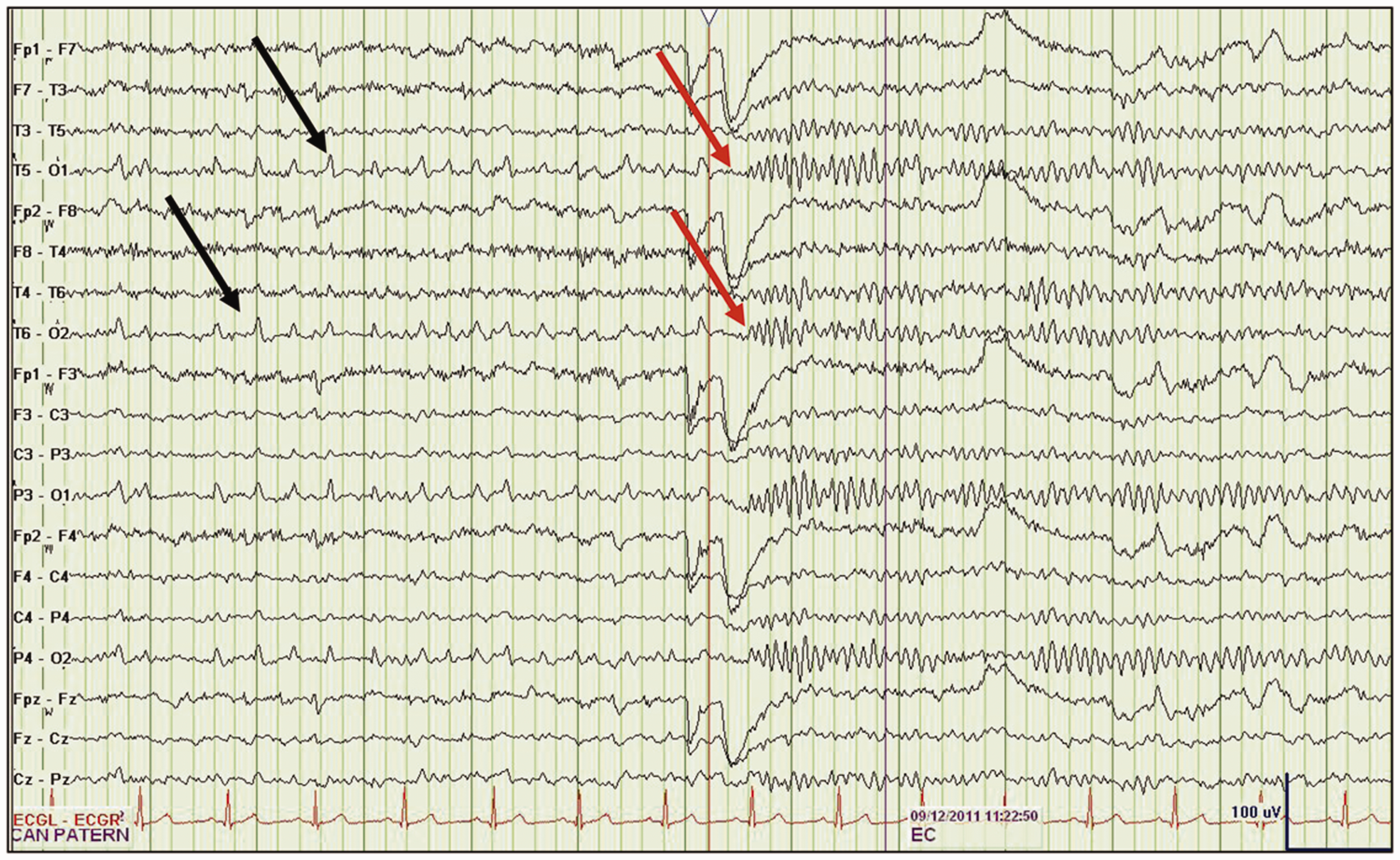
An example EEG rendered in a “bipolar montage” format. The bipolar montage displays the difference in electrical voltage between pairs of electrodes and is the most commonly used EEG viewing format. The black arrows indicate atypical sharp waves that could be indicative of seizures and the red arrows indicate points at which those waves resume normal patterns just after the patient closes their eyes. Image and explanation taken from: Tatum (2013b).
When reading EEG, neurologists distinguish between two broad categories of brain activity: normal and abnormal. Importantly, the boundary between these categories is produced and sustained using clock time. For example, examining the alpha rhythm, also called the posterior dominant rhythm, is widely considered “the starting point of clinical EEG” interpretation (Tatum, 2014: 31). The closing and opening of the eyes is said to capture the rhythm of the alpha wave in the patient’s brain. Further, “during normal development,” advises the Handbook of EEG interpretation, “an 8-Hz alpha frequency appears by 3 years of age. The alpha rhythm remains stable between 8 and 12 Hz … into the later years of life” (Tatum, 2014: 31). Eight to 12 hertz—that is, eight to twelve cycles of the alpha wave per second of clock time—thus establishes the patient’s brain’s developmental normalcy and by implication, also points out deviance from the norm. Similarly, my interlocutors identify seizures by seeking out two characteristic abnormal patterns on the EEG: the spike and the sharp wave. These patterns of abnormal brain activity too are defined in relation to clock time: “Spikes and sharp waves are … defined by their duration … A spike has a duration of 20 to 70 [milliseconds] while a sharp wave lasts 70 to 200 [milliseconds]” (Tatum, 2014: 83). Similar norms defined in relation to clock time are employed to distinguish between normal and abnormal brain activity on the EEG during sleep, wakefulness, photic stimulation, and hyperventilation.
Thus, seizure diagnosis defines normalcy and abnormalcy in relation to clock time during the process of EEG interpretation. Importantly, the EEG’s temporal norms are not intuitive but consciously and painstakingly acquired by highly trained doctors through apprenticeship under the close supervision of expert neurologists. For example, the second half of Figure 2 looks disturbed, irregular, and potentially “abnormal” to the untrained eye, whereas the first half looks evenly spaced, regular, and potentially ”normal”. In fact, it is the other way around: the first half of Figure 2 could be indicative of seizures and the second half shows normal brain waves (Tatum, 2013b). Doctors new to the EEG comment despairingly about seeing nothing but “squiggly lines” in the EEG and second-guessing themselves on everything. More experienced EEG readers acknowledge the validity of such complaints and urge the novice to keep trying, saying it took them several months to “learn to see” through the EEG, that is, to distinguish the normal from the abnormal using clock time.
Scene: Learning to see on EEG
This section depicts a pedagogical conversation between experienced epilepsy fellow Amy and neurology resident Jen to contextualize their temporal classification of, and aesthetic responses to, brain waves. Around 11 a.m. on a cold February day, Amy and Jen sit facing a computer screen showing the continuous brain waves of a routine EEG that was ordered to examine suspected seizures in an eight-year-old girl. Having discussed other patients with the supervising neurologist from 8 a.m. to 11 a.m., Amy and Jen also use this respite to snack on dried fruit and trail mix. I sit behind them, pen and notebook in hand. This is Jen’s first in-depth exposure to EEG reading. Amy, like other experienced EEG readers, scrolls forward horizontally through the EEG with her eyes focused on the brain waves even as she carries on a conversation with Jen and me. Amy: Have you heard of any epilepsy syndrome that is on the absence spectrum but has eyelid myoclonus? Jen: No. Amy: So, Jeavons syndrome is absence seizures with eyelid myoclonus. They can be hard to treat, because they tend to be more refractory than just absence seizures. Jen: Hmmm, oh! [interested tone] Amy: Eyelid myoclonus causes a funny feeling, so people wonder if the kids like doing it to themselves, unintentionally, but like maybe … [abrupt change] You guys wanna see a spike? I think maybe a little right predominant. Jen: Right predominant cause the right side looks deeper? Amy: Because it looks higher amplitude, yes. The EEG shows very large deflections in several brain waves, similar to the region with red arrows in Figure 2. Jen: Is this blinking? These frontal things … Amy: Yup, yup, you got it! Jen smiles, pleased at having correctly identified eye blinking on the EEG. Amy: So, these should all be eye blinks. I usually go for the best-formed second. Megh: The best what? Amy: What we’re looking at is something called posterior dominant rhythm or alpha rhythm. When you close your eyes, this rhythm comes out in a normal healthy kid or adult. We get a pretty clear rhythm in the posterior parts of the head, and that’s your posterior dominant rhythm or PDR. In kids, as their brain grows, their rhythms get faster. And this kid is 8 years old, so she should have about a nine hertz PDR. So, we count. Each bar on the EEG is one second, so, one, two, three, four, five, six, seven, eight, nine, ten! We’ll give her a ten. Megh: Ok. Amy: So, she’s got a nice, normal PDR and what you want to see is that it goes away when she opens her eyes. Amy uses her computer’s mouse to point out the ten consecutive peaks within one second of EEG that add up to the patient’s ten hertz alpha rhythm. We sit in silence for fifteen seconds as Amy scrolls forward through the EEG, stopping to point out a short region of smaller deflections in all brain waves. Amy: Ugh! So, this is a fragment, which you sometimes see with generalized epilepsy. It’s not clear like a beautiful spike-and-wave, but that’s what a fragment looks like. Jen nods. Amy now scrolls through a portion of the EEG showing brain waves recorded during photic stimulation, a procedure wherein a bright light is flashed repeatedly into the patient’s eyes at frequencies increasing stepwise from two to twenty hertz. This is used to see how the patient’s brain waves respond to light (Tatum, 2014). Amy: Okay, so now they’re doing photic stimulation. Each tick mark is a light flashing into the patient’s eyes: so, flash, flash, flash, flash, at two hertz. You want to see some kind of two hertz rhythm coming out in the occipital region. I see two little blips here, so I’d say that looks like driving. Now, we get to four hertz photic stimulation. Oooooooooooh! Look what we got! Jen and I are caught off-guard and give a nervous laugh in response to Amy’s sudden and nearly two-second-long “oooooooooooh.” Amy: Okay [with a visible effort to calm herself], so. Nice. This is a seizure. Amy labels the EEG with the text “sz” to mark the point in time where the seizure began. Jen: That’s like 3 hertz, right? Amy: Yup. Jen [using her fingers to point out three peaks to me]: Perfect three hertz. Amy: Yup, yup. It breaks up the background a little bit, it looks different from the rest. And this looks a little more symmetric than the fragment. Twenty-two seconds long. Jen: Yeah, that’s a beautiful … seizure?! [surprised tone] Amy: This is a good first EEG for you! Amy gives Jen a bright smile acknowledging her identification of the seizure. Jen’s giggle carries some surprise at finding a seizure beautiful. Amy [to me, with a chuckle]: And this is not even midway through a 40-minute EEG, so you can see why these longer studies can take a little time. I nod, thinking about diagnostically complex patients, who are often monitored on EEG for several days at a time. In total, Amy spends about 40 minutes viewing, interpreting, and writing up the results of this EEG in conversation with us.
Interlocking diagnostic rhythms sensationalize the abnormal and assign aesthetics
My interlocutors, both epilepsy fellows and neurologists, are careful and conscientious doctors who maintain a clear focus on patient wellbeing. They consistently favor under-diagnosis and take note of the larger picture beyond medical intervention; for instance, the kinds of resources and familial support available to the patient. However, several interlocking rhythms that intersect in the process of seizure diagnosis come together to sensationalize the abnormal in EEG interpretation.
To begin with, we have the nature of epilepsy itself: seizures are recurrent but unpredictable. EEG interpretation therefore requires my interlocutors to spend hours tuning out long swathes of “normal” brain activity as they search for the abnormal. This wait for the abnormal is particularly fraught and uncertain in diagnostically complicated seizures, where patients are at increasing risk of sedation, if not intubation, with every additional seizure. My interlocutors are often exhausted from working 80-hour weeks and managing a large caseload of clinic patients. EEG patients and caregivers are similarly exhausted from long stays in the hospital and impatient for monitoring to end so that they can return home.
For my interlocutors, therefore, the painstaking yet unrewarding quality of this second-by-second scrolling through the EEG means that when they finally notice an abnormal pattern, they break out into spontaneous exclamations, such as the “oooooooooooh” as we saw with Jen. That is, multiple rhythms and demands intersect to sensationalize the abnormal. Indeed, in making these exclamations, neurologists come to embody the occurrence of the abnormal in their patient’s body and brain: through abrupt changes in speech and mannerism, neurologists experience abnormal brain waves in their own bodies and minds. Paradoxically, however, the victorious nature of this embodiment undermines the suffering involved in experiencing the seizure and the otherwise-normalcy of the patient (because moments of note are all abnormal).
To complicate matters, abnormal activity does not necessarily indicate seizure because “artifacts are intertwined with epilepsy … They may beguile the interpreter into misidentifying waveforms … [or] obscure the recording during … seizures” (Tatum, 2013a: S12). The EEG is an old and noisy electrical device in which electromagnetic interference from faulty wiring, bone defects, chewing, static electricity, headache, pacemakers, and telephone rings can all be mistaken for seizures (Tatum, 2013a, (2014). Pointing to one such seizure-or-noise pattern in the EEG reading room, a supervising neurologist instructed an epilepsy fellow, “this is the kind of stuff that just gets your attention. That is exactly the kind of artifact you get when somebody moves.” Once my interlocutors identify an abnormal pattern, therefore, they must then carefully distinguish between a “real” seizure and a clinically irrelevant artifact. An experienced fellow said that over time, he had “trained [his] eye to look under the artifact.”
This sensational and contingent character of the abnormal (whether seizure or artifact), taken together with tedium of heavy caseloads and the imperative to diagnose as quickly as possible plays out unexpectedly and perhaps unintentionally, by assigning aesthetics to brain waves. For instance, upon seeing a "normal" 10 hertz alpha wave or 6 hertz sleep spindle, my interlocutors speak of “nice PDR” and “gorgeous sleep spindles.” Often, positive adjectives are also used to describe abnormal patterns that fit within the clock time norms of seizure diagnosis. That is, norm-conforming abnormal patterns also get called “beautiful seizure” and “pretty spike.”
Abnormal brain activity that remains unclassifiable (within the clock time norms of diagnosis) but sets off some internal discomfort in my interlocutors is associated with negative responses: “ugly diffuse delta” or very simply, “ewww!” Here is an example of epilepsy fellow Amy and supervising neurologist Nina conversing while viewing the EEG of a nine-year-old male patient whose seizures were proving difficult to diagnose. Amy: I’ve been wondering if some of these start in the medial parietal occipital region. Nina: We have wondered that too. There is a sort of nebulousness to it. He will frequently have these embedded spikes in that rhythmic slowing. But his seizure didn’t even start there. His seizure started in the electrode above. Amy: Uh-huh, uh-huh [affirmative]. Nina: Yup. This’ll be a joy to discuss in the case conference tomorrow. Amy: Ughhh, these are poly spikey. Nina: Yup. Amy: I remember was it his mom, who was like so freaked out? I can’t remember who it was… [abrupt change] Ewww! That’s just disorganized and gross. Nina: The whole thing is grossing me out. Amy: God, I feel like I’ve spent like triple the time on his EEG than the others. Nina: You could just read his EEG all day, you really could. I think I’ll stay back after 5 o’clock today to look at these, because this is impossible to do from home on a small screen. I mean, it’s not impossible, it’s very, very tedious.
Supervising neurologists like Nina often spend their entire professional careers in the same hospital and remember diagnostically complex patients. Multiple tedious and frustrating EEGs, in combination with heavy caseloads and long work hours, mean that neurologists associate and recall EEG judgments with patient names. Once Nina has finished reviewing an EEG, she typically asks the fellow, “who’s next?” The fellow recites the name and one-line medical history of the next patient on their list and opens this patient’s EEG on the computer: “Next, we have patient William, a 12-year-old with focal epilepsy.” Hearing a familiar name, Nina responds: “Oh, William – he has the worst EEGs.” Similarly, when discussing cases with colleagues, neurologists will respond to familiar patient names with EEG-related exclamations such as: “oh I remember her! She has very pretty spikes,” or, “my God, he’s the one with the ugly EEG.”
As one who has experienced the patient side of epilepsy care, I was surprised at the regularity with which my interlocutors reacted to patients’ EEGs with aesthetic judgments, because they did not employ terms of beauty and ugliness when they witness patients seize. When I asked supervising neurologist Nina why brain waves are considered beautiful or ugly, her eyes lit up with surprise and caution (in the litigious context of the United States, Nina worries about her remarks being taken out of context), but she responded with a beautiful parsing out of the factors that produce aesthetics in brain waves:
To me a seizure is beautiful because it gives me the most information. Yes, it is irreverent and perhaps tactless, but it is a way of impressing upon the trainees the relevance and gravity of the findings and to inculcate interest and excitement: you want your trainees to get excited about something. You’ve been waiting for something to happen and it finally happens. It’s not something that would ever be said to the patient. I’ve never told a patient, “oh my gosh, your seizure was beautiful.” I’ve often gone and told patients, “I have good news for you: I know where your seizures are coming from and here’s what we can do.” I’ve never said, “I have bad news for you,” but that’s because we try to avoid saying things like that.
Discussion: Towards disability-informed seizure diagnosis
As a technology of representation, the EEG shifts attention from the patient’s experience of seizures to electrical waves and clock time – from bodily experience to a detached brain representation that can be examined in the quiet confines of an EEG reading room such as the one I observe. Indeed, this displacement can be productive: by enabling diagnosis, the EEG indirectly helps patients access life-saving anti-epileptic drugs, health insurance, and even workplace accommodations.
Yet, the clock-time norms of EEG waveforms are neither natural nor intuitive (as we saw earlier), but were, in fact, constructed and adopted in particular places and times by actors with specific stakes in the epilepsy landscape: these norms originated when German physicist Hans Berger published the first ever human EEG recording in 1929, alongside initial descriptions and pictures of normal and abnormal brain activity as seen in the EEG (Berger, 1929; Jefferys, 2010). Berger’s original (and failed) intent in inventing the EEG was to detect parapsychological phenomena of the mind (Millett, 2001a; Pressman, 1988).
Circa 1930, the EEG found popularity in clinical research in the United States, where the EEG and its temporal norms were applied in eugenic projects funded by large philanthropic organizations such as the Rockefeller Foundation that sought several times to establish the hereditary basis of epilepsy and other forms of deviance using small samples of people "known" to be normal and abnormal (Millett, 2001b; Pressman, 1988). Indeed, as the most reliable and cost-effective technology of temporal representation, the EEG continues to dominate clinical and scientific discourse on epilepsy, even informing genetic research in molecular biology and neuroscience with the long-term goal of eliminating epilepsy altogether. The EEG thus has a grey past and contingent future replete with eugenic associations that cannot be ignored.
Further, the aesthetic judgments ascribed to brain waves in EEG interpretation are neither natural nor intuitive but inculcated into trainees as they learn to diagnose seizures so that they become knowledgeable, careful, and interested professionals. That is, doctors are taught to see that which is actionable and amenable to medical treatment as beautiful, and that which is difficult to classify or treat as ugly. My interlocutors would readily acknowledge that brain waves are not ugly because of any fault inherent to the patient or their brain. Brain waves are ugly because they are neither normal nor legible to the diagnostic technologies and classificatory norms of present-day biomedicine in ways that are consistent with the structural and temporal pressures faced by doctors: long working hours, heavy caseloads, the mundanity of EEG interpretation, and the imperative to act quickly (Bowker and Star, 1999; Montgomery, 2006; Stonington et al., 2018). Further, EEGs are not objectified and detached “data” but in fact the result of a relational, durational, and contingent process that is inextricably tied to the patient and their environment (Marathe, 2019). However, this objectification and disconnection means that the “ugliness” of an EEG unproblematically enters the hospital’s institutional memory and follows difficult-to-treat patients in future hospital visits.
This ascription of aesthetic labels to people who do not fit within medical or technological norms is troubling because it is incredibly reminiscent of the spectacular and ugly connotations that were historically associated with visible disabilities in public spaces. This historical association led to the creation of ugly laws that portrayed disability as an individual problem to be cured through involuntary confinement, sterilization, and institutionalization until as late as the 1970s; instead of a societal condition to be addressed through accommodations, rights-based discourses, and consensual palliative care (Davis, 1995, 2006; Kafer, 2013; Shakespeare, 2010; Wendell, 2001).
In the EEG reading room, therefore, the application of historically and geographically situated clock time norms to brain waves assigns problematic aesthetic judgments to EEGs. However, ugly EEGs, rather than being considered a manifestation of the uncertainty inherent to medical decision-making or a failing of diagnostic technology, instead come to haunt patients in future encounters with the medical system.
Epilepsy is defined by time: it is characterized by chronicity, contingency, and recurrence. Recent analyses of patient narratives show that across different kinds of seizures, the one shared experience is a disruption of clock time (Lightman et al., 2009; Ryan and Räisänen, 2012; Smith, 2012; Valeras, 2010). Geographer Niall Smith (2012) for example, finds that the lived experience of seizures varies from person to person and by type of seizure, but the perception of time is always disrupted: some feel as though time stayed still during the seizure, whereas others experience feelings of timelessness; and some experience an absence or detachment of self, whereas others experience multiple selves and times within themselves. For a condition defined by time, it is strange that clinical diagnosis defines normalcy in relation to the very thing that makes no sense to the seizing person: the EEG’s clock time. This temporal regime ascribes aesthetic labels to patients via their brain waves without regard for lived experience.
How can we avoid this disconnect in the clinical diagnosis and lived experience of seizures? More precisely, how can we broaden the aperture of diagnosis to incorporate, if not re-centre, lived experience? The EEG plays a central role in shifting doctors’ attention from the patient experience of seizures to a detached representation of brain waves against clock time. The EEG deploys a standardized and invariant imaginary of the clock: that every second is identical to the one that came before it and to the one that follows it. In essence, the EEG presumes that time is objective, universal, and forward-oriented and that clocks are independent of (or incapable of representing) subjective experience.
How could we redesign the EEG to incorporate lived experience? A redesigned EEG would still display brain waves but in relation to an X-axis capable of representing lived experience: instead of the standardized and invariant beats of the old EEG’s clock, this new X-axis would use a clock that is situated and co-produced with the patient. In asking clocks to reflect lived experience, I am not calling for biological determinism in the design of clocks. I am calling instead for mechanisms that allow clocks to be attuned to the embodied and enminded realities of marginalized people.
The heartbeat is one such mechanism: it is still discrete and divisible; that is, it is still capable of measurement and ordering, and hence of serving as a clock. But importantly, the heartbeat is also situated and co-produced: the heart beats faster in times of anxiety and slows down in times of relaxation. Indeed, this speeding up and slowing down is not based on an atomistic conception of the separate and stable self, but also varies in response to one’s surroundings; that is, it represents the interdependence of our existence.
We could redesign the EEG to use the heartbeat as a clock. What would such an EEG look like? Let’s think back to Figure 2: the first half of this EEG shows abnormal brainwaves indicative of seizure, whereas the second half shows irregular but normal brainwaves. Now, let’s reimagine Figure 2 with the patient’s heartbeat as the X-axis. With this new situated and co-produced clock, the vertical black lines that mark seconds in the EEG would be spaced apart in harmony with the patient’s heartbeat. In the first half of the EEG, the vertical black lines would be tightly compressed together to match the anxiety of experiencing a seizure. In contrast, in the second half of the EEG, the vertical black lines would be spread out to reflect a calmer, less anxious, and no longer seizing patient. This would have the additional effect of making the EEG legible to an untrained eye: tightly compressed seconds would produce irregular brain waves to indicate seizure; and spread-out seconds would produce slower brain waves to indicate calmness. Crucially, such a situated and co-produced EEG would make the lived experience of seizures an inextricable part of clinical diagnosis.
Conclusion
This paper has shown that by defining normalcy in relation to clock time, the process of seizure diagnosis assigns temporal norms to brain activity. Epilepsy doctors must internalize these definitions to successfully diagnose epileptic seizures. Multiple interlocking rhythms work together to sensationalize abnormal brain waves while making less visible the suffering of the patient, and in the process, perhaps unintentionally, assign a problematic aesthetics to brain waves: patterns conforming to clock-time norms are beautiful, while hard-to-classify patterns are ugly. Instead of being considered a failing of diagnostic technology or a manifestation of the uncertainty inherent to medical decision-making, the beauty or ugliness of brain waves instead gets assigned to the patient. Importantly, patients with difficult-to-treat seizures are haunted by ugly EEGs in future encounters with the medical system. The paper critiqued this ascription of labels to patients and situated the role of the EEG in this disregard for lived experience.
Finding that the aesthetics of medical technology are fundamental to clinical care, the paper asked: how can we broaden the aperture of diagnosis to incorporate, if not re-centre, lived experience? The paper proposed redesigning the EEG to make it situated and co-produced, thus reorienting the relationship between temporality and embodiment. Using the heartbeat as a recuperative imaginary of clock time, this paper described an EEG that is capable of representing the lived experience of seizures and making visible the labor and suffering involved in regularly and unpredictably experiencing seizures.
The paper speculates that a situated and co-produced EEG would prevent problematic aesthetic judgments from being attached to the “squiggly lines” of brain waves. Then, ugly labels would no longer haunt diagnostically complex patients in repeat hospital visits. Finally, as arbiter of normalcy in brain waves, a situated and co-produced EEG would perhaps transform the seizure from a predominantly negative phenomenon to just another facet of human experience.
Footnotes
Acknowledgements
This work would not exist without the patience, openness, and kindness of my interlocutors. I thank Elizabeth F. S. Roberts, Tiffany Veinot, Kentaro Toyama, Joyojeet Pal, Lisa Baraitser, Shriti Raj, Os Keyes, John Bell, Henry Cowles, Sriram Mohan, and the anonymous reviewers for helping improve this paper.
Declaration of Conflicting Interests
The author(s) declared no potential conflicts of interest with respect to the research, authorship, and/or publication of this article.
Funding
The author(s) received no financial support for the research, authorship, and/or publication of this article.
