
Abstract
Having excelled in histology, Dr Eugenia Cooper, following graduation in medicine in Manchester, embarked on a career spanning 44 years in anatomy and histology at Manchester University. Her inimitable character was readily remembered by those she had taught. She was the first female graduate to gain an MD with gold medal for her thesis on the histology of the endocrine organs. However, her main study was the development of the human brainstem from the early weeks of gestation, which remains the basis for anatomical understanding today. More controversial was her theory on circulation and absorption of the cerebrospinal fluid. On retiring as Reader in Histology, she expressed disappointment at not being appointed a professor, which she considered was due to her gender. Possibly to compensate for this, she had studied law as an additional interest. She continued in research for a further 10 years in reproductive pharmacology. After retirement she donated her medals to the University, three to be awarded in medicine and histology, which have now lapsed, but the medals in computer science and music continue to be important rewards.
Early and family life
Dr Eugenia Cooper was born in Chadderton on the outskirts of Oldham, Lancashire, on 22 August 1898. Her father, Clement Cooper, was the headmaster of the local Eustace Street School. 1 She had two elder sisters, Gladys born in 1894 and Winifred born in 1895, about whom little further is known. 2 Eugenia entered medical school at the Victoria University of Manchester in October 1916 qualifying MB, ChB in 1921, having been awarded the bronze medal in histology in 1918. 3 She then embarked on an academic career in Manchester that spanned more than 50 years. This was initiated by her being awarded the Leech Fellowship in 1921.
The Leech Fellowship
This was founded in 1909 in commemoration of Daniel John Leech (1840–1900), Physician at the Manchester Royal Infirmary and Professor of Therapeutics and Materia Medica at the Victoria University from 1881. Leech was reputed to be descended from John the Leech, doctor to Edward III (1312–1377). 4 The fellowship was awarded annually providing a grant of £100 to those who had graduated MB ChB in the University up to 18 months post-graduation to undertake postgraduate study or original research in a suitable institution. 5 Dr Cooper's research was in the Department of Anatomy at the Victoria University.
Histology of the endocrine organs
The research was the examination of the endocrine organs from early gestation through childhood and adolescence to old age. As she commented: ‘With the exception of the thymus gland, there seemed to be little information regarding the normal changes occurring in these glands with advancing age. In consequence, it was surmised that many physiological changes may have been wrongly ascribed to pathological processes. Furthermore, it was thought some light might be thrown on the physiological activities of the endocrine organs at important periods such at puberty, the menopause and during pregnancy’.
6
She made several original observations. In the anterior lobe of the pituitary gland she noted that eosinophilic cells arose after birth from undifferentiated cells and remained the majority of cells throughout life. Basophil cells developed during childhood from eosinophilic cells and both cells discharged their granules, becoming clear cells before repeating the cycle. Previously it had been considered that the basophil cells were the original cells and the clear cells had received little attention. 7 As regards the adrenal gland, she could find no mention in the literature of the earliest appearance of sympathetic ganglion cells in the medulla, being able to describe their presence as early as in seven-month foetuses. 8 The thyroid gland histological structure changed post-natally with acinar development. Interstitial cells that increase in adolescence persisted in adult life in situations such as pregnancy due to physiological stimulation rather than being pathological as considered previously. 9 The thyroid together with the parathyroid glands and pituitary retrogressed with advancing age and increased colloid vesicles. 10 She included a description of the thymus gland, accepting that it lacked the characteristics of the endocrine glands. It increased in size during foetal development, reaching maximum size in early adulthood, retrogressing to only a few cells by the sixth decade. Although previous authors had considered the thymus as a source of lymphocytes, she had also found numerous eosinophils and therefore doubted that proposition. 11
These studies formed the thesis for which she was awarded MD with gold medal in 1923, being the first female graduate to be awarded a gold medal.
12
The thesis was published in 1925 and favourably reviewed: ‘In the literature of endocrinology practically only the thymus has been studied from this view point; some analogous work in hibernating thyroid is available, but in the main there is lacking the type of information this little book provides’.
13
The book later formed part of her submission for the DSc degree – the external examiner, Professor A Durward (1902–1963) of Leeds University, commented: ‘At its time of publication, a very useful contribution in endocrinology and represented the results of considerable personal research and collations of much reading … when published, Miss Cooper's book was a very worthwhile contribution to our then knowledge of the endocrine organs’.
14
Mr Arthur Hughes Southam (1883–1970)
In 1923 Dr Cooper was appointed as a Demonstrator in Anatomy at Manchester University. Initially, she continued with an endocrine interest, studying the abdominally retained testis in childhood and for which she was awarded MSc in 1926. 15
This work was in collaboration with Arthur Southam, Consultant Surgeon at the Manchester Royal Infirmary. Southam was elected Hunterian Professor at the Royal College of Surgeons of England in 1926 and delivered the Hunterian Lecture on 15 February 1929 entitled ‘The Pathology and Treatment of the Retained Testis in Childhood’. Southam 16 also studied cancer of the scrotum in mule spinners from 1922 to 1928, which stimulated a Government enquiry into the condition as an industrial acquired disease.
Neuroembryological research
Dr Cooper was promoted to Lecturer in Histology in 1927. Her research now moved into the development of parts of the brain and related pathways with guidance and encouragement from the Professor of Anatomy, John Sebastian Bach Stopford (1888–1961). He had been appointed professor at the age of 30 years and had a special interest in neuroanatomy, having studied the vascular supply of the brain for his MD. He subsequently became Dean of the Medical School and, in 1932, was elected Vice-Chancellor of the University. He was Knighted in 1941 and accepted a Life Peerage in 1958. 17
Linking her previous endocrinology studies with her neurological research was a paper in 1932 on the pineal gland. This had been the result of her collecting details of four patients who had died from raised intracranial pressure due to large pineal cysts. 18 A review of the literature in 1989, citing Dr Cooper's paper, stated that large cysts of the pituitary had rarely been described. 19
Also in 1932 she published work on the relationship of brain cells oligocytes (now termed oligodendrocytes) and astrocytes in cerebral tumours. She considered that astrocytes either withdrew or lost their footplates, became hyalinised and then resembled oligocytes, 20 this theory being referred to in a textbook in 1948. 21 She was considered the first to use the term oligoastrocytoma to describe these tumours. 22 Subsequent genetic studies have suggested a common origin of oligodendroglioma and oligoastrocytoma. 23 Investigations using sophisticated technique, not then available to Dr Cooper, support the theory that certain astrocytes are the progenitors of oligodendrocytes. 24
After the Second World War she published a series of papers on the development of the brainstem, studying foetuses obtained from Manchester gynaecologists. The first paper concerned the development of the human geniculate body and demarcation of the medullary lamina and thalamic nucleus, which had previously received little attention. 25 Her description in mid-term foetuses, using a silver stain, of minute brown granules in the red nucleus later developing a diffuse brown colouration 26 was one of the first descriptions so early in foetal development of what is now known to be neuromelanin. 27 Her examination of development of the oculomotor and trochlear nerve nuclei, initially close to each other, but by the seventh week of foetal development the trochlear nucleus had moved forward. 28 A recent paper commented how little information is available on the development in humans of the trochlear nucleus. 29 She also described the development of the auditory pathway in foetuses as early as eight weeks gestation, 30 which still forms the basis for the study of auditory development. 31
In 1948 she submitted her 12 research publications together with two books to the University of Manchester for the degree of DSc, for which she was awarded with gold medal. An external examiner, Professor RD Lockwood (1894–1987) of Aberdeen University, commented: ‘Much of the work, for example the last seven papers on brain development, is upon hitherto uncharted seas’.
This was taken up by the internal examiner Professor GAG Mitchell (1906–1993): ‘Miss Cooper's most substantial contribution to the advancement of knowledge. This obtuse field of investigation has never been tackled so intensively by any other worker in the country, with the possible exception of the late Professor JE Frazer (1870–1946), and indeed few neurologists in any part of the world have had the sustained enthusiasm or specialised embryological knowledge necessary to pursue this line of research’.
32
Teaching histology
In addition to the intense research, the teaching of histology to the medical and dental students formed an important part of her work, entailing provision of histology material for examination (Figure 1). This was encapsulated in the publication of her textbook of histology for students in 1939 with a second edition in 1946.
33
Dr. Cooper in the histology laboratory 1937. Reproduced by courtesy of the University Librarian and Director, The John Rylands University Library, The University of Manchester.
Donal Sheehan (1908–1964), previously a lecturer in anatomy in Manchester, had read a draft copy of the textbook in 1937, the year of his appointment to the Chair of Anatomy, Columbia University, New York.
34
In a letter to Stopford he wrote: ‘The greatest problem with Miss Cooper's book is the number of photographic reproductions (it contained 257 monochrome illustrations and 4 colour plates) which has so far frightened away publishers in this country that I have spoken to. But I am going to do my best for her’.
Despite his efforts, an American publisher was not forthcoming. Stopford's reply suggested that there would be a limited sale in the United Kingdom but would be very happy for it to see the light of day. 35 The book became requisite reading for all students passing through her department in the subsequent years.
Her strict discipline in histology lectures was not always appreciated during wartime, and in February 1941: ‘Some students liberated some sort of tear gas rendering the lecture theatre unsuitable for the rest of the day’.
Dr Cooper was very ill from the poisoning and her friend, Dr Chisholm, rang the then Professor of Anatomy, F Wood-Jones (1879–1954), that evening informing him of the incident and that Dr Cooper would be unable to return to work for some days. 36 Catherine Chisholm (1878–1952) was one of the first two women to graduate in medicine at Manchester in 1904. She founded the Duchess of York Hospital for Babies in 1914 and was a physician for children to other Manchester hospitals. She was also a lecturer in children's diseases and vaccination at the University. 37 She had no doubt been influential in arranging for Dr Cooper lecturing on physiology and anatomy to nurses at the Duchess of York Hospital during the war as well as lecturing to nurses and health visitors at the Manchester College of Technology. 38
Research work with Frank Howarth (1922–1995)
From 1947 Dr Cooper found a valued co-worker in her neuroanatomy research in Frank Howarth. She had spotted him as a bright undergraduate and written to Stopford in 1944 asking him to advise Howarth on a potential career in experimental neurology at the University. 39
Howarth qualified MB ChB in 1945 and spent a year in neurosurgery with Sir Geoffrey Jefferson (1886–1961) at the Manchester Royal Infirmary and gained the DPM diploma. In 1947 he was appointed Demonstrator in Pharmacology and Anatomy at the University. 40 In cooperation with Dr Cooper he studied the excretion of intrathecally-injected radiolabelled local anaesthetic dibromoprocaine in cats. This appeared in the spinal and azygos veins but did not reach the cisterna of the brain. They concluded that the spinal cerebrospinal fluid (CSF) drained into the local veins. 41 This novel view was referred to in the esteemed textbook Gray's Anatomy in 195442 and continued to be quoted until 1989. 43
The research gained Howarth an MD with gold medal in 1950 but in that year he was informed by Professor Mitchell that his post would not be renewed and he left for Cambridge University as a Lecturer in Physiology and Pharmacology. This sparked off a long letter from Cooper to Stopford regretting the events and loss of a potential long-term collaborator: ‘I shall consider taking leave of absence so that we can pursue our research with the utmost vigour’.
44
She gained six months leave of absence in 1951, residing at Girton College, Cambridge.
The continuing research concentrated on the study of the passage of colloids and crystalloids from the CSF into the venous circulation in cats. They acknowledged that, although more CSF was absorbed from the cerebral arachnoid space, they considered that this was via the cerebral veins rather than the accepted view via the arachnoid granulations. 45 This was their last joint paper and in 1963, under the auspices of the British Technical Assistance Programme, Howarth was invited to become Director and then Dean of the Faculty of Medicine, The Institute of Medical Science, Haile Sellassie University, Addis Ababa, Ethiopia, for which services he received the CBE in 1969. He subsequently held similar positions in Jordan, Nigeria, Yemen and Iraq before retiring in 1988. 40
Troubled interlude
In 1947 Dr Cooper had been promoted to Reader in Histology, the University noting: ‘She is so modest and self effacing that her true standing in her own special sphere may not be appreciated’.
46
However, later that year she had to take leave of absence to look after her elderly mother, who lived with her. This was referred to in a letter to Stopford in 1950 that she had not been away from the department for more than a month since her appointment and mainly when her parents died. She wrote: ‘I want to ask your advice as a friend of 30 years standing … . I considered applying for the chair of anatomy in Liverpool, but Mitchell told me that I started at a considerable disadvantage on account of my sex, so I made no application as I felt I could not count on his support … . I never thought that my last years in the old department would be so unhappy’.
44
Whether this disappointment was the catalyst for her studying in the Faculty of Law at Manchester University is uncertain. She was subsequently admitted to the Honourable Society of the Middle Temple, London in 1956 but was not called to the Bar since she had no intention of practising as a barrister. 47
Continuing research
Despite the loss of Howarth from the department, Cooper 48 pursued her research on cerebral blood flow, considering the arachnoid granulations acted more as venous valves. The prevailing view that the granulations absorbed CSF had been gained from animal studies, and translating this into human function – other authors also considered – should be treated with caution. 49 She progressed to X-ray 50 and then cine-radiography studies in monkeys of the cerebral and spinal circulation in monkeys. 51 Using a technique she had gained from Professor G Marcozzi of Perugia, of injecting radio-opaque solution into a vertebral body, 52 she outlined the internal vertebral venous plexus. However, she could not demonstrate any flow into intracranial veins. Conversely following injections via the right carotid artery, flow into the internal vertebral veins could only be produced when both internal jugular veins were obstructed. 51 Finally, in 1963 she described three cases of hydrocephalus due to inflammation blocking the flow of CSF from the cranial subarachnoid space into the spinal theca. This, she considered, supported her research that flow of CSF distally and absorbed via the vertebral venous plexus was important and greater than believed previously. 53
This drew to a close, her research in the anatomy department from which she retired in September 1965.
Mr David Aiken (1916–1987)
From retirement in 1965 into 1966, Dr Cooper cooperated in a publication with a longstanding friend, David Aiken. She had supported him in his early surgical career in Manchester and maintained contact with him following his appointment as Consultant Surgeon in Doncaster. He had an interest in prostate surgery on which subject they published a paper in 1969, Cooper providing the histology and advice on anatomy and embryology. 54
Reproductive pharmacology
In July 1966 Cooper started in her new role as a part-time research fellow in the Unit of Reproductive Pharmacology, University of Manchester. This research unit was in an adapted house, that had been acquired through a grant of £12,000 from the Wellcome Trust. 55 This had depended on the successful bid by the University for a five year grant of $113,000 for research on antispermatogenic compounds under the direction of Dr Harold Jackson by the Ford Foundation of America for research. 56
Dr Harold Jackson (1912–2008)
A graduate in chemistry in Manchester in 1934, Harold Jackson had subsequently had qualified in medicine and joined the physiology department of the University of Manchester in 1937. 57 In 1948 he joined the staff of the Department of Radiation and Therapeutics in the Paterson Laboratories of the Christie Hospital, Manchester, as honorary lecturer funded by the Medical Research Council. He worked on drugs and radiation therapies for cancer, pioneered techniques for isotope labelling of red blood cells and was central to studies of the thyroid gland using radio-iodine. 58 By 1955 he was studying the effects of drugs, not only in relation to cancer but also to the effects on fertility. 59 He became known internationally in the field of antifertility compounds, publishing a book on the subject in 1966 in which he acknowledges Cooper's helpful criticism of the manuscript. 60 He was promoted Reader in Chemotherapy in 1966 and then Reproductive Pharmacology in 1972. 57
In 1970 the University was successful in gaining a further grant from the Ford Foundation of $179,895 from the Ford Foundation enabling Cooper to continue until retirement in 1975. 61 Her contribution to the research was analysing the histological changes in the reproductive organs of rats treated with different compounds. She was co-author with Jackson in three publications, the last being in 1975. 62
Endowment of medals
In 1982 she approached her friend Dr FB Beswick, Bursar of the University of Manchester, with a view to donating £4700 for establishing four medals to be awarded annually by the University. These were a gold medal for the dissertation for the degree of Doctor of Medicine, two silver medals in histology, one for the medical student at the time of the Second MB ChB examinations who had shown exceptional merit in the study of histology and the other similarly to the dental student at the time of the Second BDS examination, both commencing in 1983 (Figure 2).
63
Unfortunately, the award of these medals has lapsed.
Dr. Cooper with the first silver medallists in histology 1983, Miss Janet Parkinson (medical) and Dr Caroline Hacking (dental). Reproduced by kind permission of Katherine Andrews, Museum Assistant, Manchester Medical School Museum, The University of Manchester.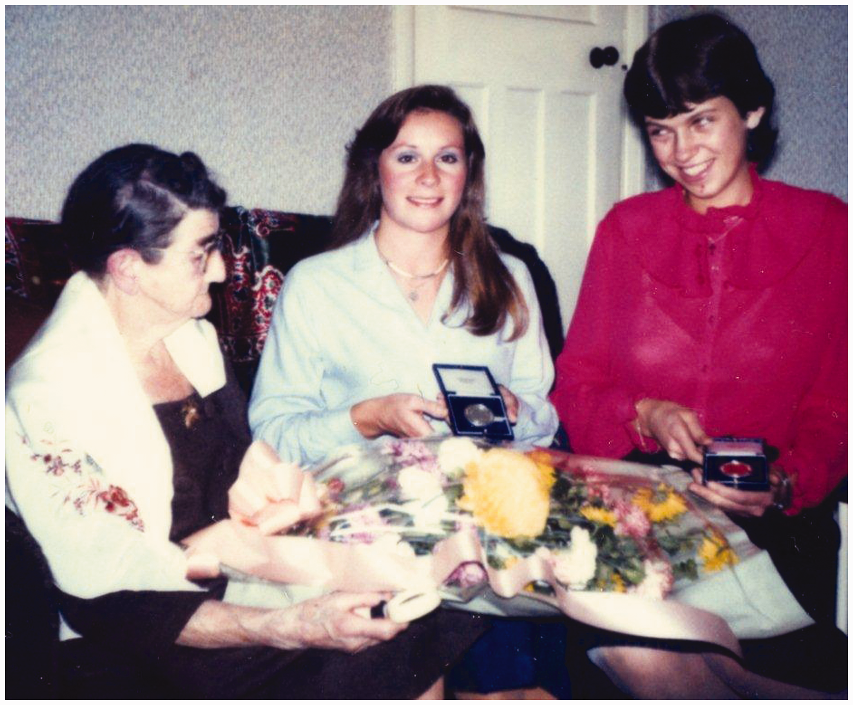
The fourth medal was silver-gilt and to be awarded to the student whose performance throughout the course in computer science for the degree of BSc had been of outstanding merit, named the Williams–Kilburn medal to commemorate the invention of the storage system and construction of the first stored programme digital computer by Professor Sir Frederick Callard Williams (1911–1979) and Professor Tom Kilburn (1921–2001). Cooper remembered attending a lecture to the academic staff at the University in the Autumn of 1948 by Williams, Professor of Electrical Engineering, and the demonstration by his then assistant Kilburn, later Professor of Computer Engineering, of the first computer known as ‘The Baby’. She donated the medal in recognition of the significance of the invention.
In her leisure time Cooper enjoyed classical music, especially opera. She was a great admirer of Sir Thomas Beecham (1869–1961), who had championed French music and especially that of Hector Berlioz (1803–1869). 64 In 1984 she endowed a medal to be awarded annually to the student whose performance through the BMus course of the University had been of outstanding merit. 65 The naming of the medal after Beecham was no doubt strongly supported by the Professor of Music, Ian Manson Kemp (1931–2011), who was an authority on Berlioz's music. 66
Conclusion
Dr Cooper devoted herself to the Department of Anatomy in Manchester for 44 years. Her special interest in histology was apparent from undergraduate days, followed by significant study of the development of the endocrine organs. However, she became recognised as an authority on the anatomical development of the human brainstem from the early weeks of gestation. In one publication of the important neurological journal Brain in 1946, four of the seven papers were by Cooper. Her painstaking and intensive study remains the basis for anatomical understanding today. Her intellectual capacity was evident with her study of law as an interest and continued to be demonstrated by the publication of research in the sphere of antifertility compounds until the age of 77 years.
The medals she donated to the University for medicine are no longer awarded due to changes in the medical curriculum. However, those in computer science and music are still coveted major awards.
In 1958 she was approached by Henry A Herbert for permission to paint her portrait (Figure 3) as part of his graduation in Art in Manchester, which was subsequently donated to the sitter. After her death the painting was presented to the University and now hangs adjacent to the dissecting room of the medical school in the Stopford Building, this being the nearest equivalent now to the anatomy department.
67
As Professor A Crossman, Professor of Anatomy, commented to this author, she was in her prime when histology was viewed as an important discipline to which she made an important contribution.
68
Portrait of Dr Cooper by HA Herbert 1958. Reproduced by kind permission of Professor Alan Crossman, Professor of Anatomy, Faculty of Life Sciences, The University of Manchester.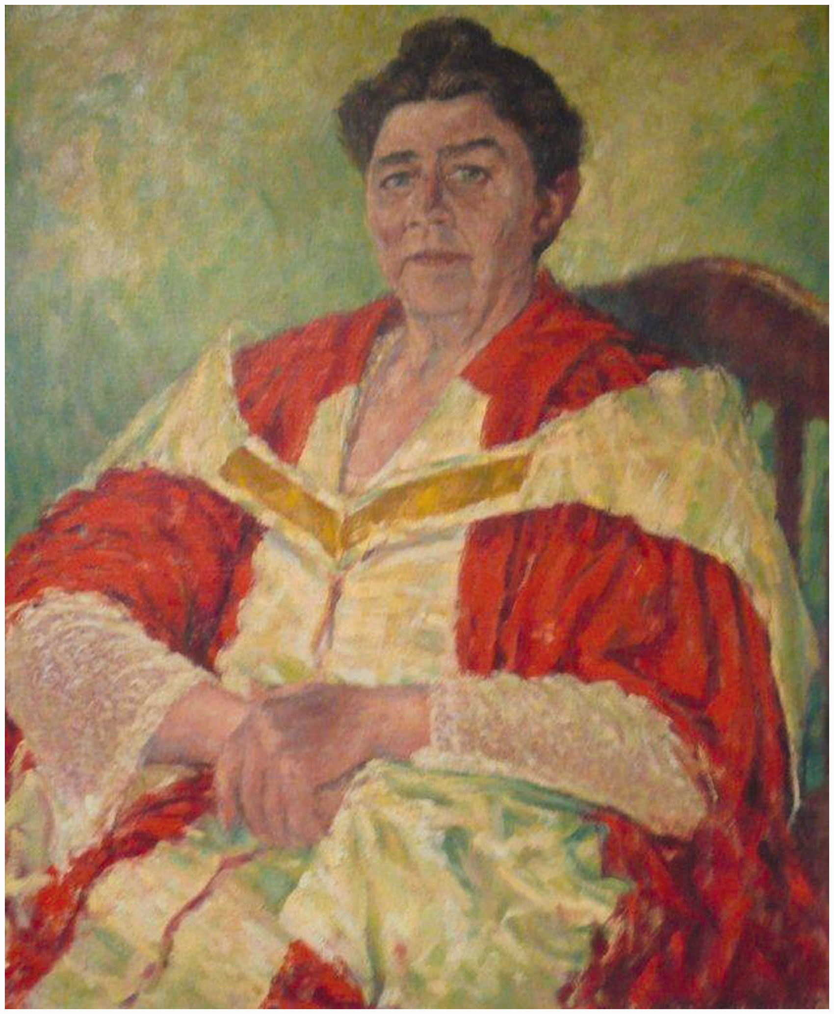
Despite her pre-eminence in her field of anatomy, Cooper was obviously disappointed at not gaining promotion. As she confided to her neighbours Professor Nairn HF Wilson and Dr Margaret Wilson in the 1980s, she felt the University lacked the courage to appoint a female Professor of Anatomy. She had however strongly encouraged female graduates in research and academic careers. 69 Cooper's career underlines the slow change in female emancipation in medicine in the universities. Manchester had only started accepting female medical undergraduates 16 years before the entry of Cooper, and less than 100 had graduated in that period. 70 The telling comment of Professor Mitchell in 1950 emphasised the predominant male attitude of the period.
Dr Cooper never married and was disappointed that her nephew failed to continue his studies in medicine in Manchester. She died from cerebrovascular disease on 12 December 1991 and was cremated at Manchester Southern Cemetery.
Footnotes
Acknowledgements
The author thanks Mrs Janet Burnett for typing the manuscript, Dr James Peters, Archivist The John Rylands University Library, The University of Manchester, for important archive access and Professor John V Pickstone, Centre for History of Science, Technology and Medicine, The University of Manchester, for reviewing the manuscript.