
Abstract
Background
Brain relaxation is attained using several techniques while sleep remains nature’s ultimate remedy. Currently, various machine learning (ML) tools are applied to identify and understand the neural correlates of relaxation from the electroencephalography (EEG) signals. Majority of earlier studies focused on comparing power in the EEG bands during eyes-open and eyes-closed resting state paradigm to train the datasets. However, several Yogic practices are performed using sitting and supine positions.
Purpose
This study was aimed to elucidate the relaxation correlates in EEG between supine and sitting position during eyes-closed condition using ML classifiers.
Methods
EEG signals were recorded on five different days from O1, OZ, O2, C3, CZ, C4, F3, FZ and F4 brain region using nine unipolar electrodes for 25 minutes during eyes-closed supine and eyes-closed sitting postures each on, along with electrocardiogram (ECG) for heart rate variability (HRV) analysis in a healthy participant. Relaxation was assessed by extracting the relative power of the alpha and theta waves from the EEG data and corroborated with the alpha and theta lateralisation index (LI) and HRV parameters. These EEG metrics were analysed by leveraging ML classifiers (K-nearest neighbours (KNN), support vector machine(SVM), random forest (RF) and XGBoost) for relaxation states under sitting and supine states.
Results
Out of all the used classifiers, performance indices of SVM excelled in classifying relaxation states from the EEG alpha and theta band data that was verified with the HRV data and correlated with LI.
Conclusion
This study demonstrates that ML especially the SVM was effective in classifying the relaxation states during different postures from the EEG. LI and HRV metrics effectively decoded the underlying message in the EEG and ECG respectively.
Keywords
Introduction
Hans Berger’s cardinal recording of human electroencephalography (EEG) for the first time in 1924 unlocked the avenues for gaining insight into functionality of brain.1, 2 Observation of alpha waves (Berger’s wave) in the resting state was historical for evoking interest in decoding the underlying message in EEG activity but the pace of research remained slow during initial four to five decades probably due to the limited availability of EEG machines at that time. However, technological advancement in the twenty-first century with digitisation, compact miniaturised machines, increased computational power and application of artificial intelligence (AI) and machine learning (ML) and EEG is becoming an important noninvasive and extensively used technique for recording electrical activities from various cortical areas of brain in clinical and research domains including consciousness, sleep diagnosis, neurological disorders, understanding various cognitive processes and overall well-being.3–12
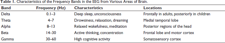
EEG is a complex signal consisting of different frequency bands with distinct functional connotations, for example, delta (0.1–3Hz) for deep sleep, basic homeostatic and motivational processes; theta (4–7 Hz) for drowsiness and meditation; alpha (8–13 Hz) for relaxed wakefulness; beta (14–30 Hz) for active thinking and concentration and gamma (30–60 Hz) for high-level cognitive functions as shown in Table 1. One of the growing concerns in the recent lifestyles is a global increase in insomnia and sleep disorders in all age groups that is associated with concomitant surge in cardiovascular diseases, hypertension, anxiety, depression, cancer and neurodegenerative disorders.13–16 It is pointed that optimal sleep each night provides natural relaxation and recuperation of the body and mind but that is a hard challenge. Various relaxation techniques including Yoga and biofeedback are becoming hope for the management of these health issues.
Characteristics of the Frequency Bands in the EEG from Various Areas of Brain.
Observation of notable increase in alpha waves in the occipital region during relaxation is a hallmark of attaining a calm and restful brain state.17, 18 Alpha power decreases during attention tasks compared to the relaxed state, particularly in the frontal and occipital regions of the brain, so the changes in alpha power can be a significant marker for distinguishing between states of relaxation and attention. 19 Theta wave activity is also often associated with drowsiness or light meditation. Conversely, alpha wave activity decreases when the eyes are open, while beta wave activity increases during alertness and cognitive engagement. The regional distribution and dominance of these individual EEG waveforms in different lobes contribute to various neural processing involved in specific functional and cognitive domains, for example, the frontal lobe in decision-making, the parietal lobe in sensory and spatial orientation, the temporal lobe auditory processing and memory and the occipital lobe for visual processing.20, 21 Hemispheric lateralisation is an interesting dimension of EEG neural processing especially for the alpha lateralisation as it enhances the processing of stimuli at relevant spatial locations while suppressing distractors.22–25 Even though no specific attentive task was planned in this study but it would be interesting to observe any changes in alpha lateralisation in resting state.
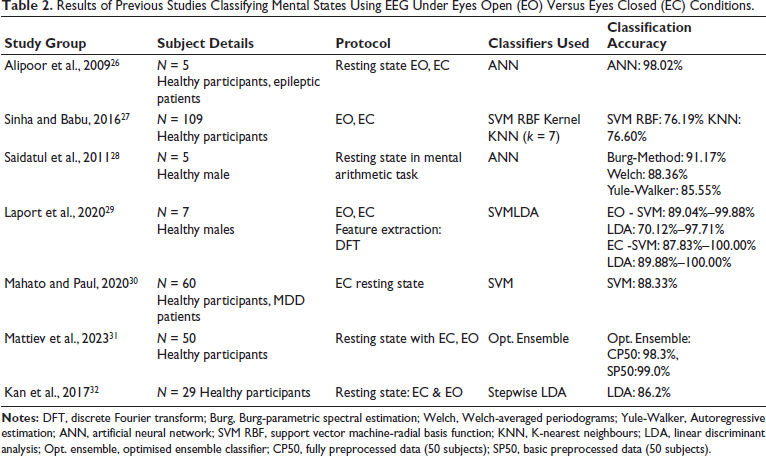
ML is a subset of AI that is broadly categorised into supervised, unsupervised or reinforced learning. In the supervised learning, the labelled dataset is used to train the algorithm for predicting the outcomes while in the unsupervised ML, the machine algorithm learns itself by identifying the patterns in the unlabelled data. In the reinforced learning technique, machine learns through trial and error as the agent learns to derive decisions by interacting with environment, and refine at each step to reach a goal. In recent years, there is surge in application of ML tool for the assessment of the neural correlates of relaxation from the EEG recordings. However, most of the studies have employed supervised ML techniques using eyes-open (EO) versus eyes-closed (EC) paradigm in the resting state (Table 2).26–32 It is pointed that the relaxation practices are conducted in both sitting and supine positions 33 but such studies are scarce. Heart rate variability (HRV) is another clinical tool that is computed from the beat-to-beat changes in the HR. It is a valuable indicator of the autonomic nervous system activity, providing a comprehensive view of sympathetic and parasympathetic balance of the body during rest and stress.34–36 Thus, this study was aimed to classify relaxation states from EEG marker (alpha and theta waves) acquired in different postures (supine and sitting position) in EC condition using ML classifiers. By combining EEG and HRV data, we aimed to deepen our understanding of relaxation across different postures through ML tools as it would be helpful in optimising relaxation techniques for clinical and therapeutic use. To accurately classify relaxation states, ML algorithms like random forest (RF), support vector machine (SVM), K-nearest neighbours (KNN) and XGBoost were used to analyse the EEG data.
Results of Previous Studies Classifying Mental States Using EEG Under Eyes Open (EO) Versus Eyes Closed (EC) Conditions.
Methods
Participant
After taking informed consent, the data were acquired from healthy right-handed subject on five different days in this study. The experimental protocol involved recording EEGs from different parts of brain using nine unipolar electrodes along with ECG under two conditions: (a) 25 minutes in a supine posture with EC and (b) 25 minutes sitting with EC in a quiet air-conditioned room using the 32-channel EEG device. The subject was asked not to get into sleep. For this study, the Institutional Ethics Committee (SCT/IEC/2074/AUGUST/2023) of Sree Chitra Tirunal Institute for Medical Sciences & Technology approval was taken.
Measures
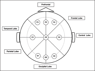
The data was acquired using the Axxonet XAmp 32-channel EEG system (Axxonet system Technologies Pvt Ltd, Bangalore, Karnataka, India) with a sampling rate of 256 Hz and a bandpass filter of 0.2–35 Hz. For recording EEGs, nine unipolar electrodes (O1, OZ, O2, C3, CZ, C4, F3, FZ, F4) were placed on scalp according to the international 10–20 system (Figure 1) and additional two ECG electrodes were placed to record HR. BESS (Brain Electro Scan Software) was used to preprocess EEG data for removing the noise and artifacts. The fast Fourier transform (FFT) was performed on these noise-free signals to extract the relative power of theta (4–7 Hz) and alpha (8–12 Hz) waves taking one-second bins.
For the ECG signals, Kubios HRV software was utilised to analyse HRV taking five minutes epoch length to obtain insights into the sympathetic and parasympathetic balance during each state. Time domain parameters (mean RR, mean HR, min HR, max HR) and frequency domain parameters (low-frequency (LF) power, high-frequency (HF) power) and LF/HF ratios) were extracted.
Data Processing and Machine Learning
Python 3.10 was used along with libraries such as NumPy, pandas, SciPy, seaborn, matplotlib, stats models, Plotly and sci-kit-learn for data curation, feature selection, exploratory data analysis, statistical analysis and model training. The study employed RF, SVM, KNN and XGBoost to classify relaxation postures. RF, SVM, KNN and XGBoost algorithms were used to classify postures based on the features derived from EEG data. Twenty per cent of the data was used for testing, while 80% was used for training. The features included relative power from all unipolar channels, and the target variable was posture (0 for sitting, 1 for supine).
ML Evaluation Parameters and Statistical Methods
Performance of the classifiers were evaluated using confusion matrix, accuracy, precision, recall, specificity, F1 score and area under the curve (AUC) and receiver operating characteristic (ROC) curves as described below:
Confusion matrix to display true positive (TP), true negative (TN), false positive (FP) and false negative (FN); Accuracy: The proportion of correct predictions (TP + TN) out of all predictions; Precision: The proportion of TPs out of all positive predictions = TP/ (TP + FP); Recall: The proportion of TPs out of actual positives = (TP/ (TP + FN); Specificity: The proportion of TNs out of actual negatives = TN/ (TN + FP); F1 Score: The harmonic mean of Precision and Recall. AUC: to measures overall performance across classification thresholds. ROC curve were also checked to compare TP rate versus FP rate.
Lateralisation index (LI) was calculated for alpha and theta power in each channel (C, O, F) by using formula (Ipsilateral power – Contralateral power/Ipsilateral power + Contralateral power) and comparison was carried out between sitting and supine state for each individual area. 25 For the ECG signals, Kubios HRV software was utilised to analyse HRV taking five minutes epoch length to obtain insights into the sympathetic and parasympathetic balance during each state. Time domain parameters (mean RR, mean HR, min HR, max HR) and frequency domain parameters (LF power, HF power) and LF/HF ratios were extracted. These data were analysed using GraphPad PRISM software (version 10). Analysis of variance (ANOVA) and t-tests were conducted to assess differences in relaxation levels between conditions to doubly check the performance of ML. Level of significance was taken as 0.05.
Results
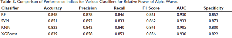
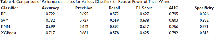
For classifying relaxation states based on the EEG data, ML models for KNN, RF, SVM and XGBoost were evaluated. For the alpha relative power, SVM achieved the highest accuracy of 84.98% and precision of 89.75%, with an AUC of 93.19% (Table 3). For theta relative power, SVM has the highest accuracy of 74.29% and AUC 81.58% in Table 4.
The ROC curves and AUC values showed that all models performed well in classifying brain activity for both alpha and theta power. For alpha power, performance of the SVM model was highest with an AUC of 0.933, indicating it could distinguish between different states very effectively. XGBoost and RF were close behind, each with an AUC of 0.931 and 0.930, proving to be reliable, while KNN, with an AUC of 0.906 (Figure 2A).
Comparison of Performance Indices for Various Classifiers for Relative Power of Alpha Waves.
Comparison of Performance Indices for Various Classifiers for Relative Power of Theta Waves.
Receiver Operating Characteristic Curve of All Classifiers to Classify Relaxation State Using Relative Alpha Power (A) and Theta Power (B).
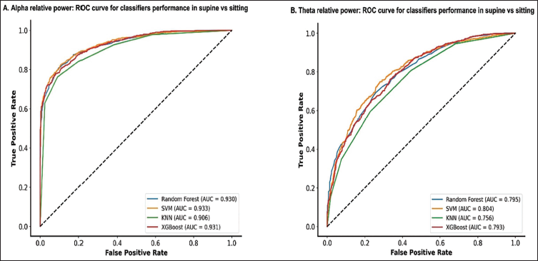
For theta power, SVM performed better with an AUC of 0.804, showing it could pick up differences between states well. RF and XGBoost followed with AUCs of 0.795 and 0.793 respectively and KNN, with an AUC of 0.793, also performed decently (Figure 2B). Overall, the high AUC values across all models suggest they are effective at this task, with SVM standing out as the top performer, especially in recognising subtle changes in brain activity.
Among various ML models evaluated for classifying relaxation states using EEG data, SVM stood out as the most effective classifier for alpha and theta relative power (Tables 2 and 3; Figure 2). Both models achieved the highest AUC values of 0.933 for alpha power, highlighting their superior discriminative abilities. In theta power classification, SVM had also led with AUC values of 0.804. Overall, SVM demonstrated the best balance of precision, recall and AUC, establishing them as the top choices for EEG-based relaxation state classification.
Changes in the relative power of alpha and theta EEG waves from different regions of brain were analysed under two different states EC supine and EC sitting postures (Figure 3). In the supine posture, relative alpha power of EEG was significantly higher in all the recorded regions in comparison to sitting state (Figure 3A). Highest changes in alpha power were evident in the occipital channels (O1, OZ, O2) compared to other channels, whereas lowest change was observed in the CZ channel (p < .05) as depicted in Figure 3A.
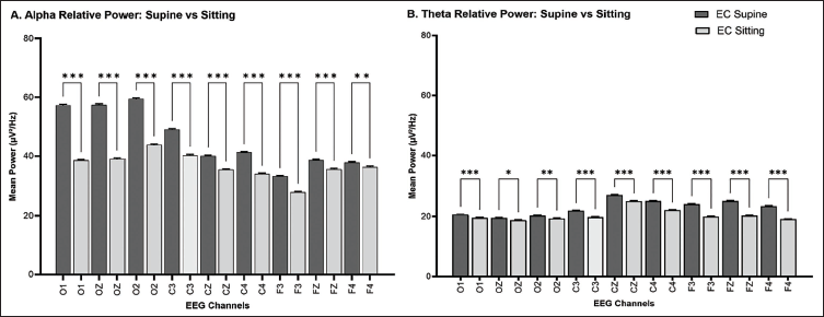
It is noted that the overall relative EEG theta power was lower than the alpha powers during both these EC states (Figure 3). Within recorded brain areas, significant increases in theta powers were found in the occipital channels O1 (p < .05), OZ, central and frontal area (p < .001) and during the supine posture (Figure 3B).
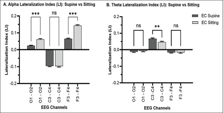
The LI for alpha power band significantly decreased in the occipital and frontal regions, while it increased for the theta power band during transition from the EC supine to EC sitting posture (Figure 4). LI was negative in central region for alpha band, occipital and frontal region for theta band without showing any significant changes between supine to sitting state.
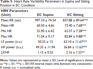
Changes in HRV between the EC supine and EC sitting positions are summarised in Table 5. The mean RR interval increased significantly in the supine position, indicating a slower HR. Both the mean HR and the minimum and maximum HRs were lower in the supine position (p < .001). LF power was significantly higher in the sitting position, while HF power was significantly higher in the supine position (p < .001).
Heart Rate Variability Parameters in Supine and Sitting Position in EC Condition.
Discussion
This study illustrated effects of different body postures on our body and brain relaxation when in resting in supine and sitting in EC condition. Using ML tools and statistical analysis on data from EEG and HRV, we gained valuable insights into how these postures influence our relaxation levels and physiological states.
In this pilot study, the used ML models, that is, RF, SVM, KNN and XGBoost, were effective in classifying relaxation states based on EEG signal. SVM performed best, with high accuracy and strong discriminative capabilities. The successful implementation of ML in this study highlights that by leveraging these advanced models, we can enhance our understanding and treatment of various brain-related conditions for broader applications in EEG analysis.
Significant increase in the absolute alpha power during supine position compared to sitting in EC condition suggests achievement of a deeper state of relaxation and less mental engagement, with a reduction in the visual and sensory processing. Similarly, significant increased theta power while in EC supine, indicated this state promotes profound relaxation akin to meditative states.
Observation of significant changes in alpha and theta relative power between the EC supine and EC sitting postures ensured achieving relaxation states without getting into sleep. 37 In the EC supine posture, consistent higher alpha power in the central, frontal and occipital regions is indicative of relaxation and reduced mental engagement suggests that the supine position promotes greater relaxation compared to sitting.30, 37 In the occipital region, increased alpha power reflects reduced visual processing demands, while in the central region, it suggests decreased somatosensory and motor processing. The frontal region showed reduced mental effort, aligning with increased calmness and relaxation. The consistent rise in alpha power across all EEG channels in the supine position highlights a general state of relaxation, with implications for activities requiring mental and physical rest, such as meditation.
Increased in the EC supine posture increased theta power, associated with relaxation, drowsiness and meditative states support the idea that lying down inhibits cortical activity, enhances relaxation and may benefit cognitive activities by promoting a more restful state even reported in other mental states.38–42 This study provided important insight into positional changes in the EEG in the EC states even though comparison of EC and EO in states provides role of visual stimulus only.31, 32, 42–46 The Yogic practices like Yoga Nidra are generally carried out in corpse position, but it can be performed in sitting position as well; similarly Zen and Vipassna practices can be performed in multiple positional states.47–50 This study provides broader framework to test ML tools to explore neural correlates of relaxation in various condition of practices. 51
In the occipital lobe channel, decreased alpha LI during EC supine condition suggested reduction in cognitive functions in comparison to EC sitting which may be consistent with attention processing demands even in EC state. The negative alpha LI values during both postures of EC sitting and EC supine indicated minimal hemispheric dominance, implying a balanced cognitive state. Similarly, reduced alpha LI in frontal lobe during supine position compared to sitting suggests left hemisphere dominance, often linked to emotional processing and cognitive engagement thereby reflecting increased relaxation. The theta LI values are near zero in occipital and frontal channels for both postures suggesting no significant hemispheric dominance, which aligns with a relaxed cognitive state.
In the central lobe, the negative LI values for alpha in both EC sitting and EC supine suggested a slight right hemisphere dominance, indicating consistent cognitive processing or attentional focus. The positive LI values for theta in both postures indicate balanced engagement between hemispheres, with a slight left hemisphere engagement during EC sitting and a more balanced state in EC supine. These patterns of increased alpha and theta power across both hemispheres could guide the development of relaxation techniques tailored to individual neural activity and hemispheric dominance.
The potential application of AI–ML tools is fast growing with availability of large digital data sets and thus opening new avenues and holds future for real-time monitoring systems for relaxation and stress management in healthy population as well as for therapeutic well-being assistance and also early detection of various neurodegenerative diseases including dementia, Alzheimer’s disease, depression, epilepsy, Parkinson’s disease, schizophrenia and even for forensic age mapping.52–62 These techniques can also be applied to other neuroimaging data derived from functional magnetic resonance imaging (fMRI) and positron emission tomography (PET) scans, offering more precise and personalised insights into brain activity. These findings suggest that brain states differ significantly between the two postures: EC sitting promotes more left hemisphere engagement linked to emotional processing and cognitive activity, while EC supine facilitates a more balanced or relaxed state, as indicated by the generally lower LI values across channels. Moreover, recent studies on role of alpha hemispheric power during various condition including normal sleep, aging and under condition of stimulus suggest for its broader implication in attention control that may not entirely depend on a particular task.63–65
Conclusion and Limitations
This pilot study emphasises the intricate connection between body posture, brain responses and relaxation levels. This research contributes to a better understanding of posture influences on our physical responses and cognitive processes, with potential implications for healthcare, neurology, and practices promoting wellness/Yoga programs.
One limitation of this study was small sample size and repeated data from the same participant due to limited time study for thesis work. Larger sample size from various age groups for generalisability, less bias, more channel (high density EEG for propagation study) would be appropriate. Beyond the models used in this study, other ML algorithms like decision trees, gradient boosting machines, naive Bayes and ensemble methods can be explored for EEG analysis. Each of these classifiers offers unique strengths and could contribute to more robust and accurate classification systems. Exploring these algorithms could further enhance the detection and monitoring of relaxation states and other cognitive conditions.
Footnotes
Acknowledgements
KKG thanks SCTIMST, Trivandrum, an Institute of National Importance under Department of Science and Technology (DST), Government of India for providing the infrastructure and facility for conducting this pilot study. KKG acknowledges the research grant DST/SATYAM/2020/303 dated 29/01/2024 from DST. This research study was carried out at SCTIMST, Trivandrum towards the MSc thesis work of CG (Student, Department of Computational Biology & Bioinformatics, University of Kerala, Thiruvananthapuram, India).
Authors’ Contributions
KKG conceived the idea, recruitment of subject, conduction of study, interpretation of results, analysis and drafting of manuscript, overall supervision of the study and approved the final version of the manuscript.
CG helped in EEG analysis and drafting of manuscript and approved the final version of the manuscript.
Data Availability Statement
The datasets generated during and/or analysed during this study are available from the corresponding author on reasonable request.
Statement of Ethics
This study was approved by the Institutional Ethics Committee the Sree Chitra Tirunal Institute for Medical Sciences and Technology, Trivandrum (SCT/IEC/2074/AUGUST/2023) and written informed consent was taken from the subject.
Declaration of Conflicting Interest
The authors declared no potential conflicts of interest with respect to the research, authorship and/or publication of this article.
Funding
The authors received following financial support for the research, authorship and/or publication of this article: The author (KKG) utilised learning resource allowance from her institute for publication of this article.