






Abstract
Background
Liver cirrhosis, a chronic and progressive liver disease, represents the end-stage of several hepatic injuries, characterized by widespread fibrosis, nodule formation, and ultimately, liver dysfunction and failure. Existing treatments often focus on managing complications rather than addressing the underlying pathological processes, which underscores the need for safer, potentially alternative therapeutic agents.
Purpose
The present work was performed to assess the ameliorative mechanisms and hepatoprotective properties of rosiridin against thioacetamide-induced liver cirrhosis in experimental rats.
Materials and Methods
Liver cirrhosis was experimentally induced in rats by administering 200 mg/kg of thioacetamide thrice per week for 2 months. Rosiridin and silymarin were administered to the rats for 2 months. After treatments, liver dysfunction markers and biochemical parameters were evaluated in the experimental rats. The concentrations of inflammatory and oxidative stress markers were evaluated in the experimental rats. The liver tissues were obtained and underwent histopathological assessments.
Results
The present findings demonstrate that rosiridin treatment markedly reduced liver dysfunction markers and regulated serum biochemical markers in thioacetamide-induced rats. Furthermore, the treatment with rosiridin considerably reduced the pro-inflammatory cytokine levels and oxidative stress markers, while enhancing the antioxidant concentrations in the thioacetamide-induced rats. In addition, histopathological analysis revealed that rosiridin ameliorated histopathological alterations in thioacetamide-induced rats.
Conclusion
The findings of this work indicated that rosiridin markedly ameliorated the development of thioacetamide-triggered liver cirrhosis in rats. These findings highlight that rosiridin may serve as a promising treatment agent for liver cirrhosis.
Introduction
The liver, a vital organ, performs a myriad of essential functions, including metabolism, detoxification, and protein synthesis. Its complex architecture and dual blood supply from the hepatic artery and portal vein facilitate these diverse roles, making it highly susceptible to injury from various endogenous and exogenous insults. Liver cirrhosis represents the end-stage appearance of chronic liver disease, characterized by persistent inflammation, extensive hepatocyte necrosis, and the development of advanced fibrosis (GBD 2017 Cirrhosis Collaborators, 2020). This condition is histologically defined by the development of regenerative nodules enclosed within dense fibrous bands, which invariably results in portal hypertension and end-stage liver failure. Despite the significant global health burden posed by cirrhotic disease, pharmacological interventions to prevent or reverse hepatic fibrosis remain limited, thereby underscoring the critical need for a more comprehensive elucidation of the molecular mechanisms driving hepatic fibrogenesis (Devarbhavi et al., 2023). The sustained liver injury, regardless of its etiology, drives chronic inflammation and progressive fibrosis, which are the hallmarks of cirrhosis. While the etiology of cirrhosis is multifactorial, including chronic viral hepatitis, alcohol, and non-alcoholic steatohepatitis, the common pathway converges on a progressive accumulation of fibrotic tissue. This advanced stage of liver disease is a foremost global cause of mortality, accountable for nearly 1.3 million mortalities (Huang et al., 2023).
The intricate biochemical conversions occurring in the liver, particularly those involving xenobiotics, significantly increase reactive oxygen species (ROS) production, which are critical mediators of hepatocellular damage. These liberated radicals, often produced by hepatotoxins such as thioacetamide, can induce severe cellular damage, progressing from centrilobular hepatic necrosis to widespread cirrhosis with chronic exposure. Drug-induced liver injury is a significant subset of acute liver injury, often resulting from the liver’s attempt to metabolize drugs into more excretable forms, a process that can inadvertently generate toxic metabolites (Ginès et al., 2021). This phenomenon can manifest as an unpredictable idiosyncratic reaction or as a predictable consequence of intrinsic drug toxicity, encompassing a wide range of hepatic lesions. Such injuries can range from mild, asymptomatic increases in liver enzymes to serious liver failure, often necessitating a thorough understanding of the underlying mechanisms to identify risk factors and develop effective therapeutic strategies (Tapper et al., 2022).
While liver transplantation remains the definitive treatment for advanced cirrhosis, its widespread application is limited by organ scarcity and the intensive post-operative care required. Despite improvements in managing complications, current pharmacological interventions largely address symptoms rather than the underlying fibrotic process. Despite significant advances in understanding the molecular mechanisms underlying hepatic fibrogenesis, effective pharmacological strategies to prevent or reverse this process remain elusive (Zheng et al., 2024). Existing treatments often focus on managing complications rather than directly addressing the underlying pathological processes of fibrosis and nodule formation. Therefore, there is an urgent need to develop safer, more effective alternative therapeutic agents that can specifically target fibrotic pathways and potentially reverse the damage in cirrhotic livers (Yoshiji et al., 2021). Rosiridin is a bioactive monoterpene compound derived from the plants Rhodiola rosea and Rhodiola sachalinensis, exhibiting significant monoamine inhibitory properties and being advantageous for the treatment of depressive episodes and early-stage dementia (van Diermen et al., 2009). Several previous studies reported that rosiridin showed neuroprotective properties against a Parkinson’s disease model (Rafeeq et al., 2025), anti-Huntington’s disease effects (Afzal et al., 2022), and ameliorated memory impairments in AlCl3-induced rats (Alqarni et al., 2024). Furthermore, rosiridin also demonstrated anti-diabetic activity (Kazmi et al., 2025a), antioxidant activity (Lee et al., 2000), and nephroprotective activity against cisplatin-induced renal toxicity (Kazmi et al., 2025b). Nevertheless, no study has been undertaken on the effect of rosiridin on liver toxicity and liver cirrhosis induced by thioacetamide. This research aims to examine the hepatoprotective potential of rosiridin against thioacetamide-induced liver injury and cirrhosis in rats.
Materials and Methods
Chemicals
The major chemicals used in this study, such as rosiridin (≥90%), thioacetamide, and others, were obtained from Sigma-Aldrich, USA. The assay kits for estimating biochemical markers were purchased commercially from Abcam and Elabscience, USA, respectively.
Experimental Rats
Six- to eight-week-old Sprague Dawley rats weighing 180–200 g were acquired from the institutional animal house. Animals were treated humanely and kept in accordance with the Animal Research: Reporting of In Vivo Experiments (ARRIVE) guidelines, and their regular circadian rhythms were maintained during the investigation. All animals were caged in wire-bottomed cages at 25°C ± 2°C, on a 12-h light–dark cycle, fed a standard pellet diet, and provided tap water. They were also kept in a room with a humidity range of 50%–60%. The present study was approved by the ethical committee of Affiliated Hospital of Hebei University, China.
Experimental Groups
Rats were distributed randomly into five experimental groups of six animals each. The control group (Group I) was given distilled water (5 mL/kg) daily and received intraperitoneal administrations of 10% Tween-20 as a vehicle thrice a week for 2 months. To create the liver cirrhosis model (Group II), thioacetamide (200 mg/kg) was given intraperitoneally three times a week for 2 months. They were also given an oral dose of vehicle Tween-20 daily. Groups III and IV rats received rosiridin orally at a dosage of 10 and 20 mg/kg daily, and three intraperitoneal administrations of 200 mg/kg of thioacetamide each week for 2 months. Group V rats received daily oral treatment of silymarin at a 50 mg/kg dosage, along with thioacetamide administration, as described in Group II. Rats were given ketamine (50 mg/kg) and xylazine (5 mg/kg) anesthesia at the end of the treatments after fasting for 24 h. The jugular vein was used to extract blood, which was then utilized for biochemical assays. The liver tissues were collected immediately and utilized for further biochemical and histological studies.
Analysis of Biochemical Marker Levels
A commercial test kit was used to assess the liver dysfunction marker enzymes, namely, alkaline phosphatase (ALP), alanine transaminase (ALT), aspartate transaminase (AST), and gamma-glutamyl transferase (GGT), in the serum of experimental rats, following the manufacturer’s instructions (Abcam, USA). The concentrations of albumin, globulin, total bilirubin, and total protein in rat serum were assessed using commercial test kits according to the manufacturer’s specifications (Elabscience, USA).
Analysis of Oxidative Stress Markers
Using commercial test kits, the levels of oxidative stress biomarkers such as catalase (CAT), glutathione peroxidase (GPx), superoxide dismutase (SOD), and malondialdehyde (MDA) were examined in the liver tissue homogenates of the experimental rats. In accordance with the manufacturer’s recommended procedures, the tests were carried out in triplicate (Abcam, USA).
Analysis of Inflammatory Marker Levels
The concentrations of inflammatory cytokines, including interleukin (IL)-10, IL-6, and tumor necrosis factor-alpha (TNF-α), in the serum of experimental rats were examined using commercially available test kits. The tests were conducted in triplicate as per the manufacturer’s specifications (Elabscience, USA).
Histopathological Analysis
The collected liver tissue samples were processed with a 10% neutral formalin solution to evaluate the changes in their tissue architecture. Following paraffinization, the liver tissues were cut into 5-µm-diameter slices and stained with eosin-hematoxylin. Finally, a microscope was used to examine liver tissue to assess histopathological alterations.
Statistical Analysis
GraphPad Prism software was used to analyze the values, and the results are presented as the mean ± SD of triplicates. One-way analysis of variance (ANOVA) and Duncan’s Multiple Range Test (DMRT) analyses are used to examine the data, with a significance threshold of p < .05.
Results
Effect of Rosiridin on Liver Dysfunction Markers in the Serum of Thioacetamide-induced Rats
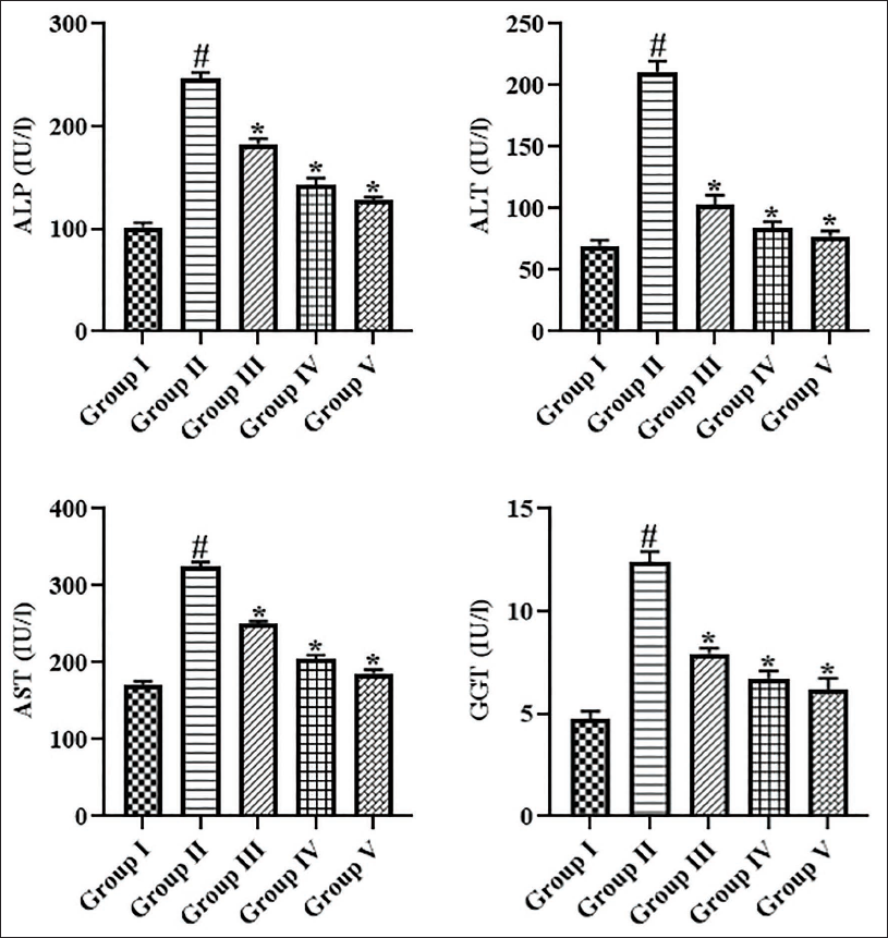
Figure 1 shows the results of the rosiridin’s effects on the liver dysfunction marker enzymes in the serum of experimental rats. In comparison to control, the ALT, AST, ALP, and GGT levels were significantly increased in the thioacetamide-induced rats, which evidences the onset of liver tissue damage. Surprisingly, rosiridin at doses of 10 and 20 mg/kg considerably decreased these marker enzymes in the serum of thioacetamide-treated rats. These results are further corroborated by findings from standard drug treatment with silymarin.
Effect of Rosiridin on Biochemical Marker Levels in the Serum of Thioacetamide-induced Rats
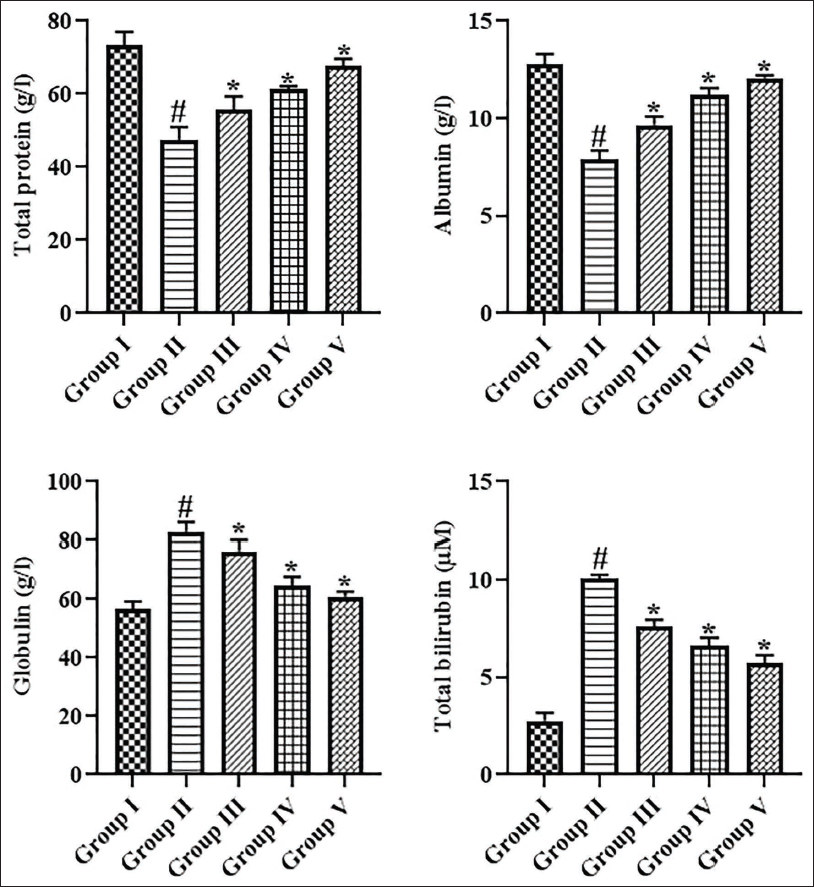
The levels of biochemical markers, including globulin, albumin, total protein, and total bilirubin, in the serum of rats were measured, and the results are shown in Figure 2. The serum levels of globulin and total bilirubin were elevated, and subsequently albumin and total protein were reduced in the thioacetamide-treated rats. On the other hand, these alterations were significantly prevented and reversed by rosiridin treatment at 10 and 20 mg/kg (Figure 2). The outcomes of the silymarin treatment further supported the effectiveness of rosiridin.
Effect of Rosiridin on Oxidative and Antioxidant Marker Levels in the Liver Tissues of Thioacetamide-induced Rats
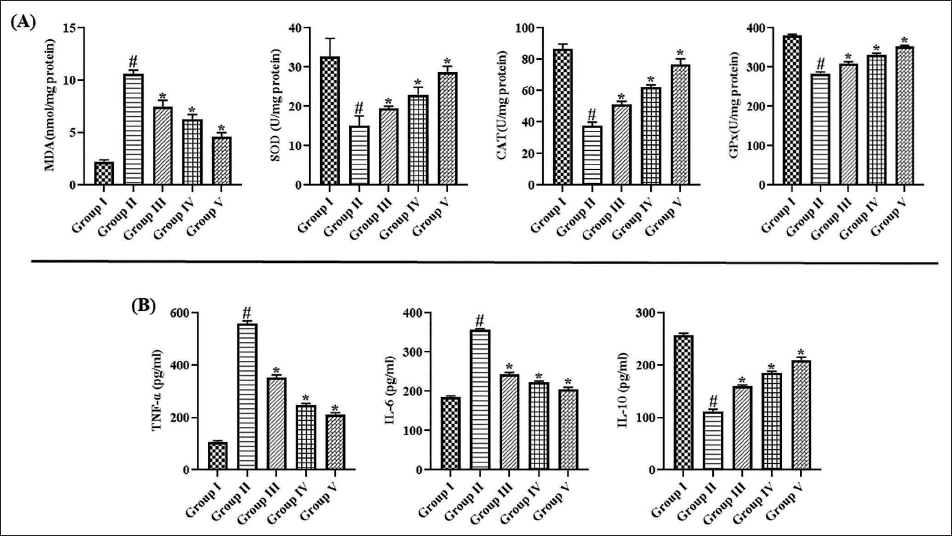
Figure 3(A) illustrates the outcomes of the rosiridin treatment on the alterations in oxidative and antioxidant biomarkers in the liver of thioacetamide-induced rats. The thioacetamide-induced rats exhibited a substantial elevation in MDA, while reduced antioxidants CAT, GPx, and SOD in their liver tissues in comparison to the control. Interestingly, the rosiridin treatment at dosages of 10 and 20 mg/kg, respectively, led to a reduction in MDA and elevations in antioxidants in the liver tissues of thioacetamide-treated rats, which evidences its antioxidant effects. These results are corroborated by findings from standard drug treatment with silymarin.
Effect of Rosiridin on Inflammatory Markers in the Serum of Thioacetamide-induced Rats
The inflammatory cytokine concentrations in the serum of experimental rats are assessed and presented in Figure 3(B). The thioacetamide-treated rats revealed an increase in TNF-α and IL-6 levels, while reducing the IL-10 level in their serum when compared with the controls. Captivatingly, the treatment with 10 and 20 mg/kg dosages of rosiridin to the thioacetamide-induced rats resulted in a significant reduction in TNF-α and IL-6 and elevated the IL-10 levels in their serum. These results are found similar to the findings of silymarin treatment, which supports the anti-inflammatory activity of rosiridin.
Effect of Rosiridin on Liver Histopathology of the Thioacetamide-induced Rats
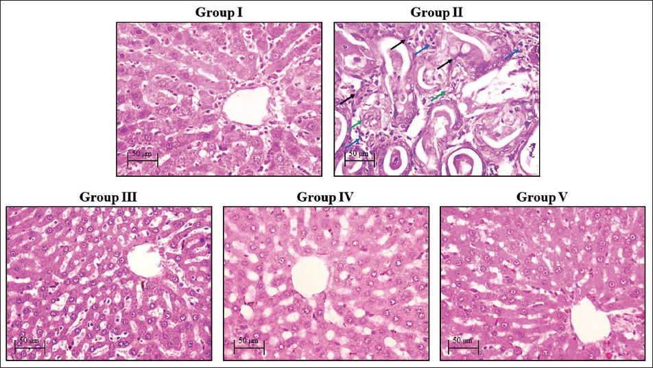
Figure 4 illustrates the findings of histopathological analysis of liver tissues from experimental rats. The liver tissues of rats in the control group exhibited smooth surfaces devoid of nodules. However, the liver tissues of thioacetamide-induced rats had a pronounced presence of fibrous septa, proliferating bile ducts, and inflammatory cell infiltrations when compared with the control. Interestingly, both silymarin- and rosiridin-treated rats (10 and 20 mg/kg, respectively) exhibited a reduced number of micronodules, diminished septa, and an increased presence of normal hepatocytes in their liver tissues.
Discussion
Liver cirrhosis is a chronic hepatic condition, defined by widespread fibrosis and the formation of regenerative nodules, which fundamentally disrupts liver architecture and function, leading to progressive hepatocellular decompensation. Annually, liver cirrhosis accounts for approximately 2 million deaths globally, representing 4% of all mortalities, with a disproportionately higher incidence in men. This condition arises from the irreversible scarring of liver tissue, in which fibrotic tissue progressively replaces healthy hepatocytes, impairing hepatic function (Nie et al., 2023). This pathological transformation critically compromises the liver’s capacity for detoxification, protein synthesis, and metabolic regulation, ultimately leading to severe systemic complications. This extensive scarring is the culmination of chronic liver injury, which activates hepatic stellate cells and promotes the deposition of extracellular matrix proteins that gradually replace functional liver parenchyma. This structural distortion precipitates portal hypertension and subsequent systemic complications (Xiao et al., 2023). The long-term pathological consequence of this progressive distortion, particularly from chronic viral hepatitis, is the evolution into severe cirrhosis, which manifests through a range of life-threatening problems. Given the escalating prevalence and complexity of cirrhosis, the need for novel therapeutic methods is imperative to alleviate disease progression and improve patient outcomes. Current therapeutic approaches often focus on symptom management and complication prevention rather than addressing the underlying fibrotic process or reversing established liver damage (Kisseleva & Brenner, 2021). Consequently, there is a need for innovative and safer alternative therapeutic agents that can effectively halt or reverse the fibrosis, thereby addressing the fundamental pathological drivers of cirrhosis. While liver transplantation remains the primary treatment for end-stage cirrhosis, its application is restricted by organ availability and patient suitability. Consequently, there is a critical need for less invasive pharmacological interventions that can therapeutically target the mechanisms of fibrosis, with broader applicability for patients who are not candidates for transplantation. Therefore, this work addressed the therapeutic effects of rosiridin against thioacetamide-induced liver toxicity cirrhosis (Romanelli & Stasi, 2016).
The liver dysfunction marker enzymes play a crucial role in the diagnosis of liver cirrhosis, as their fluctuating levels are indicators of disease progression and severity. Specifically, ALT and AST enzymes are commonly referred to as transaminases and serve as crucial indicators of hepatocellular injury, necessitating further investigation upon elevated levels. The ratio of AST to ALT can provide additional clinical insight into the etiology and severity of liver disease, with AST often exceeding ALT in advanced cases such as cirrhosis (Iluz-Freundlich et al., 2020). Furthermore, GGT and ALP are cholestatic enzymes, and their elevated levels typically signify biliary obstruction or cholestasis, although GGT can also reflect broader liver pathology and even indicate tumorigenesis. Additionally, assessing these enzymes provides a holistic view of liver function and injury. Such a comprehensive evaluation allows clinicians to monitor disease progression, predict prognosis, and guide therapeutic interventions for patients suffering from liver cirrhosis. It has already been indicated that the elevated levels of these enzymes are commonly associated with liver damage. Moreover, the dynamic changes in these enzyme levels over time offer valuable prognostic information, often correlating with the stage of fibrosis and the onset of portal hypertension or hepatic encephalopathy (Cheng et al., 2020). The intricate interplay between these enzymes and other systemic factors can further elucidate the underlying pathogenic mechanisms driving cirrhotic transformation and its associated sequelae. Given the multifaceted roles of these biomarkers, a deeper understanding of their individual kinetic profiles and interactions within the complex milieu of liver metabolism is essential to refine diagnostic accuracy and therapeutic approaches in cirrhosis. For instance, GGT has shown particular sensitivity in detecting hepatic disease, often more so than ALP, and its activity can be disproportionately elevated in cases of biliary obstruction compared to viral hepatitis (Kwo et al., 2017). Therefore, it is essential to evaluate the changes in these liver enzymes to diagnose and predict the stages of liver damage and cirrhosis. The present findings revealed that the ALT, AST, ALP, and GGT were increased considerably in the thioacetamide-induced rats due to the onset of liver tissue damage. However, the rosiridin treatment markedly decreased the serum levels of these liver marker enzymes in thioacetamide-treated rats. Therefore, it was evident that rosiridin treatment mitigated the liver tissue damage in the thioacetamide-induced rats.
The pathophysiology of liver cirrhosis is intricately linked to alterations in hepatic protein synthesis and metabolism, which are commonly reflected in the levels of total protein, albumin, globulin, and total bilirubin, serving as critical indicators of liver function and disease severity. These biochemical markers provide valuable insights into the liver’s synthetic capacity, its inflammatory state, and the degree of cholestasis, thereby offering prognostic value and guiding clinical management in cirrhotic patients (Sharma, 2022). Specifically, albumin levels reflect the liver’s synthetic function, as it is produced exclusively by hepatocytes, whereas changes in globulin concentrations often reflect the immune response and inflammatory processes associated with chronic liver injury. In cirrhosis, compromised liver function leads to reduced albumin synthesis and often a diminished quality of the existing albumin due to increased oxidative stress and inflammation, further impairing its physiological roles. This reduction in both quantity and quality of albumin exacerbates fluid imbalances, compromises drug pharmacokinetics, and impairs immune responses in cirrhotic patients (Chen et al., 2020).
Moreover, bilirubin, a product of heme catabolism, serves as a crucial marker of hepatic excretory function and cholestasis, with elevated levels often signifying impaired bile flow or hepatocellular dysfunction. The detailed examination of these biochemical parameters is therefore paramount for assessing the extent of hepatic compromise, predicting clinical outcomes, and optimizing therapeutic interventions in patients with liver cirrhosis (López-Velázquez et al., 2013). The focus on these specific biochemical markers, such as total protein, albumin, globulin, and total bilirubin, is rooted in their established utility as non-invasive indicators of the dynamic pathophysiological processes occurring within the cirrhotic liver, providing a readily accessible means to monitor disease progression and treatment efficacy (Newsome et al., 2018). The findings of this study exhibited that the serum levels of globulin and total bilirubin were increased, while albumin and total protein were diminished in the thioacetamide-induced rats. Interestingly, these alterations were considerably ameliorated by rosiridin treatment in thioacetamide-induced rats.
Oxidative stress is increasingly recognized as a pivotal factor in the pathogenesis of several chronic liver diseases, including cirrhosis. This condition can result in significant oxidative damage to hepatic tissues, thereby exacerbating liver injury and contributing to disease progression. Specifically, oxidative stress induces hepatic damage through the changes of crucial biological molecules like DNA, proteins, and lipids, while also modulating gene transcription, protein expression, and cellular apoptosis (Allameh et al., 2023). This cellular damage can precipitate mitochondrial dysfunction, further intensifying oxidative stress and perpetuating liver injury. The liver’s inherent metabolic activities and its role in xenobiotic biotransformation make it particularly susceptible to ROS accumulation, leading to this disruptive redox imbalance. This pro-oxidant state is further exacerbated in decompensated cirrhosis, leading to systemic inflammation, mitochondrial dysfunction, and impaired pleiotropic functions of albumin, all contributing to multiorgan dysfunction and failure (Mooli et al., 2022).
The sustained oxidative stress observed in cirrhotic patients also triggers the activation of redox-sensitive transcription factors, such as Nuclear factor-kappa B (NF-κB), which subsequently upregulate the expression of pro-inflammatory mediators in Kupffer cells, thereby amplifying hepatic inflammation and fibrosis. This persistent inflammatory state, coupled with direct oxidative damage, accelerates the fibrotic process, ultimately leading to the structural distortion and functional impairment characteristic of advanced liver disease (Li et al., 2015). Understanding the interplay between these oxidative stress markers and antioxidant enzymes is crucial for developing novel salutary methods focused on mitigating hepatic damage and enhancing patient outcomes. Under physiological conditions, the liver is equipped with robust antioxidant defenses to neutralize the adverse effects of ROS and maintain cellular homeostasis (Han et al., 2016). However, in the context of liver diseases and particularly cirrhosis, this delicate balance is profoundly disrupted, leading to an overwhelming accumulation of pro-oxidants that overwhelm the endogenous antioxidant capacities. This imbalance contributes significantly to hepatocellular damage, perpetuating inflammation and fibrogenesis (Salomone et al., 2016). In a similar manner, the present findings also revealed the increased oxidative stress marker MDA levels and reduced antioxidant levels in the livers of the thioacetamide-induced rats. Interestingly, the rosiridin treatment markedly reduced MDA levels and increased antioxidant levels in the liver tissues of thioacetamide-induced rats, suggesting its antioxidant activity.
Inflammation and inflammatory cytokines, such as TNF-α, IL-6, and IL-10, were profoundly influenced by the intricate cellular interactions driving hepatic fibrogenesis. The dysregulation of these cytokines can either promote or inhibit fibrogenesis, critically influencing the trajectory of liver disease progression. The persistent inflammatory state in chronic liver diseases drives the progression from initial injury to fibrosis, ultimately leading to cirrhosis by promoting the excessive deposition of extracellular matrix components (Taru et al., 2024). TNF-α, a potent pro-inflammatory cytokine, significantly contributes to hepatic injury and fibrogenesis by promoting the recruitment of inflammatory cells, inducing hepatocyte apoptosis, and activating hepatic stellate cells, thereby accelerating collagen production and extracellular matrix deposition. Conversely, IL-6, while having a dual role, primarily acts as a pro-inflammatory mediator that sustains the acute phase response and contributes to hepatocyte proliferation and survival, but can also exert pro-fibrogenic effects by promoting stellate cell activation (Scarlata et al., 2024). In contrast, IL-10, an anti-inflammatory cytokine, generally attenuates liver fibrosis by inhibiting pro-inflammatory cytokine production, suppressing immune cell activation, and reducing hepatic stellate cell proliferation and collagen synthesis, thus offering a protective effect against disease progression. This intricate interplay among pro-inflammatory and anti-inflammatory cytokines dictates the delicate balance between liver regeneration and fibrotic progression (Clària et al., 2023). Understanding the precise roles of these cytokines in the context of chronic liver inflammation is paramount for developing targeted therapeutic strategies to inhibit or reverse the progression from liver fibrosis to cirrhosis and improve patient outcomes. Further elucidation of these pathways is crucial for designing precision medicine approaches that can effectively modulate the inflammatory milieu and counteract fibrogenesis, offering new avenues for therapeutic intervention. Furthermore, therapeutic strategies targeting these cytokine pathways, such as blocking pro-fibrotic cytokines or augmenting anti-fibrotic ones, hold promise for attenuating disease progression and improving clinical outcomes (Kaps et al., 2023). In this study, the results exhibited that thioacetamide-induced rats demonstrated a considerable elevation in TNF-α and IL-6 levels, while reducing the IL-10 in their serum. Whereas, the treatment with rosiridin in thioacetamide-induced rats effectively reduced the TNF-α and IL-6 levels and augmented the IL-10 levels in their serum, which supports the anti-inflammatory activity of rosiridin.
Conclusion
The results of this study elucidated the hepatoprotective effects of rosiridin against thioacetamide-treated liver injury and cirrhosis. The findings of our study indicated that rosiridin treatment restored liver marker enzyme levels, regulated biochemical markers, decreased inflammatory responses, and mitigated the oxidative stress response in thioacetamide-induced rats. These findings indicated the advantageous hepatoprotective effects of rosiridin. However, further study is necessary to provide additional evidence on the hepatoprotective characteristics of rosiridin, thereby establishing it as a talented therapeutic agent for hepatotoxicity and cirrhosis.
Footnotes
Abbreviations
ALP: Alkaline phosphatase; ALT: Alanine transaminase; AST: Aspartate transaminase; CAT: Catalase; GGT: Gamma-glutamyl transferase; GPx: Glutathione peroxidase; IL: Interleukin; MDA: Malondialdehyde; SOD: Superoxide dismutase; TNF-α: Tumor necrosis factor-alpha.
Declaration of Conflicting Interests
The authors declared no potential conflicts of interest with respect to the research, authorship, and/or publication of this article.
Ethical Approval
The present study was approved by the ethical committee of Affiliated Hospital of Hebei University, China.
Funding
The authors received no financial support for the research, authorship, and/or publication of this article.
Informed Consent
Not applicable.
