






Abstract
Background
Breast cancer remains the most widely diagnosed cancer among women worldwide and a major cause of cancer-associated deaths, posing a substantial and escalating burden on public health systems worldwide.
Purpose
This study was intended to evaluate the anti-cancer effects of albiflorin against breast cancer cells.
Materials and Methods
The effect of albiflorin treatment at different dosages (1–50 µM) on the growth of MDA-MB-231 cells was evaluated using the MTT assay. The mitochondrial membrane potential (MMP) level and apoptosis in the albiflorin-treated breast cancer cells were assessed using fluorescent staining assays. The apoptotic proteins Bax, Bid, and Bcl-2 levels in the albiflorin-treated breast cancer cells were studied using kits.
Results
The findings of the MTT test revealed that albiflorin treatment significantly diminished the MDA-MB-231 cell viability. The albiflorin treatment diminished the MMP levels and triggered apoptosis in the breast cancer cells, as shown by fluorescent staining studies. Additionally, the albiflorin-treated MDA-MB-231 cells showed augmented pro-apoptotic Bax and Bid protein levels and reduced anti-apoptotic Bcl-2 levels.
Conclusion
The present study suggests that albiflorin can inhibit viability and promote apoptosis in breast cancer cells. Thus, the current data suggest albiflorin as a promising candidate that can be utilized for breast cancer management in the future.
Introduction
Breast cancer is characterized by uncontrolled cellular proliferation and tumor formation within the mammary glands, representing a complex and multifaceted disease. This abnormal cellular growth occurs when the body’s normal regulatory signals, which typically halt cell division, are ignored, leading to the continuous multiplication of damaged or aged cells (Bray et al., 2024). It is the most commonly diagnosed cancer and the major factor in cancer-associated deaths among women globally. The global burden of breast cancer has been gradually increasing over the past years, necessitating a comprehensive understanding of its prevalence, causes, and associated burdens to inform effective intervention strategies. Globally, cancer ranks as a primary or secondary cause of death worldwide, underscoring its profound impact on public health (Iacoviello et al., 2021). Specifically, breast cancer alone accounts for one in eight cancer patients globally, making it the most widespread type of cancer globally. Its growing burden is particularly notable in developing countries, where rising incidence and persistent high mortality rates demand urgent global intervention. This alarming statistic underscores the critical need for enhanced diagnostic capabilities and treatment modalities, especially given projections that the incidence will exceed 3 million new incidences and over 1 million mortalities by 2040 (Giaquinto et al., 2022). These projections highlight the escalating crisis, necessitating advanced research into early detection, personalized therapies, and preventative measures to mitigate the substantial human and economic costs. Therefore, understanding the underlying mechanisms of breast cancer, from genetic predispositions to environmental factors, is paramount for developing targeted interventions and enhancing patient outcomes (Heer et al., 2020).
Apoptosis, a form of programmed cell death, is a critical physiological mechanism that removes damaged or unwanted cells, thereby sustaining tissue homeostasis. In the context of tumors, particularly breast cancer, dysregulation of apoptosis is a hallmark that permits tumor cells to evade normal cellular constraints and proliferate uncontrollably (Singh et al., 2019). This evasion often stems from alterations in intrinsic apoptotic pathways, characterized by the activation of intracellular proteins leading to cellular demise, or extrinsic pathways initiated by external death signals. Consequently, understanding the intricate mechanisms by which breast cancer cells subvert apoptotic pathways is crucial for developing effective therapies. This dysregulation frequently manifests as an imbalance between pro- and anti-apoptotic proteins, fundamentally altering the cellular response to cytotoxic stimuli and promoting tumor survival (Campbell & Tait, 2018). Moreover, the aberrant regulation of apoptosis is not only central to tumorigenesis but also profoundly impacts the efficacy of various breast cancer therapies, many of which rely on inducing programmed cell death to eliminate malignant cells. Therefore, targeting these dysregulated apoptotic pathways represents a promising avenue for novel therapeutic interventions aimed at restoring apoptotic sensitivity and enhancing treatment responses in breast cancer patients (Carneiro & El-Deiry, 2020).
Contemporary breast cancer treatments, including chemotherapy and radiotherapy, frequently encounter limitations such as dose-limiting toxicities, the emergence of multidrug resistance, and considerable financial burdens on patients (Lustberg et al., 2023). These conventional methods often lead to severe adverse effects, encompassing debilitating fatigue, persistent nausea, anemia, and significant emotional distress, thereby diminishing patients’ quality of life. Such limitations and resistance mechanisms necessitate the exploration of novel therapies that provide enhanced efficacy and fewer side effects (Pan et al., 2024). Consequently, plant-based compounds, or phytochemicals, have gained considerable interest due to their biocompatible nature and ability to induce selective cancer cell destruction through diverse mechanisms, presenting a hopeful avenue for alternative cancer therapies (Wu et al., 2025). Albiflorin is a bioactive monoterpenoid glycoside, which is extensively found in the roots of Paeonia lactiflora. The various biological activities exerted by albiflorin have already been reported, including its anti-Parkinson’s disease (Gao et al., 2025), anti-depressant (Zhao et al., 2022), anti-diabetic retinopathy (Xie et al., 2025), anti-liver fibrosis (Song et al., 2024), anti-ulcerative colitis (Wang et al., 2022), and antioxidant and anti-inflammatory (Fang et al., 2023) activities. However, the anti-cancer activities of albiflorin, especially against breast cancer, have not been assessed yet. Therefore, this study was intended to assess the growth inhibitory and apoptosis-inducing potentials of albiflorin on breast cancer cells.
Materials and Methods
Chemicals
Albiflorin, Dulbecco’s Modified Eagle Medium (DMEM), antibiotics, fetal bovine serum (FBS), dimethyl sulfoxide (DMSO), and others were procured commercially from Sigma-Aldrich, USA. The kits to assess biochemical markers were purchased from MyBioSource, USA.
Cell Culture
The MDA-MB-231 cells were procured from ATCC, USA, and cultivated in DMEM media with 10% FBS and 1% antibiotics in a 5% CO2 incubator. The grown cells were recovered at achieving 80% confluency and employed for subsequent fluorescent staining and biochemical assays.
MTT Cytotoxicity Assay
The impact of albiflorin at various dosages on the MDA-MB-231 cell growth was evaluated utilizing the MTT assay (Mosmann, 1983). The cells were cultured on a 96-well plate for 24 h and then subjected to various dosages (1, 5, 10, 15, 20, 25, and 50 µM) of albiflorin treatment for 24 h. Following incubation, MTT (20 µL) and DMEM (100 µL) were mixed to the wells and incubated for 4 h. Subsequently, DMSO (100 µL) was mixed to the wells to solubilize the formazan deposits, and subsequently absorbance was taken at 570 nm.
JC-1 Staining
The alterations in the mitochondrial membrane potential (MMP) of untreated and albiflorin-treated cells were studied using JC-1 fluorescence staining method (Perelman et al., 2012). A 24-well plate was utilized to culture MDA-MB-231 cells at 2 × 10⁵ cell population per well for 24 h. Then, cells were exposed to 15 µM of albiflorin for 24 h. Subsequently, JC-1 fluorescent dye (1 µg/mL) was applied for 20 min, after which cells were cleansed with saline and examined under fluorescent microscope.
DAPI Staining The DAPI fluorescence staining was utilized to study the apoptosis in untreated and albiflorin-treated MDA-MB-231 cells (Cummings & Schnellmann, 2004). After cultivation of cells in 24-well plate, they were administered with albiflorin at a dosage of 15 µM for 24 h. The cells were then fixed in paraformaldehyde (4%) for 30 min and then stained with DAPI (200 µg/mL) for 20 min. The influence of albiflorin on apoptosis in breast cancer cells was further investigated utilizing a fluorescent microscope.
Analysis of Apoptotic Protein Levels The untreated and albiflorin-treated breast cancer cells were collected, lysed using lysis solution, and the resulting homogenate was centrifuged at 5,000 rpm for 15 min. The cell resultant lysate was then utilized for the assessment of apoptotic protein levels. The concentrations of B-cell lymphoma 2 (Bcl-2), Bcl-2-associated X protein (Bax), and BH3 interacting domain death agonist (Bid) were evaluated using the relevant test kits. The tests were conducted in triplicate according to the manufacturer’s recommended specifications (MyBioSource, USA).
Statistical Analysis
The data were given as mean ± standard deviation (SD) of triplicates, following analysis using GraphPad Prism software (version 10.2.0). One-way analysis of variance (ANOVA) and Tukey’s post hoc test were utilized to scrutinize the changes in the values of treatment groups, with significance threshold of p < .05.
Results
Effect of Albiflorin on the Viability of Breast Cancer MDA-MB-231 Cells
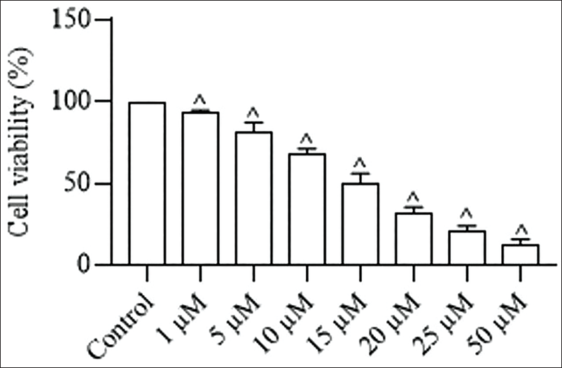
Figure 1 illustrates the results of MTT assay, demonstrating the cytotoxicity of albiflorin on the proliferation of breast cancer cells. The treatment with albiflorin considerably diminished the MDA-MB-231 cell growth at increasing dosages (1–50 µM). The increasing concentrations of albiflorin markedly inhibited the breast cancer cell viability, as depicted in Figure 1. The IC50 concentration of albiflorin was determined to be 15 µM, and this concentration was selected for additional studies.
Effect of Albiflorin on the MMP Level in MDA-MB-231 Cells
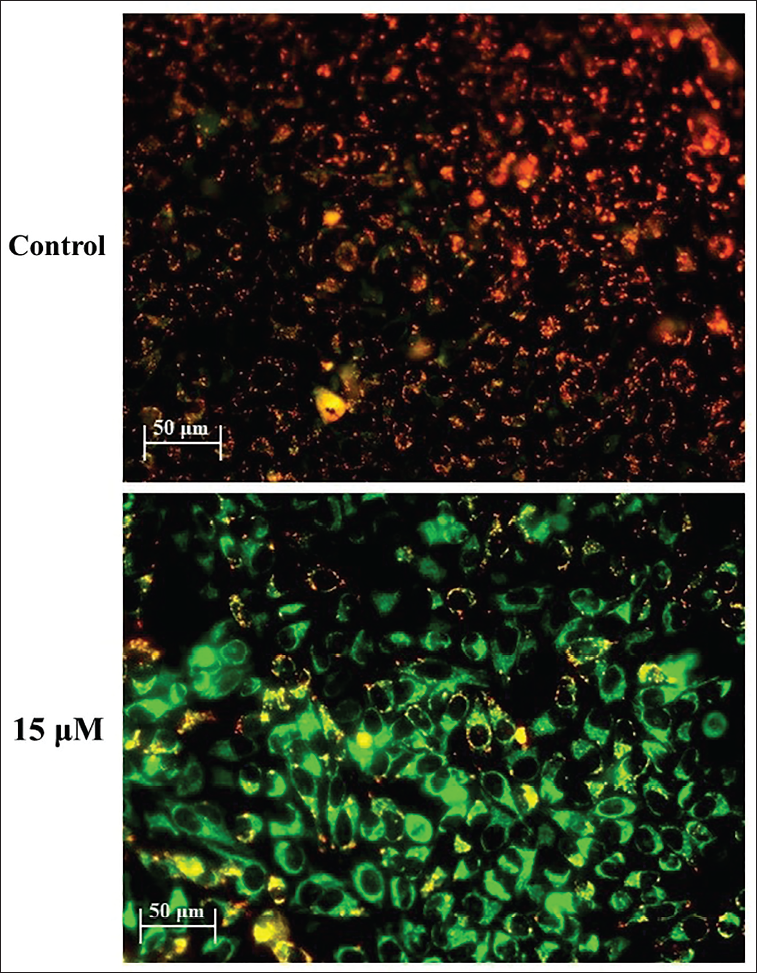
The effect of albiflorin on the MMP levels in MDA-MB-231 cells was assessed using JC-1 staining method. Figure 2 demonstrates that the JC-1 emits red/orange fluorescence in untreated cells, signifying a normal and undamaged MMP level. In contrast, the MDA-MB-231 cells, upon exposure to 15 µM of albiflorin for 24 h, exhibit high green fluorescence. The transition of fluorescence from red to green signifies that albiflorin treatment reduced the MMP level in MDA-MB-231 cells.
Effect of Albiflorin on the Apoptotic Cell Death in MDA-MB-231 Cells
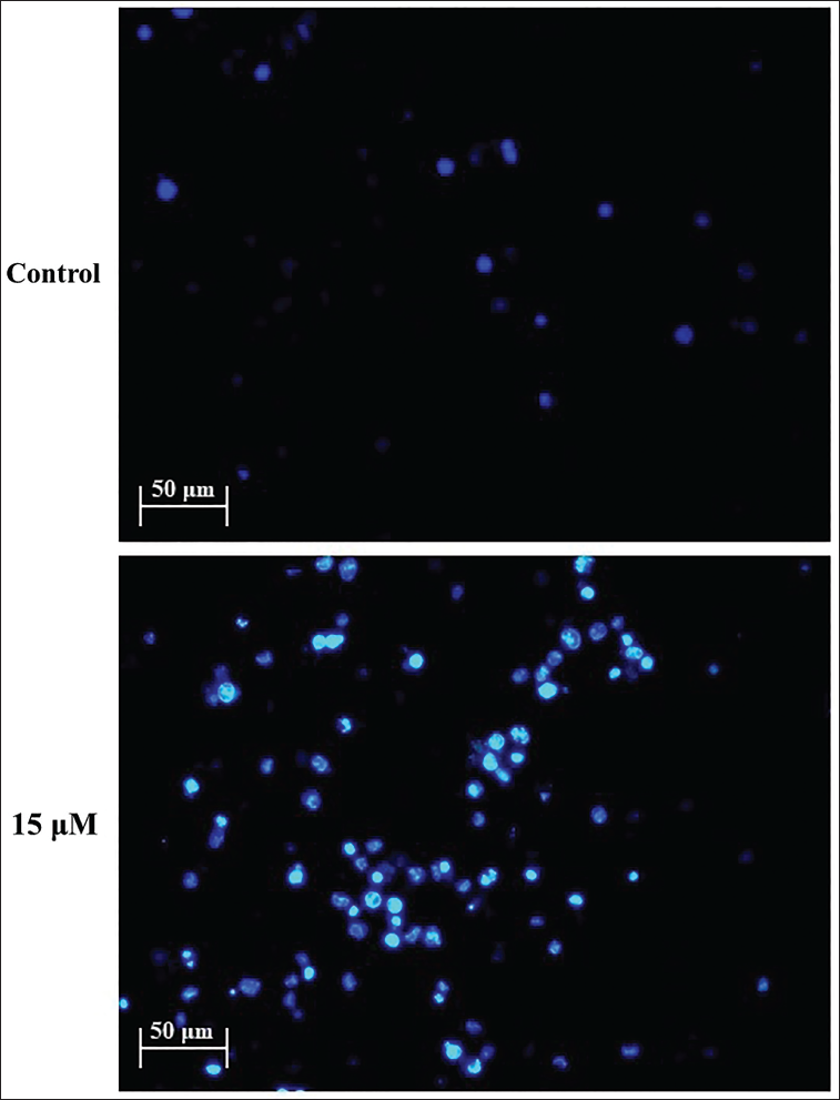
The extent of apoptotic levels in untreated and albiflorin-treated breast cancer cells was assessed by DAPI staining method. The results indicate that the 15 µM of albiflorin treatment significantly enhanced cell morphological alterations, nuclear damage, and apoptotic body development in the breast cancer cells (Figure 3). Consequently, it was evident that albiflorin might induce nuclear damage and enhance apoptosis in the breast cancer cells.
Effect of Albiflorin on the Apoptotic Protein Levels in the MDA-MB-231 Cells
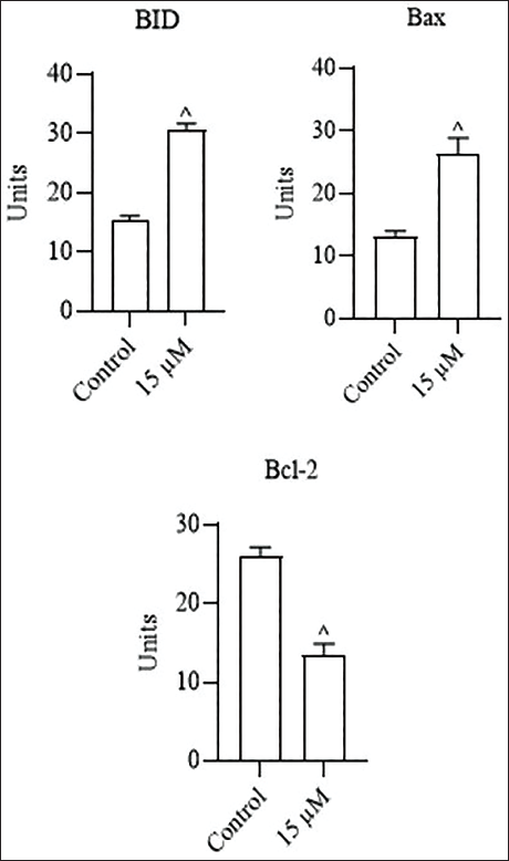
Figure 4 depicts the levels of apoptotic proteins in both untreated and albiflorin-treated breast cancer cells. In the untreated cells, elevated Bcl-2 levels, alongside decreased Bax and Bid levels, were observed. Simultaneously, cells treated with 15 µM of albiflorin demonstrated a marked decrease in Bcl-2 and elevated Bax and Bid protein levels relative to the control. These findings demonstrate that albiflorin can influence the apoptotic protein levels, thereby facilitating apoptosis in breast cancer cells.
Discussion
Despite advancements in therapeutic and diagnostic procedures, breast cancer remains a significant challenge, necessitating the exploration of novel treatment modalities due to various complications such as drug resistance, severe adverse effects, and metastasis. Specifically, breast cancer is the second major cause of cancer-associated deaths and the foremost cause of mortality among women aged 45–55, underscoring the critical need for more potential and less toxic interventions (Sung et al., 2021). The cases and death rates associated with breast cancer have unfortunately risen in recent years, solidifying its position as a major factor in cancer-associated fatalities in women worldwide. In developing nations, the situation is particularly dire, with high mortality rates often attributed to late-stage diagnoses, contributing to an overall survival rate that frequently does not exceed 60%. Although modern treatment methods such as radiation, chemotherapy, and surgeries have been shown to be successful, they often come with crippling adverse effects and challenges such as treatment resistance (Sun et al., 2017). The multifaceted nature of cancer, with over a hundred distinct forms, necessitates individualized treatment approaches, as a universally applicable therapy remains elusive. This pervasive health crisis underscores the critical demand for innovative therapeutic strategies that not only enhance efficacy but also mitigate the severe side effects associated with conventional treatments, which often significantly diminish patients’ quality of life (Lin et al., 2024).
The MMP is a crucial indicator of cellular bioenergetics and plays a pivotal role in sustaining cellular homeostasis, with its dysregulation frequently observed in various pathologies, including cancer. In mammalian cells, mitochondria are responsible for generating a substantial portion of ATP through oxidative phosphorylation, a process intrinsically linked to the MMP. Specifically, the electrochemical potential across the mitochondrial inner membrane, also known as the MMP, directly drives ATP synthesis and reflects the cell’s metabolic state (Nóbrega-Pereira et al., 2023). Cancer cells, in particular, often exhibit a hyperpolarized MMP compared to normal cells, suggesting a unique metabolic adaptation that supports their rapid proliferation and survival. This aberrant hyperpolarization is critical for tumor initiation and sustained growth, making the MMP a promising therapeutic target in oncology. This hyperpolarization is particularly pronounced in multidrug-resistant cancer cells, where it contributes to sustained cellular viability even after chemotherapy, highlighting its importance in therapeutic resistance. Consequently, analyzing MMP levels in drug-treated cancer cell samples can provide valuable insights into therapeutic efficacy and resistance mechanisms (Sandoval-Acuña et al., 2021). The comprehensive evaluation of mitochondrial functional parameters, beyond merely morphology, is essential for a thorough understanding of mitochondrial health in cancer, encompassing ATP synthesis, ROS production, and ion transport. Furthermore, the inherent metabolic plasticity of mitochondria and their involvement in critical cellular processes like apoptosis and ROS generation underscore their significance as a therapeutic target in cancer (Lin et al., 2019). In this work, the MMP level in the albiflorin-treated breast cancer cells was evaluated using JC-1 cells. The present findings suggested that albiflorin treatment considerably diminished the MMP levels in the MDA-MB-231 cells. These findings highlight that albiflorin treatment might influence the hyperpolarization of MMP, thereby disrupting the cellular functions and facilitating apoptosis in breast cancer cells.
Apoptosis, a form of programmed cell death, is crucial for upholding tissue homeostasis by removing injured, senescent, or unwanted cells without causing inflammation in the surrounding microenvironment. This highly regulated cellular mechanism involves a series of biochemical events that lead to characteristic morphological alterations and eventually cell death that are subsequently cleared by phagocytes (Qian et al., 2024). Dysregulation of apoptosis is implicated in various pathological conditions, most notably cancer, where a reduction in programmed cell death contributes to tumor development and therapeutic resistance. Specifically, impaired apoptotic mechanisms allow for the prolonged survival of neoplastic cells, leading to the accumulation of detrimental mutations that drive uncontrolled proliferation, evade immune surveillance, and foster metastatic dissemination (Qi et al., 2025). Crucially, therapeutic interventions in oncology often leverage the promotion of apoptosis to remove malignant cells. The dysregulation of apoptosis is a hallmark of tumor progression, enabling uncontrolled cellular proliferation and resistance to therapeutic interventions. Many genetic alterations in malignant cells contribute to a reduced apoptotic rate, thereby extending cell survival. This deregulation often stems from the activation of anti-apoptotic systems within cancer cells, complicating treatment strategies and leading to tumor recurrence (Pistritto et al., 2016). Consequently, investigating the apoptosis-inducing potential of various pharmacological agents within cancer cell lines is critical for identifying novel therapeutic strategies. Understanding the molecular mechanisms underpinning apoptosis induction, whether through extrinsic death receptor pathways or intrinsic mitochondrial pathways, is paramount for developing targeted therapies that overcome resistance (Kabore et al., 2004). In this work, the apoptosis-inducing potential of albiflorin on breast cancer cells was evaluated using DAPI staining technique. The current results showed an increase in apoptotic incidences in breast cancer cells treated with albiflorin, suggesting the apoptosis-inducing capacity of albiflorin in breast cancer cells.
The intricate balance between pro- and anti-apoptotic proteins of Bcl-2 family plays an essential role in determining cell fate, especially in the context of cancer progression and therapeutic response. This family comprises anti-apoptotic members like Bcl-2, pro-apoptotic effectors like Bax, and BH3-only proteins including Bid, which initiate apoptosis by detecting cellular stress (Yoshino et al., 2006). Dysregulation, often involving the overexpression of anti-apoptotic proteins, confers survival advantages to malignant cells, enhancing their resistance to therapies. Consequently, targeting these anti-apoptotic proteins to restore apoptotic pathways represents a hopeful strategy for novel therapeutic development in oncology. A key mechanism by which cancer cells evade death involves the overexpression of Bcl-2, which binds and neutralizes pro-apoptotic Bax, thereby suppressing intrinsic apoptosis (Bessou et al., 2020). This anti-apoptotic dominance shifts the Bax/Bcl-2 ratio, a key marker of apoptotic vulnerability, toward cell survival and impedes the mitochondrial permeabilization necessary for the discharge of pro-apoptotic factors. The intrinsic apoptotic cascade, governed by the Bcl-2 family proteins, is critical for triggering apoptosis through the mitochondrial membrane permeabilization and the discharge of apoptotic factors. This pathway is triggered by diverse stimuli, including DNA damage or oxidative stress, which activate BH3-only proteins to either stimulate pro-apoptotic Bax or neutralize anti-apoptotic Bcl-2 proteins (Yamaguchi et al., 2019). Ultimately, the dysregulation of this fine balance, particularly an elevated Bcl-2/Bax ratio, is a hallmark of many cancers, enabling uncontrolled proliferation and resistance to programmed cell death. Therefore, analyzing the precise effects of novel therapeutic agents on these protein levels within cancer cells is paramount for developing effective strategies that re-sensitize malignant cells to apoptotic signals (Merino et al., 2016). This analytical approach facilitates the identification of compounds capable of modulating these critical regulatory proteins, thereby restoring the delicate equilibrium required for programmed cell death in cancerous tissues. Furthermore, understanding the specific mechanisms by which various compounds alter the Bcl-2 family protein expressions, particularly how they influence the critical balance between pro- and anti-apoptotic members, is crucial for optimizing therapeutic regimens and overcoming drug resistance (Qian et al., 2022). In the present work, the influence of albiflorin on the apoptotic proteins Bcl-2, Bax, and Bid levels in the breast cancer cells was assessed. The results exhibited the increased Bax and Bid levels and diminished Bcl-2 in the albiflorin-treated breast cancer cells. These findings highlight that albiflorin can promote apoptosis in breast cancer cells by influencing Bcl-2 family proteins.
While our study demonstrates albiflorin’s promising anti-cancer effects against MDA-MB-231 breast cancer cells, it has several limitations. First, the study’s in vitro nature restricts its direct applicability to in vivo systems. Additionally, the molecular mechanisms underlying albiflorin’s effects, particularly its interactions with other cellular pathways, require further elucidation. The study’s focus on a single cell line also limits its generalizability to other breast cancer subtypes. Future directions include investigating albiflorin’s efficacy in vivo, exploring its effects on other breast cancer cell lines, and deciphering its interactions with conventional therapies. Further research is also needed to identify potential biomarkers for albiflorin’s therapeutic response and to optimize its dosage and delivery for clinical applications. Addressing these limitations will provide a more comprehensive understanding of albiflorin’s therapeutic potential and facilitate its translation to clinical settings. Ultimately, this may contribute to the development of novel, effective treatments for breast cancer.
Conclusion
The present study suggests that albiflorin can inhibit viability and promote apoptosis in breast cancer MDA-MB-231 cells. The apoptosis-inducing ability of albiflorin may be attributed to the depletion of MMP levels, an elevation in Bax and Bid levels, and a reduction in Bcl-2 in the MDA-MB-231 cells. Thus, the current data suggest albiflorin as a promising candidate that can be utilized for breast cancer treatment. Moreover, future investigations are highly recommended to comprehensively elucidate the precise mechanisms of albiflorin’s anti-cancer effects on breast cancer.
Summary
Breast cancer is characterized by uncontrolled cellular proliferation and tumor formation within the mammary glands, representing a complex and multifaceted disease. Despite advancements in therapeutic and diagnostic procedures, breast cancer remains to pose a significant challenge. The present study suggests that albiflorin can inhibit viability and promote apoptosis in breast cancer MDA-MB-231 cells. Thus, the current data suggest albiflorin can be a promising candidate to treat breast cancer.
Footnotes
Abbreviations
Bax: Bcl-2-associated X protein; Bcl-2: B-cell lymphoma-2; BID: BH3 interacting domain death agonist; DMEM: Dulbecco’s Modified Eagle Medium; DMSO: Dimethyl sulfoxide; FBS: Fetal bovine serum; MMP: Mitochondrial membrane potential.
Data Availability Statement
Data will be made available on request.
Declaration of Conflict of Interests
The authors declared no potential conflicts of interest with respect to the research, authorship, and/or publication of this article.
Ethical Approval and Informed Consent
Not applicable.
Funding
The authors disclosed receipt of the following financial support for the research, authorship, and/or publication of this article: The project was supported by Health Commission of Hebei Province (Grant No. 20210407).
