






Abstract
Background
Oral squamous cell carcinoma (OSCC) represents a considerable worldwide health challenge due to its elevated incidence, morbidity, and fatality rates. Despite extensive research into therapies, the 5-year survival rate for oral cancer has remained poor over the past decades, highlighting the limitations of existing therapies.
Purpose
The present work assessed the anti-cancer effects of rhoifolin against oral cancer KB cells.
Materials and Methods
The cytotoxic effect of rhoifolin at various doses (1–50 µM) on oral cancer KB cells was evaluated using an MTT assay. The DAPI and dual staining techniques were employed to assess apoptotic cells in rhoifolin-treated KB cells. The concentrations of caspase enzymes in the rhoifolin-exposed KB cells were evaluated using the kits.
Results
The MTT assay results revealed that diverse dosages of rhoifolin considerably inhibited the KB cell proliferation in a dose-dependent manner. The findings of both DAPI and dual staining assays demonstrated that rhoifolin treatment significantly increased apoptotic cell death in oral cancer KB cells. Furthermore, the rhoifolin treatment enhanced the caspase enzyme activities in KB cells.
Conclusion
The current study shows that rhoifolin demonstrates anti-cancer effects against oral cancer cells by diminishing cell growth and triggering caspase-dependent apoptosis in KB cells. Thus, the current data suggest that rhoifolin may be a promising therapeutic agent for future oral cancer treatment.
Introduction
Oral cancer, predominantly oral squamous cell carcinoma (OSCC), develops from the mucosal epithelium of the oral cavity and constitutes nearly 90% of all oral cancers, significantly impairing essential functions such as appearance, pronunciation, swallowing, and taste perception. Worldwide, it ranks among the top 10 most common cancers, with an estimated 378,500 new cases of intraoral cancer reported annually. The global incidence of newly diagnosed lip and oral cavity cases reached 389,485 in 2022, positioning it as the 16th most prevalent malignancy globally, with an associated mortality of 188,230, making it the 15th major cause of cancer-associated fatalities (Bray et al., 2024). This considerable mortality has not substantially decreased over the past four decades, with a 5-year mortality rate for OSCC remaining close to 50%. This persistent high mortality rate is largely attributed to diagnoses often occurring at advanced stages, which complicates treatment and reduces the likelihood of successful intervention. This delay in diagnosis is frequently a result of limited public awareness regarding precancerous lesions, early symptoms, and established risk factors, which subsequently contribute to diminished survival rates (Barsouk et al., 2023). Though the oral cavity is readily accessible for visual assessment, the majority of OSCCs are detected at advanced stages, which significantly contributes to the observed low 5-year survival rate of nearly 62%, in contrast to other cancers. This highlights the crucial need for improved diagnostic strategies, as early detection significantly enhances prognosis and treatment outcomes (Kolegova et al., 2022).
Despite advancements in therapies, the 5-year survival rate for oral cancer remains distressingly low, often attributed to late diagnosis and aggressive disease progression. This aggressive pathology is often multifactorial, stemming from isolated or combined risk factors like tobacco and alcohol drinking, and human papillomavirus infection (Machiels et al., 2020). Furthermore, the recurring challenge of treatment resistance and severe adverse effects associated with conventional chemotherapy underscores the urgent need for novel, safer, and more effective therapeutic strategies. Specifically, the dysregulation of apoptosis, a fundamental process of apoptosis, plays an essential role in the initiation and development of oral cancers by enabling uncontrolled cellular proliferation and survival. This dysregulation contributes significantly to therapeutic resistance, a critical impediment to successful oral cancer treatment (Satgunaseelan et al., 2021). Consequently, understanding the molecular processes underlying apoptosis dysregulation in oral cancer is essential for developing targeted interventions that can circumvent resistance and improve patient outcomes. Recognizing the complex etiology and high intratumoral heterogeneity of OSCC further emphasizes the necessity for innovative therapeutic approaches (Tarle & Lukšić, 2024).
The global cases of oral cancer are predicted to increase significantly in the coming decades, further underscoring the urgency for improved therapeutic interventions. This alarming trend necessitates a concerted focus on chemoprevention, which involves the systemic administration of agents designed to revert, halt, or delay the carcinogenic process, as a vital strategy to mitigate the rising incidence and improve the prognosis of oral cancer (Mitea et al., 2024). This pressing demand has spurred intensive research into novel therapeutic targets and agents, with particular emphasis on phytochemicals for their multifaceted anti-cancer properties and relatively low toxicity profiles. These natural compounds, derived from plants, offer a promising avenue for developing alternative and adjunctive therapies for several diseases, including cancers (Chou et al., 2018; Huang et al., 2021). Rhoifolin, a well-known bioactive flavone glycoside compound extracted from various citrus plants, including lemons and grapes, has demonstrated significant pharmacological properties (Barreca et al., 2020). Several studies have already reported that rhoifolin exhibits antioxidant and anti-inflammatory properties (Al-Shalabi et al., 2022), anti-arthritis activity (Peng et al., 2020), anti-colitis activity (Xu et al., 2025), and nephroprotective effects. Furthermore, it showed anti-cancer (Kiliç et al., 2025) and anti-proliferative (Eldahshan, 2013) properties. Particularly, rhoifolin effectively inhibited cell viability and promoted apoptotic cell death in pancreatic cancer (Zheng et al., 2022) and hepatocellular carcinoma (Chen et al., 2025). However, there are no such reports on the anti-cancer effects of rhoifolin against oral cancer. Therefore, this study evaluated the anti-cancer properties of rhoifolin against oral cancer KB cells.
Materials and Methods
Chemicals
The primary chemicals used in this work, including rhoifolin, dimethyl sulfoxide (DMSO), fetal bovine serum (FBS), and antibiotics, were obtained commercially from Sigma-Aldrich, USA. The biochemical markers were evaluated utilizing standard kits procured from Elabscience, USA.
Maintenance of Cell Culture
The oral cancer KB cells were obtained from ATCC, USA, and cultured in DMEM media supplemented with 10% FBS and 1% anti-mycotic mixture in a 5% CO2 chamber. The grown cells were obtained upon achieving 80% confluency and employed for subsequent fluorescence staining and biochemical assays.
Cytotoxicity Assay
The cytotoxicity of rhoifolin on KB cells was assessed using the MTT assay. Cells were placed into a 96-well plate with DMEM medium at a density of 5 × 10³ cells/well for 24 h at 37 °C. Subsequently, the wells containing cells were added with varied concentrations (1, 5, 10, 15, 20, 25, and 50 µM) of rhoifolin and incubated for 24 h. Consequently, MTT (20 µL) and DMEM (100 µL) were mixed in each well and incubated for 4 h. The formazan deposits in the well were dissolved with DMSO (100 µL), and absorbance was then measured at 570 nm.
Acridine Orange/Ethidium Bromide Staining
The dual acridine orange/ethidium bromide (AO/EB) staining experiment was done on untreated and rhoifolin-treated KB cells to assess apoptotic levels. The KB cells were grown for 24 h and then treated with rhoifolin (15 µM) for an additional 24 h. A mixture of AO/EB stains (100 µg/mL) was then applied to the wells for 5 min in a shady place. The presence of apoptotic cells was then evaluated using a fluorescent microscope.
DAPI Staining
The nuclear morphology of apoptotic cells in control and rhoifolin-treated KB cells was studied using DAPI staining. After seeding KB cells in a 24-well plate, they were treated with rhoifolin (15 µM) for 24 h. Following a 30-min fixation in 4% paraformaldehyde, cells were stained with DAPI (200 µg/mL) for 15 min. The impact of rhoifolin on nuclear damage and apoptosis in oral cancer KB cells was further evaluated utilizing a fluorescent microscope.
Analysis of Caspase Activities
The KB cells treated with rhoifolin and the control groups were lysed using a cell lysis solution to prepare cell lysate for the evaluation of caspase activities. The levels of caspase-3, caspase-8, and caspase-9 were quantified in both untreated and rhoifolin-treated KB cells using commercial kits according to the manufacturer’s specifications (Elabscience, USA).
Statistical Analysis
The statistical tests were conducted using GraphPad Prism software, and the values are presented as the mean ± SD of triplicate measurements. One-way analysis of variance (ANOVA) and Duncan’s multiple range test (DMRT) were used to analyze the data, with a significance level set at p < .05.
Results
Effect of Rhoifolin on the Viability of Oral KB Cells
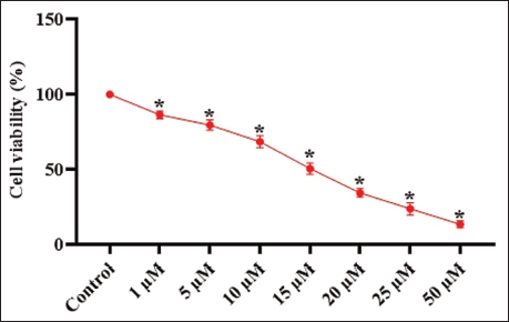
Figure 1 displays the results of the MTT assay, demonstrating the cytotoxic effect of rhoifolin on the proliferation of oral cancer KB cells. Treatment with rhoifolin significantly reduced the proliferation of KB cells at various concentrations (1–50 µM). The increased concentrations of rhoifolin demonstrated greater cytotoxicity against KB cell viability, as depicted in Figure 1. The IC50 of rhoifolin against KB cells was determined to be 15 µM, and this concentration was selected for the following studies.
Effect of Rhoifolin on the Viability of Oral KB Cells. The Administration of Different Concentrations of Rhoifolin (1–50 µM) Markedly Diminished the Viability of KB cells. The IC50 Value for Rhoifolin in KB Cells Was Determined to be 15 µM. The Data Are Expressed as Mean ± SD of Triplicates, Analyzed via One-way Analysis of Variance (ANOVA) and Duncan’s Multiple Range Test (DMRT) Assays Using GraphPad Prism Software. * Indicates that the Values Differ Significantly from the Control at p < .05.
Effect of Rhoifolin on Apoptotic Level in Oral Cancer KB Cells
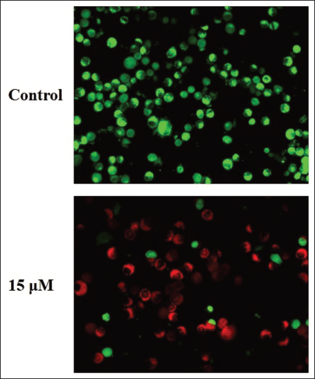
The dual staining method was utilized to examine rhoifolin-induced apoptotic level in KB cells, with results depicted in Figure 2. After treatment with 15 µM rhoifolin, KB cells showed elevated levels of red fluorescent cells, indicating both early- and late-stage apoptotic cell death. Therefore, it was evident that rhoifolin can induce apoptosis in oral cancer cells.
Effect of Rhoifolin on the Apoptotic Level in the Oral Cancer KB Cells. The Dual Staining Method Was Utilized to Examine Rhoifolin-induced Apoptosis in KB Cells. The Rhoifolin (15 µM)-treated KB Cells had Elevated Levels of Red Fluorescent Cells, Signifying the Occurrence of Apoptotic Cell Death.
Effect of Rhoifolin on Apoptosis in Oral Cancer KB Cells
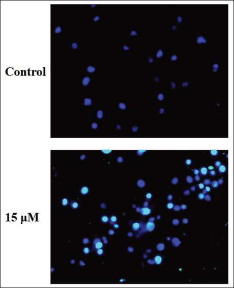
The apoptotic levels in both untreated and rhoifolin-treated KB cells were assessed by DAPI staining, as depicted in Figure 3. KB cells treated with 15 µM rhoifolin exhibited heightened apoptotic events, characterized by chromatin condensation, membrane and nuclear disruptions, apoptotic body formation, and decreased cell density, indicating the onset of apoptosis.
Effect of Rhoifolin on the Apoptosis in the Oral Cancer KB Cells. The Apoptotic Levels in Both Control and Rhoifolin-treated KB Cells Were Assessed by DAPI Staining. The KB Cells Treated with 15 µM of Rhoifolin Exhibited Heightened Apoptotic Events.
Effect of Rhoifolin on the Caspase Activities in the Oral Cancer KB Cells
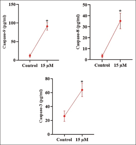
Figure 4 represents the activities of caspase enzymes, evaluated in both untreated and rhoifolin-treated KB cells. In control cells, moderate activities of caspase-3, caspase-8, and caspase-9 were observed. On the other hand, KB cells treated with 15 µM of rhoifolin demonstrated a considerable elevation in the activities of caspase-3, caspase-8, and caspase-9 relative to the control cells. Therefore, it was evident that rhoifolin can facilitate apoptosis in KB cells via enhancing caspase activities.
Effect of Rhoifolin on the Caspase Activities in the Oral Cancer KB Cells. The Data Are Expressed as Mean ± SD of Triplicates, Analyzed via One-Way Analysis of Variance (ANOVA) and Duncan’s Multiple Range Test (DMRT) Assays Using GraphPad Prism Software. * Indicates that the Values Differ Significantly from the Control at p < .05.
Discussion
OSCC represents a significant global health burden due to its high incidence, morbidity, and mortality rates. Despite considerable research into treatment modalities, the 5-year survival rate for oral cancer has remained largely stagnant over the past four to five decades, a fact that underscores the limitations of current therapeutic approaches (Sung et al., 2021). This stagnation in survival rates, despite advances in therapies such as radiotherapy and chemotherapy, underscores the need to explore novel therapeutic agents that can overcome inherent or acquired resistance mechanisms and mitigate severe side effects. Despite its accessibility for direct visual examination, a substantial proportion of OSCCs are diagnosed at advanced stages, which significantly contributes to the low survival rates observed (Chamoli et al., 2021). This delayed diagnosis is a critical factor limiting the efficacy of current treatments, as early-stage OSCC is largely curable through surgery alone, whereas advanced cases necessitate multidisciplinary approaches involving surgery, radiotherapy, and chemotherapy. The persistent challenges, including tumor recurrence and the adverse effects of conventional chemotherapeutic agents, further underscore the need to develop more effective and less toxic treatment options (Barsouk et al., 2023).
The assessment of cytotoxicity is pivotal in drug discovery, serving as a primary indicator for the therapeutic potential of novel compounds against neoplastic diseases. This analytical approach distinguishes between compounds that induce specific biomolecular interactions at low concentrations and those that exert broader cytotoxic effects, often mediated by cell stress pathways, physico-chemical disruptions, or non-covalent interactions. Consequently, accurate quantification of these cytotoxic effects is crucial for identifying promising drug candidates and optimizing their therapeutic indices, while accounting for their mechanisms of action and the specific cellular context (Sazonova et al., 2022). This becomes especially relevant in the context of tumor cells, where the objective is to selectively eliminate malignant cells while sparing healthy tissues, thereby minimizing off-target toxicities. Furthermore, the emergence of drug resistance in tumor cells necessitates continuous investigation into novel agents and combination therapies to circumvent these adaptive mechanisms. The metabolic vulnerabilities of cancer cells, which differ significantly from those of healthy cells, offer promising avenues for targeted therapeutic intervention (Kamiloglu et al., 2020). The MTT assay serves as a cornerstone in pharmacological and toxicological research, providing a reliable and high-throughput method for evaluating the growth and metabolic activity of cells in response to various agents, particularly in the context of cancer drug discovery and biocompatibility evaluation. This method is further valued for its ease of execution, rapid results, and accurate quantification, making it a preferred choice for high-throughput screening in drug development (Ghasemi et al., 2021). In this study, the cytotoxicity of rhoifolin against oral cancer KB cells was assessed using the MTT assay. The findings of the MTT assay demonstrated that the rhoifolin treatment markedly reduced the growth of KB cells. These findings showed that rhoifolin has cytotoxic effects against oral cancer cells.
The intricate balance between cellular proliferation and apoptosis is fundamental for maintaining tissue homeostasis and preventing uncontrolled cellular expansion. However, dysregulation of this finely tuned process, particularly the escape from apoptosis, is a hallmark of tumors, contributing significantly to tumorigenesis, progression, and resistance to therapy. This evasion confers a survival advantage to malignant cells, enabling them to bypass crucial cellular checkpoints and develop multidrug resistance (Jan & Chaudhry, 2019). This highlights the critical need for a detailed understanding of the regulatory mechanisms governing cell death and survival pathways in human cancers, as such knowledge is pivotal for developing novel therapeutic strategies. Apoptosis is essential for eliminating damaged or unwanted cells, thus preventing abnormal cellular accumulation that could lead to neoplastic transformation. This tightly regulated process involves two principal pathways: The extrinsic pathway, triggered by death signals, and the intrinsic pathway, triggered by intracellular stress. Both pathways converge on the activation of caspases that execute the cellular dismantling process (Carneiro & El-Deiry, 2020).
A major mechanism by which cancer cells evade apoptosis involves the dysregulation of the BCL-2 proteins, essential regulators of the intrinsic apoptotic pathway. Specifically, an overexpression of anti-apoptotic proteins is frequently observed in various cancers, enhancing tumor cell survival and conferring resistance to conventional chemotherapy and radiation (Singh et al., 2019). Conversely, a reduction in the expression or function of pro-apoptotic proteins further contributes to the apoptotic resistance observed in malignant cells. This imbalance shifts the cellular regulator toward survival, enabling cancerous cells to evade the physiological cues that would normally trigger their demise. This dysregulation of apoptotic pathways not only fosters tumor growth but also presents a significant challenge in cancer treatment, as many chemotherapeutic agents induce their cytotoxic effects by activating apoptotic mechanisms (Qian et al., 2022).
Understanding the intricate mechanisms that govern the apoptotic mechanism is paramount in the advancement of novel therapies against malignancies. This is particularly essential given that the escape from apoptosis is a hallmark of tumors, contributing to tumor progression and resistance to conventional therapies (Zheng et al., 2020). Consequently, inducing apoptosis in cancer cells using targeted drug compounds represents a highly promising avenue for cancer therapy, focusing on restoring the delicate balance between cell proliferation and death within the tumor microenvironment. This approach leverages the fact that defects in apoptosis can lead to treatment resistance, necessitating the development of new agents that activate or reactivate the intrinsic cell death machinery in malignant cells (Sun et al., 2019). Deregulation of these apoptotic programs is a significant factor in therapeutic resistance, underscoring the necessity for a comprehensive understanding of death and survival pathways to inform the development of targeted interventions. The inherent sensitivity of cancer cells to apoptosis, despite their often-specific defects in apoptotic pathways, provides numerous opportunities for therapeutic manipulation. This has led to extensive research into strategies designed to selectively induce apoptosis in cancer cells, often by targeting dysregulated apoptotic modulators (Kim & Kim, 2018). In the present study, the findings of both DAPI and dual staining techniques revealed that rhoifolin treatment enhanced the apoptosis in KB cells. Therefore, it was evident that rhoifolin may inhibit oral cancer progression via inducing apoptosis in oral cancer cells.
Caspases, a family of cysteine-dependent aspartate-directed proteases, play a crucial role in both the initiation and execution phases of apoptosis, serving as critical regulators of cellular homeostasis and genomic integrity. Apoptosis is a tightly regulated process that involves a cascade of molecular events, with caspases serving as the central executor (Dho et al., 2025). These proteases are synthesized as inactive zymogens and undergo proteolytic cleavage to become active enzymes, initiating a self-amplifying cascade that dismantles cellular components and leads to the formation of apoptotic bodies. Alterations in apoptotic control are frequently observed in various pathologies, including cancer, where the evasion of apoptosis is a hallmark of malignancy. The central apoptotic machinery can be broadly categorized into two major signaling pathways, such as extrinsic and intrinsic pathways (Van Opdenbosch & Lamkanfi, 2019). Both pathways are dependent on the activation of executioner caspases, such as caspase-3, which then cleave several cellular substrates, leading to the morphological and biochemical alterations characteristic of apoptosis. The escape from apoptosis is a recognized hallmark of cancer, contributing to tumor development and resistance to various therapies by allowing cancer cells to circumvent apoptosis (Fritsch et al., 2019).
Understanding the intricate mechanisms governing apoptosis, particularly the roles of key executioner and initiator caspases, is essential for developing potential therapies to combat cancer. The intricate molecular pathways governing programmed cell death, particularly those involving caspases, represent a crucial area of investigation within oncology due to their direct implications for therapeutic strategies. Dysregulation of apoptotic processes is frequently observed in malignant transformation and contributes to tumor growth, metastasis, and resistance to conventional treatments (Tian et al., 2024). Consequently, understanding the processes by which cancer cells evade apoptosis and, conversely, identifying strategies to reinstate or enhance caspase-mediated cell death is paramount for developing effective anti-cancer therapies. The targeted activation of caspase cascades offers a promising avenue for therapeutic intervention, aiming to re-establish the apoptotic machinery in cancer cells. To this end, significant research efforts have focused on developing novel therapeutic agents that either directly activate caspases or modulate upstream regulatory proteins to restore apoptotic susceptibility in resistant tumors (Huet et al., 2014). Similarly, the present findings revealed that rhoifolin treatment significantly enhanced caspase-3, caspase-8, and caspase-9 activity in KB cells. These findings suggest that rhoifolin can induce and mediate apoptosis in KB cells via increasing caspase activities.
Conclusion
The findings demonstrate that rhoifolin exhibits anti-cancer effects against oral cancer by reducing cell viability and inducing caspase-dependent apoptosis in KB cells. The rhoifolin treatment markedly decreased cell viability, increased apoptosis, and enhanced caspase enzyme activities in KB cells. Thus, the current data suggest that rhoifolin may be a promising therapeutic agent for future oral cancer treatment. Moreover, future studies are highly recommended to comprehensively elucidate the precise therapeutic functions of rhoifolin’s anti-cancer effects on oral cancer.
Abbreviations
AO/EB: Acridine orange/ethidium bromide; DMSO: Dimethyl sulfoxide; FBS: Fetal bovine serum; OSCC: Oral squamous cell carcinoma.
Footnotes
Acknowledgments
The authors would like to express their sincere gratitude to the Department of ENT (West), Baoding First Central Hospital, Baoding, China, the Department of Rehabilitation Medicine, the Department of Anesthesiology, and the Department of Pediatrics, Affiliated Hospital of Hebei University, Baoding, China, for their valuable support and collaboration in this study.
Declaration of Conflicting Interests
The authors declare no potential conflicts of interest with respect to the research, authorship, and/or publication of this article.
Ethical Approval and Informed Consent
The project was approved by the Animal Ethics Committee of the Affiliated Hospital of Hebei University (No. HBU-2024-18).
Funding
The authors received no financial support for the research, authorship and/or publication of this article.
