






Abstract
Background
Diabetic nephropathy, a severe microvascular complication of diabetes, is a primary cause of end-stage renal disease worldwide, significantly contributing to morbidity and mortality. Despite advancements in glycemic and blood pressure control, a substantial portion of diabetic patients still progress to diabetic nephropathy, underscoring the need for more effective therapeutic strategies.
Purpose
The current work was devoted to examining the anti-diabetic nephropathy properties of α-hederin against streptozotocin (STZ)-induced rat model.
Materials and Methods
The rats were treated with 65 mg/kg of STZ to develop diabetic nephropathy. The STZ-treated diabetic rats were subsequently administered α-hederin for a duration of 60 days. After the conclusion of treatments, body weight, blood glucose, kidney weight, and food and water consumption levels of the rats were examined. The levels of renal dysfunction markers, like uric acid, creatinine, and blood urea nitrogen, were determined. The levels of inflammatory cytokines and anti-oxidants were assessed using assay kits.
Results
Treatment with 25 and 50 mg/kg of α-hederin significantly increased body weight and insulin levels and subsequently decreased glucose levels in rats with diabetic nephropathy. Moreover, α-hederin treatment reduced the levels of renal dysfunction markers in the serum of STZ-treated rats. The α-hederin treatment significantly decreased inflammatory cytokine levels and subsequently increased anti-oxidant levels in STZ-induced rats.
Conclusion
The present data highlight that α-hederin may ameliorate diabetic nephropathy in the STZ-induced rat model. In conclusion, our findings indicate that α-hederin could serve as a beneficial therapeutic agent for diabetic nephropathy.
Introduction
Diabetic nephropathy is a significant microvascular consequence of diabetes mellitus, marked by increasing renal impairment that ultimately results in end-stage renal disease (ESRD). It is a prominent sequel to diabetes mellitus, characterized by glomerular enlargement, extracellular matrix deposition, and glomerular basement membrane thickening. This condition is a pivotal cause of ESRD in developed nations, affecting nearly one-third of individuals with diabetes (Zhang et al., 2025). Globally, over 460 million people are affected by diabetes, positioning diabetic nephropathy as a significant public health challenge. The substantial rise in diabetes incidence, projected to reach 642 million by 2040, further exacerbates the burden of diabetic nephropathy, which accounts for nearly 50% of ESRD incidence. This escalating prevalence contributes significantly to increased morbidity and mortality among diabetic patients (Saeedi et al., 2019). These pathological alterations are primarily induced by sustained hyperglycemia, leading to structural and functional changes within the renal tubules and glomeruli. These include the loss of endothelial windows, expansion of the mesangial matrix, podocyte deletion, and ultimately segmental mesangial dilatation. These morphological abnormalities manifest clinically as a persistent elevation in the urinary albumin-to-creatinine ratio and a diminution in the glomerular filtration rate (Selby & Taal, 2020).
The intricate pathogenesis of diabetic nephropathy involves a complex interplay among metabolic, hemodynamic, and inflammatory cascades, further complicated by hyperglycemia-induced oxidative stress. Indeed, these factors collectively contribute to cellular injury and fibrosis within the renal parenchyma (Jin et al., 2023). Specifically, elevated glucose levels adversely impact hemodynamics, hormone production, and various metabolic pathways, alongside inducing oxidative stress and inflammation. Key interconnected metabolic factors, including polyol, hexosamine, and advanced glycation end-products (AGEs) pathways, become hyperactive due to hyperglycemia, fueling a feedback loop that exacerbates kidney damage (Moreno et al., 2018). Sustained exposure to high glucose levels also leads to overproduction of mitochondrial superoxide, which is considered a primary driver of hyperglycemia-induced apoptosis in renal cells. This chronic oxidative stress, along with increased renal polyol formation and AGE aggregation, frequently triggers substantial injury in the diabetic kidney (Wang et al., 2019). Moreover, the persistent oxidative stress and inflammation related to diabetic nephropathy contribute significantly to renal endothelial dysfunction and damage, thereby compromising renal hemodynamics and exacerbating fibrotic and sclerotic changes within the glomeruli and tubules. These molecular and cellular perturbations ultimately lead to characteristic renal structural impairments, including glomerular mesangial hypertrophy, podocyte dysfunction, and extracellular matrix protein accumulation (Ruiz-Ortega et al., 2020).
Despite advancements in glycemic and blood pressure control, a substantial portion of diabetic patients still progress to diabetic nephropathy, underscoring the need for more effective therapeutic strategies (Tuttle et al., 2022). Conventional treatments, while managing risk factors, often have limitations, including adverse effects and limited efficacy in completely halting disease progression. The complex interplay of glucose metabolism disorder, inflammation, and oxidative stress further complicates its pathogenesis, necessitating comprehensive treatment approaches (Van Raalte et al., 2024). Thus, identifying novel therapeutic agents that can interrupt these interconnected pathophysiological mechanisms is crucial for developing effective interventions to prevent or delay kidney damage. This persistent challenge necessitates the exploration of novel therapeutic avenues, and one promising approach involves plant-based therapies, which often exhibit multifaceted pharmacological activities with fewer adverse effects than synthetic drugs (Efiong et al., 2025).
Triterpenoid saponins have emerged as a particularly promising class of natural compounds for diabetes management due to their ability to modulate insulin secretion, improve glucose uptake, and alleviate oxidative stress (Kaushik et al., 2024). However, despite the known anti-diabetic potential of structurally related triterpenoid saponins (Nazaruk & Borzym-Kluczyk, 2015), the specific impact of α-hederin on diabetic nephropathy remains largely unexplored in the current literature. α-Hederin is a bioactive monodesmosidic triterpenoid saponin compound mostly found in the leaves of Nigella sativa and Hedera helix plants, and it has been thoroughly investigated for its various pharmacological properties. The numerous pharmacological properties of α-hederin were already reported in several previous studies, including its anti-inflammatory (Gepdiremen et al., 2005), anti-tumor (Meng et al., 2024), neuroprotective (Wu et al., 2017), anti-sepsis (Zeng & Zhao, 2023), anti-oxidant (Gülçin et al., 2004), and chemopreventive (Bahr et al., 2021) effects. However, the anti-diabetic and anti-diabetic nephropathy properties of α-hederin have not been described yet. Consequently, the current study is devoted to examining the anti-diabetic nephropathy properties of α-hederin against a Streptozotocin (STZ)-induced rat model.
Materials and Methods
Chemicals
The primary chemicals employed in this work, such as α-hederin, STZ, and glibenclamide, were sourced commercially from Sigma–Aldrich, USA. The commercial test kits for biochemical marker estimations were sourced from Abcam, MyBioSource, and Elabscience, USA, respectively.
Experimental Rats
Healthy male Wistar rats weighing about 210–230 g were employed in this study. All experimental protocols were conducted in accordance with the guidelines of the institutional ethics committee for animal care and use and were approved by the institutional animal ethics committee. All animals were caged in infection-free confines inside a regulated laboratory environment, with a temperature of 22°C–26°C and humidity levels of 40%–60%. A 12-h cycle of alternating illumination and darkness was instituted. During the entire study period, all rats were provided with unrestricted access to conventional rodent chow and filtered drinking water. Before the initiation of the study, all rats underwent a 7-day acclimation phase in a laboratory environment.
Treatment Groups
After a 1-week acclimatization phase, animals were grouped randomly into five treatment groups with six rats (n = 6) in each using a simple randomization method (computer-generated random numbers). To minimize bias, investigators responsible for biochemical analyses and histological assessments were blinded to the treatment assignments until the data collection was complete. Group I was to control animals, which received standard rodent chow and were administered only the vehicle (0.5% carboxymethyl cellulose in distilled water) via oral gavage. Group II rats were administered a single dosage of STZ (65 mg/kg) to trigger diabetic nephropathy. The rats were administered glucose (0.5%) to avert death caused by sudden hypoglycemia. After a 3-day administration of STZ, blood glucose was evaluated, and animals with over 250 mg/dL glucose level were designated as diabetic and chosen for subsequent investigations. Groups III and IV consisted of rats with STZ-induced diabetic nephropathy, which were administered a dosage of 25 and 50 mg/kg of α-hederin (Bahr et al., 2021), respectively, every day for a duration of 60 days. Group V animals were induced with diabetic nephropathy as specified in Group II, and administered 5 mg/kg of the standard anti-diabetic drug glibenclamide for a duration of 60 days. The body weight, food, and water consumption levels of each rat were rigorously measured during the investigation. The glucose level of the rats was measured using a commercial Accu-Chek glucometer (Roche, Switzerland). Upon the conclusion of treatments, animals were sacrificed under pentobarbital sodium anesthesia. The blood was collected and centrifuged to prepare serum. The serum was frozen for further analysis. The kidney tissues were swiftly excised and weighed. One segment was preserved in 4% paraformaldehyde for histological examination, while the remaining kidney portions were quickly frozen for further investigations.
Analysis of Insulin and Homeostatic Model Assessment for Insulin Resistance Markers
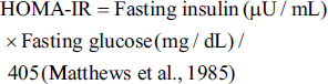
The insulin level was measured using a Rat Insulin enzyme-linked immunosorbent assay (ELISA) Kit (Cat. No. E-EL-R3034; Elabscience, USA). The experiment was conducted in triplicate using the manufacturer’s recommended specifications. The homeostatic model assessment for insulin resistance (HOMA-IR) level was determined using a formula:
Analysis of Renal Dysfunction Markers
The levels of renal dysfunction markers, including blood urea nitrogen (BUN) (Cat. No. MBS292018), creatinine (Cat. No. MBS3809095), and uric acid (Cat. No. MBS3808193), in the serum of both untreated and treated rats were analyzed using specialized ELISA kits (MyBioSource, USA). The experiments were conducted in triplicate using the manufacturer’s specifications.
Analysis of Anti-oxidant Levels
The renal tissues were swiftly removed from the rats, homogenized, and centrifuged at 10,000 rpm for 20 min. The resultant supernatant was employed to analyze the renal anti-oxidant levels, including glutathione (GSH) (Cat. No. ab65322) and superoxide dismutase (SOD) (Cat. No. ab65354), utilizing commercial test kits in accordance with the manufacturer’s specifications (Abcam, USA).
Analysis of Inflammatory Cytokine Levels
The amounts of pro-inflammatory cytokines interleukin (IL)-6 (Cat. No. MBS355410) and tumor necrosis factor-alpha (TNF-α) (Cat. No. MBS2507393) in the serum of control and treatment rats were analyzed using test kits. The assays were conducted in triplicate following the manufacturer’s specified recommendations (MyBioScience, USA).
Statistical Analysis
All experimental data were analyzed using GraphPad Prism software (Version 9.0, USA). Results are expressed as mean ± standard deviation (SD). For each experimental group, n = 6 represents the number of biological replicates (individual rats). To ensure technical precision, all biochemical assays were performed in technical triplicate, and the average of these replicates was used for the final statistical calculation for each animal. Statistical comparisons between the treatment groups were evaluated using one-way analysis of variance (ANOVA), followed by Tukey’s post hoc test for multiple comparisons to determine significant differences between specific groups. A p value of less than .05 (p < .05) was considered statistically significant.
Results
α-Hederin Treatment Decreases the Fasting Blood Glucose Levels in the Streptozotocin-induced Diabetic Nephropathy Rats
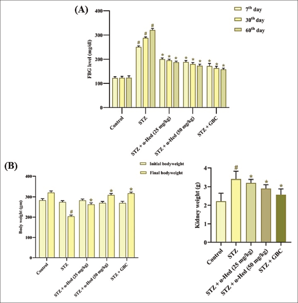
The anti-hyperglycemic effects of α-hederin were assessed in STZ-induced rats, and the results are illustrated in Figure 1(A). The STZ-induced rats revealed a significant elevation in the fasting blood glucose (FBG) levels in comparison to the control group, which are evaluated at the intervals of 7th, 30th, and 60th days of the experiment. Conversely, the treatment with α-hederin at a dosage of 25 and 50 mg/kg, respectively, remarkably diminished the FBG level in rats with diabetic nephropathy. Similar findings were also found in the standard drug glibenclamide treatment, which corroborates the anti-hyperglycemic activity of the α-hederin treatment.
α-Hederin Treatment Increases Body Weight and Decreases Kidney Weight in the Streptozotocin-induced Diabetic Nephropathy Rats
The changes in the body weight and kidney weight of control and treatment rats were analyzed, with the results depicted in Figure 1(B). The diabetic nephropathy rats showed a reduction in their body weight and an elevation in their kidney weight in comparison to the control. Whereas, the 25 and 50 mg/kg of α-hederin markedly increased the body weight and decreased the kidney weight of the STZ-induced nephropathy rats. These results are further validated by the outcomes of glibenclamide treatment, which also increased the body weight and reduced kidney weight of the diabetic rats.
α-Hederin Treatment Regulates the Food and Water Intakes, Insulin, and Homeostatic Model Assessment for Insulin Resistance in Streptozotocin-induced Diabetic Nephropathy Rats
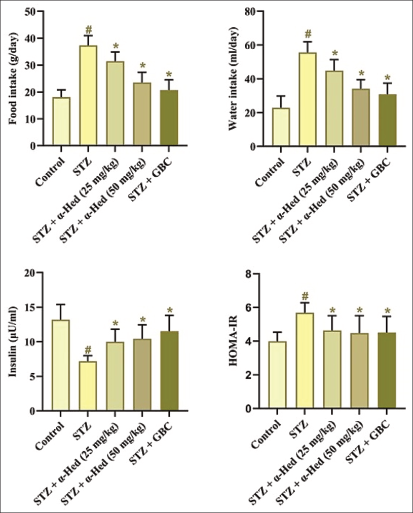
Figure 2 depicts the levels of food and water intake, insulin, and HOMA-IR levels of the experimental animals. The STZ-induced rats showed a significant upsurge in both food and water intakes and HOMA-IR levels, and subsequently decreased insulin levels when compared with the control. Whereas, the α-hederin treatment at dosages of 25 and 50 mg/kg, respectively, displayed a marked decrease in the food and water intakes and HOMA-IR in comparison to the STZ-treated rats. The α-hederin treatment also elevated insulin in diabetic rats. The treatment of glibenclamide also regulated these marker levels in diabetic rats, hence supporting the anti-diabetic efficacy of α-hederin.
α-Hederin Treatment Decreases the Renal Dysfunction Markers in the Serum of Streptozotocin-induced Diabetic Nephropathy Rats
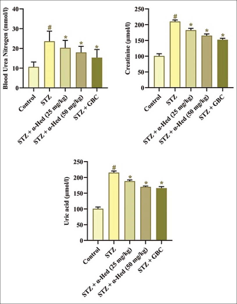
The effect of α-hederin on renal dysfunction markers, including BUN, creatinine, and uric acid, was evaluated, with the results displayed in Figure 3. The STZ-induced nephropathy rats exhibited a remarkable elevation in the BUN, creatinine, and uric acid levels in relation to the control group. However, the administration of 25 and 50 mg/kg of α-hederin, respectively, to the STZ-induced rats showed a reduction in the BUN, creatinine, and uric acid levels. These findings are further verified by the results of glibenclamide, which also decreases these renal dysfunction markers in the diabetic rats.
α-Hederin Treatment Increases Anti-oxidant Levels in the Renal Tissues of Streptozotocin-induced Diabetic Nephropathy Rats
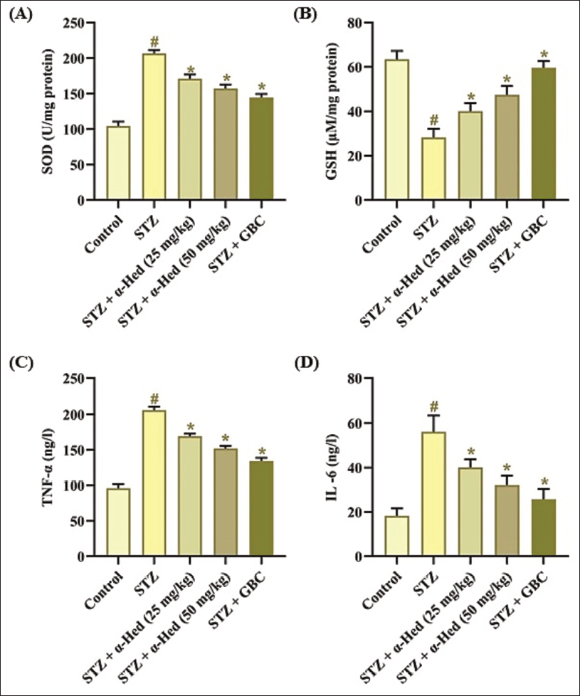
The impact of α-hederin treatment on anti-oxidant levels was evaluated in kidney tissues, with the results shown in Figure 4(A). The reduction in the anti-oxidants GSH and SOD levels was noted in the kidney tissues of STZ-treated rats relative to the control. Captivatingly, the administration of 25 and 50 mg/kg of α-hederin significantly augmented the SOD and GSH in the kidneys of STZ-treated rats. These anti-oxidant effects of α-hederin were validated by the outcomes of glibenclamide, which also increased these anti-oxidants in diabetic rats.
α-Hederin Treatment Decreases Pro-inflammatory Cytokines in the Serum of Streptozotocin-induced Diabetic Nephropathy Rats
The inflammatory cytokines in the serum of experimental animals were assessed, and the results are depicted in Figure 4(B). The considerable elevation in the IL-6 and TNF-α levels was noted in the serum of diabetic nephropathy rats. However, the 25 and 50 mg/kg of α-hederin, respectively, in the STZ-induced rats, led to a notable decrease in both IL-6 and TNF-α in their serum. Furthermore, glibenclamide treatment also reduced these cytokine levels in the serum of STZ-induced rats, consequently underscoring the anti-inflammatory properties of the α-hederin.
Discussion
Diabetic nephropathy signifies a formidable microvascular complication of diabetes, profoundly impacting renal function and significantly contributing to global morbidity and mortality. This complex disorder is characterized by persistent hyperglycemia, which instigates structural changes within the kidney, including glomerular enlargement, extracellular matrix deposition, and basement membrane thickening (de Boer et al., 2022). These pathological alterations culminate in glomerulosclerosis and tubulointerstitial fibrosis, eventually resulting in a progressive decrease in kidney function and, if undiagnosed, ESRD. The societal and economic burden associated with diabetic nephropathy and ESRD is immense, given the escalating global incidence of diabetes (Sheng et al., 2020). Furthermore, diabetic nephropathy necessitates costly interventions such as dialysis or kidney transplantation. Despite its prevalence and severe consequences, current therapeutic strategies for diabetic nephropathy are limited and often accompanied by side effects, underscoring the immediate need for new, safer, and more effective interventions (Danta et al., 2021). Specifically, present treatment modalities focused on decreasing glucose levels do not consistently avert the advancement of diabetic nephropathy, highlighting a critical gap in treatment efficacy. The transition of diabetic nephropathy to ESRD is often irreversible, underscoring the critical need for specific therapeutic strategies that can mitigate or even reverse renal damage (Warren et al., 2019). The present findings offer substantial evidence for the therapeutic activity of α-hederin in ameliorating nephropathy in STZ-treated rats.
The analysis of physiological and metabolic changes is crucial for evaluating the efficacy of potential therapeutic interventions targeting diabetic nephropathy, as these parameters are direct indicators of metabolic disruption and renal pathology in STZ-induced diabetic models (Ghasemi & Jeddi, 2023). For instance, significant changes in body weight and food consumption often reflect the systemic metabolic derangements characteristic of diabetes, while kidney weight provides a direct morphological assessment of renal hypertrophy or atrophy. Changes in water intake and FBG levels further elucidate the severity of diabetes and the potential impact of the drug compound on glycemic control (Cheng et al., 2013). Furthermore, monitoring these metrics allows for the assessment of the drug compound’s ability to mitigate or reverse the pathological progression of diabetic nephropathy, providing insights into its potential mechanistic actions. Body weight reduction in diabetic rats, often linked to STZ-induced cachexia, can be effectively prevented by anti-diabetic treatments, while an elevated kidney weight serves as a critical marker for diabetic nephropathy progression (Chen et al., 2020). Moreover, food and water intake measurements are vital for understanding metabolic compensatory mechanisms and the drug’s influence on appetite regulation and fluid balance, while FBG levels remain the primary benchmark for assessing glycemic control. The fluctuations in these parameters across different experimental groups, including normal controls and diabetic groups treated with various compounds, are instrumental in discerning the specific therapeutic benefits of the investigational drug (Dey et al., 2022). Specifically, STZ induction in rats typically leads to weight loss, increased blood glucose, and elevated kidney-to-body weight ratios, all of which are key indicators for assessing the progression of nephropathy (Kishore et al., 2017). Here, the influence of α-hederin treatment on the physiological parameters in the STZ-induced rats was evaluated. The present findings clearly showed that the α-hederin treatment substantially increased body weight and decreased the FBG levels, kidney weight, and food and water consumption in the STZ-treated rats. These findings offer evidence that α-hederin can mitigate the physiological and metabolic changes in the STZ-treated rats.
Assessing insulin and HOMA-IR levels provides crucial insights into the efficacy of novel therapeutic agents in ameliorating insulin resistance and improving glycemic control in diabetic nephropathy models. Specifically, insulin levels reflect pancreatic β-cell function, while HOMA-IR serves as a robust surrogate marker for insulin sensitivity in these STZ-induced models (Huang et al., 2024). Therefore, analyzing insulin and HOMA-IR levels is essential for understanding the drug compounds’ potential to restore metabolic balance and mitigate the progression of renal damage in these animal models. Such analysis allows for the elucidation of mechanisms by which compounds might improve insulin signaling pathways and reduce the metabolic burden on pancreatic beta cells. Fasting serum insulin and insulin resistance, as quantified by HOMA-IR, are routinely measured to assess the insulin-sensitizing potential of drug compounds in diabetic nephropathy models (Shang & Mohideen, 2023). A reduction in HOMA-IR values in drug-treated animals signifies an improvement in insulin sensitivity and a potential restoration of β-cell function. This restoration is particularly critical in diabetic nephropathy, where chronic hyperglycemia and insulin resistance contribute significantly to renal damage progression. Consequently, evaluating these parameters is critical for discerning whether potential drug compounds alleviate insulin resistance and enhance glucose utilization in nephropathy (Shang et al., 2024). The present findings illustrated that the α-hederin treatment substantially increased insulin levels and subsequently reduced the HOMA-IR level in the STZ-treated nephropathy rats. These findings highlight that the α-hederin treatment can improve insulin sensitivity and restore the β-cell function in STZ-treated rats.
The renal dysfunction markers, including BUN, creatinine, and uric acid, are crucial indicators of kidney function, with elevated levels often correlating with progressive kidney disease and damage. Specifically, BUN and creatinine are metabolic waste products whose accumulation in the serum directly reflects impaired glomerular filtration and renal clearance (Gutierrez et al., 2019). Furthermore, uric acid, a purine catabolite byproduct, also accumulates in the serum when renal excretion is compromised, contributing to the overall pathology of diabetic nephropathy. The assessment of these biochemical parameters provides a comprehensive evaluation of the therapeutic compound’s impact on preserving renal function and integrity in diabetic nephropathy (Dugbartey et al., 2022). This approach allows for a direct correlation between biochemical alterations and the morphological changes observed in the renal parenchyma, offering a robust assessment of treatment efficacy. This includes monitoring changes in creatinine clearance rates, which provide a more dynamic measure of glomerular filtration compared to static serum levels alone. Such analyses are critical for distinguishing STZ-induced diabetes and evaluating treatment effects, as evidenced by significant changes in renal cortex and medulla values in diabetic models (Deshmukh & Manjalkar, 2021). The quantitative measurement of these markers allows for precise monitoring of renal health and the therapeutic compound’s effect on mitigating diabetic nephropathy progression. The elevation of these markers is consistently observed in STZ-induced diabetic nephropathy models, indicating significant renal impairment, which can be ameliorated by various therapeutic interventions (Singh et al., 2022). In this work, the effect of α-hederin on the renal dysfunction markers was evaluated in the STZ-treated rats. The current results suggested that the α-hederin treatment markedly decreased BUN, creatinine, and uric acid in the serum of STZ-treated rats. These results highlight the nephroprotective effects of α-hederin treatment on the diabetic condition, which may facilitate the mitigation of nephropathy progression.
Diabetic nephropathy is defined by a complex interplay of metabolic changes, including hyperglycemia, inflammation, and heightened oxidative stress, all of which participate in progressive renal damage. Central to this pathological process is the overproduction of reactive oxygen species (ROS) and a concurrent reduction in anti-oxidants. Oxidative stress is an imbalance between the ROS accumulation and the capacity to detoxify these reactive intermediates, which plays an imperative role in cellular and tissue damage observed in diabetic nephropathy (Lodhi et al., 2021). Specifically, hyperglycemia exacerbates oxidative stress through multiple biochemical pathways, leading to an increased generation of ROS. This heightened oxidative burden subsequently impairs cellular function and contributes to the structural and functional alterations characteristic of diabetic kidney disease. This imbalance between pro-oxidant and anti-oxidant processes diminishes the efficacy of endogenous anti-oxidant enzymes, further exacerbating renal injury (Kashihara et al., 2010). These reactive species can originate from various enzymatic and non-enzymatic sources within the diabetic kidney, including glucose auto-oxidation, AGEs formation, and elevated flux through the polyol pathway. The resultant oxidative damage contributes to glomerular hypertrophy, extracellular matrix deposition, and glomerular membrane thickening, all hallmarks of diabetic nephropathy. Indeed, the inhibition of oxidative stress has been shown to ameliorate features associated with STZ-induced nephropathy, highlighting its essential role in disease advancement (Kao et al., 2010). Thus, evaluating the efficacy of novel compounds in ameliorating oxidative stress via activating anti-oxidant mechanisms, like GSH and SOD, is crucial for advancing potential techniques to combat diabetic nephropathy. Furthermore, the restoration of GSH and SOD levels in renal tissue serves as a crucial indicator of reduced oxidative damage, directly correlating with improved renal function and attenuated nephropathic progression. This approach provides insights into the intricate interplay between anti-oxidant defenses and drug efficacy in mitigating the severe renal complications associated with diabetes (Kang & Yang, 2020; Liu et al., 2021). Therefore, this work investigated the anti-oxidant mechanisms of α-hederin in the STZ-induced rats. The current outcomes revealed that the α-hederin treatment remarkably elevated the anti-oxidants GSH and SOD levels in the kidneys of STZ-treated rats. These findings highlight that α-hederin can ameliorate oxidative stress response in the kidneys under a diabetic condition. Hence, it was evident that these anti-oxidant properties of α-hederin can aid in ameliorating diabetic nephropathy.
Inflammation is the crucial driver of the pathogenesis of diabetic nephropathy, driven by an intricate interplay of several inflammatory cytokines, with TNF-α and IL-6 playing pivotal roles. Elevated levels of these cytokines have been consistently noted in diabetic individuals, correlating with the initiation and development of renal damage (Rohm et al., 2022). Specifically, these inflammatory mediators are often found in higher concentrations in individuals with diabetic nephropathy compared to those with uncomplicated type 2 diabetes or healthy controls. This sustained inflammatory state contributes to local renal tissue damage through mechanisms of oxidative stress. Emerging evidence highlights that activated innate immunity and subsequent inflammatory responses significantly contribute to the onset of diabetic nephropathy (Perez-Gomez et al., 2016). This inflammatory milieu is characterized by the influx of immune cells into the kidneys and the discharge of additional inflammatory markers, worsening renal injury. Chronic inflammatory state subsequently instigates a cascade of cellular and molecular events, ultimately compromising renal function (Tziastoudi et al., 2017). It has been shown that IL-6 and TNF-α are augmented in the serum of individuals with diabetes and diabetic nephropathy, appearing at early stages of the disease and correlating with the degree of albuminuria. This sustained inflammatory activation, particularly involving TNF-α, is implicated in the progression from chronic hyperglycemia to significant renal lesions (Donate-Correa et al., 2015). Therefore, the novel therapeutic approaches that target and mitigate the inflammatory response are of great interest to treat diabetic nephropathy. The present results illustrated that the α-hederin treatment considerably decreased the TNF-α and IL-6 in the serum of STZ-treated rats. These findings evidenced the anti-inflammatory properties of the α-hederin in a diabetic condition, which may help in ameliorating diabetic nephropathy.
Conclusion
The present data highlight that α-hederin may ameliorate diabetic nephropathy in the STZ-induced rat model. The α-hederin treatment markedly decreased the physiological changes in the diabetic rats. Moreover, the treatment with α-hederin showed a reduction in renal dysfunction markers, inflammatory response, and oxidative stress by enhancing anti-oxidants in the STZ-induced rats. In conclusion, our findings indicate that α-hederin may serve as a beneficial therapeutic candidate for diabetic nephropathy. Moreover, it is essential to perform additional studies in the future to attain a precise understanding of the therapeutic efficacy of α-hederin against diabetic nephropathy.
Summary
Diabetic nephropathy, a severe microvascular complication of diabetes, is a primary cause of end-stage renal disease worldwide. Despite advancements in glycemic and blood pressure control, a substantial portion of diabetic patients still progress to diabetic nephropathy, underscoring the need for more effective therapeutic strategies. The present data highlight that α-hederin may ameliorate diabetic nephropathy in the STZ-induced rat model. In conclusion, our findings indicate that α-hederin may be a promising therapeutic candidate for diabetic nephropathy.
Abbreviations
AGEs: Advanced glycation end-products; BUN: Blood urea nitrogen; ESRD: End-stage renal disease; GSH: Glutathione; HOMA-IR: Homeostatic model assessment for insulin resistance; IL: Interleukin; SOD: Superoxide dismutase; STZ: Streptozotocin.
Footnotes
Declaration of Conflicting Interests
The authors declared no potential conflicts of interest with respect to the research, authorship, and/or publication of this article.
Ethical Approval
The experiment was approved by the Animal Ethics Committee of the Affiliated Hospital of Hebei University.
Funding
The authors received no financial support for the research, authorship, and/or publication of this article.
Informed Consent
Not applicable.
