
Abstract
Fibrolipoma is a well-recognized benign neoplasm composed of adipocytes transected by fibrous connective tissue, but large population studies on these neoplasms are lacking in veterinary medicine. We retrospectively searched for canine cutaneous and subcutaneous fibrolipomas submitted to the surgical diagnostic pathology service of the Athens Veterinary Diagnostic Laboratory (Athens, GA, USA) between 2017 and 2024. We retrieved 236 neoplasms from 64,560 biopsies. The median age of affected patients was 8 y (±2.8 y), with an average tumor size of 2.9 cm (±2.3 cm). The head was the location affected most commonly (31.9% of cases), followed by the carpal area (10.3%), elbow (9.9%), thorax (6.9%), and abdominal wall (5.6%). Most cases were classified as cutaneous (90%). Compared with the total number of biopsy submissions, castrated male (46.2%; p = 0.013) and Labrador Retriever (23%; p = 0.047) dogs were affected most often.
Keywords
Fibrolipomas are benign neoplasms that have a unique histologic appearance consisting of mature adipose tissue (similar to a lipoma) interspersed with fibroblasts and thick collagen bundles arranged in a branching pattern.4,12 The occurrence of oral 2 and intraarticular 13 fibrolipomas has been reported in dogs. However, the prevalence of cutaneous and subcutaneous fibrolipomas has not been well documented in dogs. 8 Here we describe the signalment, clinical signs, and prevalence of cutaneous and subcutaneous fibrolipomas in canine submissions to the surgical biopsy service of the Athens Veterinary Diagnostic Laboratory (AVDL; University of Georgia, Athens, GA, USA) between 2017 and 2024.
We searched the web-based database of the AVDL for biopsy cases of cutaneous and subcutaneous fibrolipoma in dogs diagnosed between January 1, 2017 and December 31, 2024 using the keyword “fibrolipoma”. Only cases with a final diagnosis were included. Submission forms and pathology reports were reviewed for patient signalment, clinical signs, anatomic location of lesions, pathology findings, and diagnosis. Descriptive statistics were used to analyze continuous data (age, tumor dimension); frequencies and percentages were calculated for categorical data (breed, sex, neuter status, anatomic location, duration) using statistics software (SPSS v.29.0.2.0; IBM). A chi-square test was performed to assess the statistical significance of associations among variables. Binary logistic regression was used to evaluate the relationship between breed and the occurrence of neoplasia in all biopsy samples received during the study period. For all statistical analyses, p ≤ 0.05 was considered statistically significant. For the literature comparative review, we searched Google, PubMed, CAB Direct, Web of Science, and Scopus for cases of fibrolipoma and lipofibroma.
We retrieved 64,560 canine biopsy cases during the study period, and found 236 (0.4%) cutaneous and subcutaneous fibrolipomas. The age of affected patients was available in 98.3% of the reports, with a median age of 8 y (±2.8 y). Tumor size, documented by the referring veterinarian in 65.7% of the reports, averaged 2.9 cm (±2.3 cm). The duration of the lesions was reported in 38.1% of the cases, with an average duration of 36 wk (±36.9 wk). A higher percentage of males (55.6%) were diagnosed with fibrolipomas (Table 1). This difference was primarily driven by a higher proportion of castrated males with fibrolipomas (46.2%) compared to castrated males among the total number of received biopsies (37.9%), which was statistically significant (p = 0.013).
Cutaneous fibrolipomas in 236 dogs at the AVDL (2017–2024), by sex and neuter status.
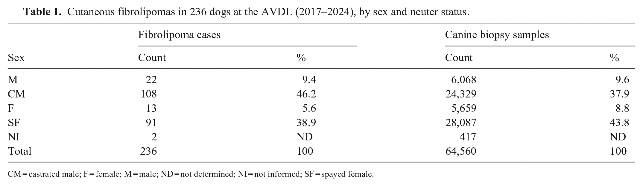
CM = castrated male; F = female; M = male; ND = not determined; NI = not informed; SF = spayed female.
Breed information was missing for only 1 of the 236 cases (Table 2). Labrador Retrievers were the most commonly reported breed (23%), followed by mixed-breed dogs (10.2%), American Pit Bull Terriers (9.4%), Boxers (5.6%), Siberian Huskies (3.8%), German Shepherds (3%), Beagles (2.6%), Australian Shepherds, Golden Retrievers, Great Pyrenees, and Rottweilers (2.1% each), and Chihuahuas and Yorkshire Terriers (1.7% each). Other breeds accounted for <1.3% of the cases.
Cutaneous fibrolipomas diagnosed in 236 dogs at the AVDL, 2017–2024.
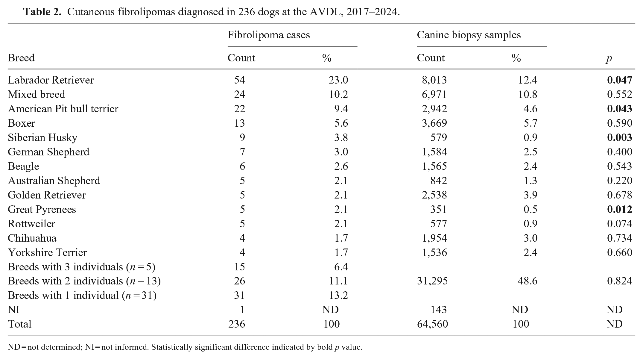
ND = not determined; NI = not informed. Statistically significant difference indicated by bold p value.
The anatomic location of tumors was available for 98.3% of cases (Fig. 1; Table 3). The head was the most common location, with periocular (6.9%), submandibular (6.5%), chin (6%), forehead, ear, and lip (3.5% each), and temporal area (2.2%) accounting for 31.9% of cases. The thoracic limb was involved in 28.2% of cases; the pelvic limb in 23.5%; and the abdomen, thorax, neck, and tail in 16.4% of cases. More specific locations included the carpal area (10.3%), elbow (9.9%), thorax (6.9%), abdominal wall (5.6%), thigh (4.3%), digits, hip, and perineum (3.9% each), hock (3.5%), and stifle (3%). Other locations were described in <2.5% of cases.
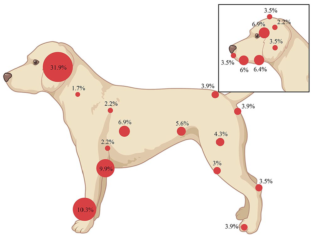
Anatomic location of 236 cutaneous fibrolipomas diagnosed in dogs at the AVDL, 2017–2024. Inset: breakdown by locations on the head.
Cutaneous fibrolipomas diagnosed in 236 dogs at the AVDL, 2017–2024, by location.
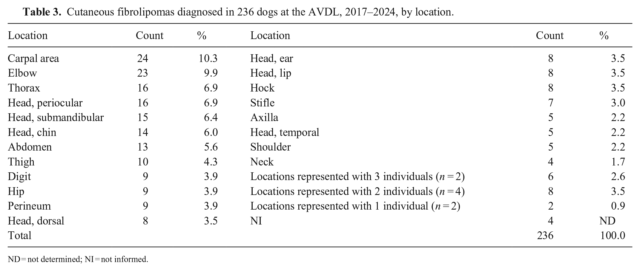
ND = not determined; NI = not informed.
Histologically, proliferations of adipocytes were intersected by a moderate-to-large amount of mature fibrous connective tissue (Fig. 2A, 2B). Tumors containing ~20–60% of connective tissue admixed with the adipocytes were categorized as fibrolipoma. Six cases (2.5%) were classified as infiltrative fibrolipoma. Neoplasms were classified as infiltrative when they infiltrated muscle, including the panniculus carnosus. In all other cases (230; 97.5%), the neoplasm was expansive and well demarcated. The mitotic count was zero in 2.37 mm2 (10 FN22/40× fields) in all samples. Among the 236 cases, 212 (89.8%) were cutaneous fibrolipomas, and only 24 (10.2%) were subcutaneous fibrolipomas.
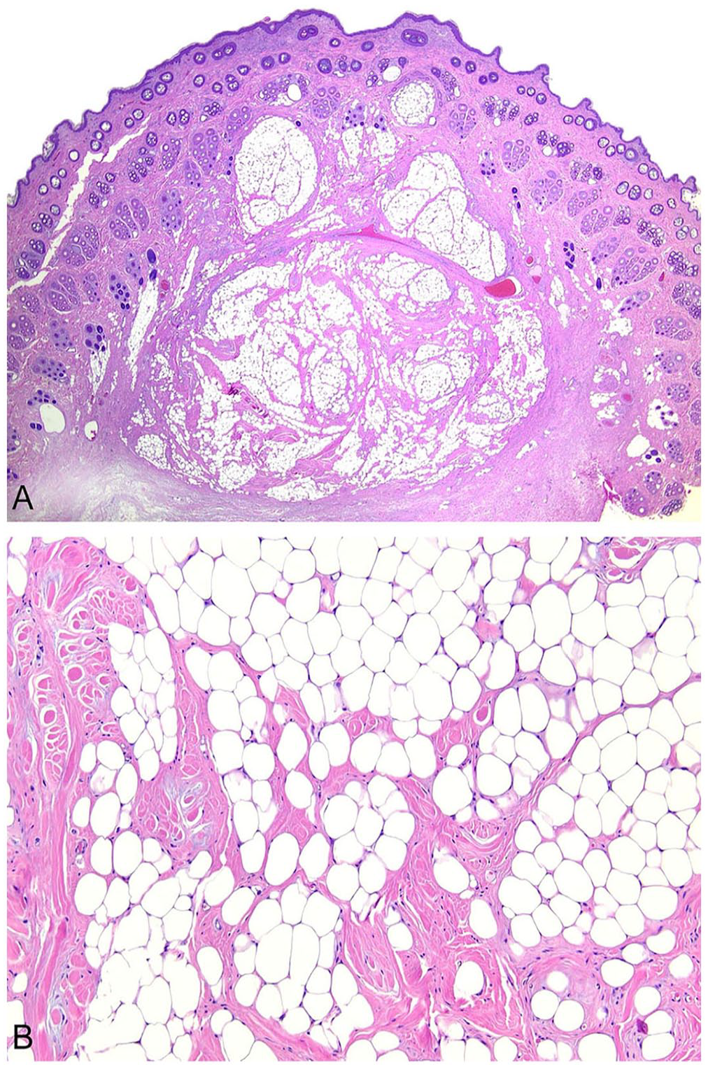
Canine cutaneous fibrolipoma.
The World Health Organization classifies benign lipomatous neoplasms in human beings into classic lipoma, lipoblastoma, lipomatosis, angiolipoma, spindle cell or pleomorphic lipoma, angiomyolipoma, myelolipoma, hibernoma, and atypical lipoma. 18 Fibrolipoma is not included in this classification. On the other hand, under the fibroblastic or myofibroblastic tumors, lipofibromatosis is listed but is a dense population of spindle cells infiltrating the adipose tissue with a locally aggressive behavior. None of these terms fit the cases in our study. Some human pathologists may consider fibrolipomas as a rare subtype of lipoma. 14 In fact, human textbooks describe that when a significant fibrous tissue component is present between adipocytes in a tumor, it may be termed fibrolipoma. 16
Cutaneous and subcutaneous lipofibromas are solitary lesions in older humans with no site predilection or recurrence after surgical excision. 8 They are uncommon in the dermatologic field, being rarely mentioned in reports involving skin tumors or lipomas, 19 and are most commonly referred to as subcutaneous.10,14 It has been proposed that the term fibrolipoma should be replaced with lipofibroma because the bulk of the nodule is fatty tissue and not fibrous connective tissue. 1 In veterinary medicine, the term fibrolipoma is preferred and used in the most relevant literature.2,4,6,12
The veterinary literature is scant on canine fibrolipomas, which have been primarily documented as isolated case reports 8 and non-cutaneous tumors.2,13 A search of Google Scholar, PubMed, CAB Direct, Web of Science, and Scopus using search terms “fibrolipoma” or “cutaneous fibrolipoma”, and “dog” and/or “canine”, yielded one case of canine cutaneous fibrolipoma, 8 confirming that this condition is scarcely reported in veterinary medicine.
According to the American Kennel Club, the French Bulldog became the most common dog breed in the United States in 2022, followed by the Labrador Retriever. 5 Between 1991 and 2021, the Labrador Retriever was the most popular breed in America. 5 Our study identified a higher prevalence of fibrolipomas in Labrador Retrievers (23%). Although this could initially be attributed to a “presentation bias,” whereby common breeds are often perceived as predisposed to certain conditions due to their frequency in clinical practice, the statistical analysis of our sample proportions (12.4% for Labrador Retrievers) suggests that this finding is not biased. In fact, our statistical analysis revealed a significant association between Labrador Retriever and the occurrence of fibrolipomas (p = 0.047). Other breeds with significant associations included American Pit Bull Terriers (p = 0.043), Siberian Huskies (p = 0.003), and Great Pyrenees (p = 0.012).
Lipomas are benign tumors of adipose tissue that are most often subclinical but may cause concern for pet owners.4,12 The prevalence of cutaneous lipomas in dogs has been reported as 27.4% 15 among all cutaneous neoplasms and 0.6% among a population of more than one million dogs. 15 Other authors described a prevalence of 0.7 9 –4.3% 17 among populations of 384,000 and 43,000 dogs, respectively. Certain breeds have been identified as having a higher risk of developing cutaneous lipomas, including Weimaraner (7.8%), Doberman Pinschers (6.9%), German Pointers (5.2%), Springer Spaniels (5.2%), and Labrador Retrievers (5.1%) 9 ; mixed-breed dogs have been described as having a higher percentage of lipomas (27.4 9 –34.1% 7 ). Some of these findings align with our results, in which Labrador Retrievers (22.9%) were identified as the breed with the highest prevalence of fibrolipomas. A genetic component in the development of lipomas has been hypothesized. 3 Furthermore, certain purebreeds have been shown to have a predisposition to lipomas. 9
Previous studies have shown that castrated males and spayed females have a higher risk of developing lipomas. 9 Similarly, we found a higher risk of fibrolipomas in castrated males and spayed females, although the difference was statistically significant only for castrated males. Similar results in castrated males and spayed female dogs were described for oral fibrolipomas. 2
More than half (54.3%) of canine cutaneous lipomas in one study 11 were found on the abdomen and thorax, with the head being affected in only 0.4% of cases. In contrast, we found a higher prevalence of fibrolipomas on the head (31.9%) and a significant occurrence of fibrolipoma on the thoracic limbs (28.2%), primarily the carpal area (10.3%) and elbow (9.9%). This contrasts with the 5.7% prevalence of lipomas on the thoracic limbs reported elsewhere. 11
The diagnosis of fibrolipoma in human and veterinary medicine relies on the straightforward histologic features of the tumors.6,12 Our study is limited by its retrospective nature. No data related to neoplasm progression was available. Evaluating the efficiency of surgical excision and rates of tumor recurrence was not possible. However, given their similarity to lipomas, these neoplasms are likely amenable to surgical excision, and recurrence should not be anticipated if excision is complete.6,11
Footnotes
Declaration of conflicting interests
The authors declared no potential conflicts of interest with respect to the research, authorship, and/or publication of this article.
Funding
The authors received no financial support for the research, authorship, and/or publication of this article.