
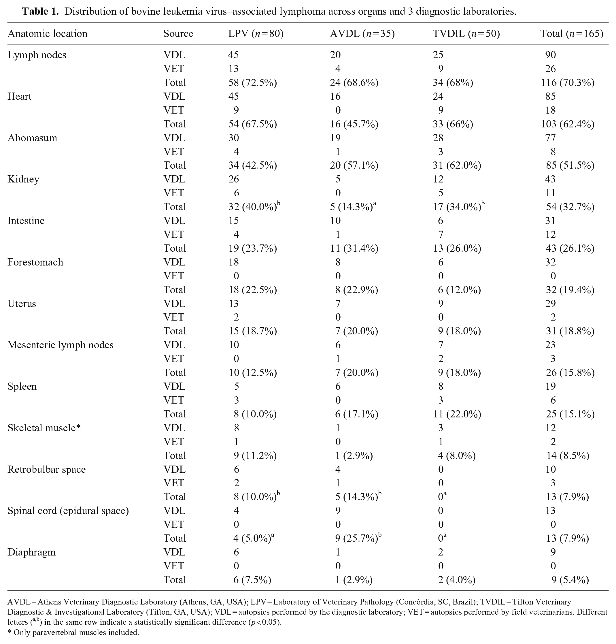
Abstract
Bovine leukemia virus–associated lymphoma (BLV-AL) is a significant neoplastic disease in cattle globally, resulting in substantial economic losses. Here we describe the anatomic distribution of lymphoma in adult cattle in 3 veterinary diagnostic laboratories (VDLs). Between 2001 and 2024, we retrieved 5,290 reports of bovine autopsies performed by these VDLs and 2,200 autopsies submitted by field veterinarians for diagnosis. We selected 165 bovine lymphoma cases, of which 122 (73.9%) originated from VDL autopsies and 43 (26.1%) from autopsies performed by field veterinarians. The most affected organs were lymph nodes (70.3%), followed by the heart (62.4%), abomasum (51.5%), kidney (32.7%), intestinal wall (26.1%), and forestomach wall (19.4%). Among adult cattle lymphoma cases diagnosed at these VDLs, lymphoma was observed more frequently in kidney and intestinal wall than in uterus or spinal cord epidural space.
Bovine lymphoma is most often associated with infection by bovine leukemia virus (BLV; Retroviridae, Deltaretrovirus bovleu) in adult cattle, but sporadic cases are also reported in young animals. The disease was originally described in Germany in 1871, 12 and although bovine lymphoma has been eradicated in some countries, it continues to rise in South 4 and North America, 11 Japan, 15 and China. 14 BLV-associated lymphoma (BLV-AL) affects almost any organ system, but historically, tumors are most often reported in lymph nodes, abomasum, heart, epidural space of the spinal cord, kidney, and uterus.1,18 Other investigations describe tumors more commonly affecting the abomasum, heart, lymph nodes, 3 intestine,5,19 skeletal muscle13,19 and, rarely, the ureter, kidney, and retrobulbar space. 3
The acronym HAULS (heart, abomasum, uterus, lymph nodes, spinal cord) is commonly used to identify the most common locations of bovine lymphoma in cattle. 2 However, our experience suggests that these organs may not always be the most affected sites for bovine lymphoma. Here we describe the anatomic distribution of lymphoma in adult cattle in 3 veterinary diagnostic laboratories, based on gross and histologic lesions.
We searched the database of the Athens Veterinary Diagnostic Laboratory (AVDL; Athens, GA, USA), Tifton Veterinary Diagnostic and Investigational Laboratory (TVDIL; Tifton, GA, USA), and the Laboratório de Patologia Veterinária (LPV; Instituto Federal Catarinense, Concórdia, SC, Brazil) for autopsy cases of bovine lymphoma from 2001–2024 (AVDL), 2009–2024 (TVDIL), and 2013–2024 (LPV). For the AVDL and TVDIL, the search was performed on our web-based system using the keywords “lymphoma”, “leukosis”, and “lymphosarcoma”. For the LPV cases, the search was performed manually by reviewing 1,769 autopsies performed by the LPV and 393 autopsies performed by field veterinarians (referring veterinarians, rDVMs) and reported during the study period. Cases were selected if gross and histologic descriptions were consistent with bovine lymphoma. Submission forms and pathology reports were reviewed for signalment, clinical signs, and pathology findings.
Data on lesion distribution were obtained from gross and histologic examinations of each case. Established laboratory protocols of these 3 diagnostic institutions required a description of all gross lesions and subsequent histologic analysis. Given that some autopsies were performed by rDVMs, and the set of tissues submitted may not represent all affected organs, we decided to keep these data separately. Cases of sporadic lymphoma in cattle <2.5-y-old were not included.
Data were analyzed using SPSS Statistics (v.29.0.2.0). Descriptive statistics were used to summarize the characteristics of the study population. The chi-square test was used to compare the distribution of variables among laboratories and anatomic locations. The Tukey HSD test was used to perform pairwise comparisons to identify specific differences in the distribution among laboratories. A p-value of ≤0.05 was considered statistically significant.
We retrieved 5,290 bovine autopsy cases performed by the VDLs and 2,200 autopsy cases from rDVMs during the studied periods. At the AVDL, there were 1,935 autopsies (72.9%) and 719 field autopsies (27.1%) for a total of 2,654 cases. At the TVDIL, there were 1,586 autopsies (59.3%) and 1,088 autopsies performed by rDVMs (40.7%) for a total of 2,674 cases. At the LPV, 1,769 cases were autopsies conducted by this diagnostic laboratory (81.8%), and 393 cases were autopsies performed by rDVMs (18.2%), with a total of 2,162 diagnoses.
We retrieved 165 bovine lymphoma cases, including 122 (73.9%) VDL autopsies and 43 (26.1%) rDVM autopsies (Table 1). At the AVDL, bovine lymphoma was diagnosed in 35 cases (1.3%), comprising 30 AVDL autopsies (1.5%) and 5 autopsies received from rDVMs (0.7%). At the TVDIL, there were 50 diagnoses (1.9%), including 31 TVDIL autopsies (1.9%) and 19 rDVM autopsies (1.7%). At the LPV, there were 80 bovine lymphoma cases (3.7%), with 61 cases performed by this VDL (3.4%) and 19 by rDVMs (4.8%).
Distribution of bovine leukemia virus–associated lymphoma across organs and 3 diagnostic laboratories.
AVDL = Athens Veterinary Diagnostic Laboratory (Athens, GA, USA); LPV = Laboratory of Veterinary Pathology (Concórdia, SC, Brazil); TVDIL = Tifton Veterinary Diagnostic & Investigational Laboratory (Tifton, GA, USA); VDL = autopsies performed by the diagnostic laboratory; VET = autopsies performed by field veterinarians. Different letters (a,b) in the same row indicate a statistically significant difference (p < 0.05).
Only paravertebral muscles included.
The anatomic location of the tumors based on gross and histologic evaluation of tissues collected at the time of diagnosis varied slightly according to the laboratory (Table 1). Overall, the most affected organs were lymph nodes (116 of 165 cases; 70.3%), followed by the heart (103 cases; 62.4%), abomasum (85 cases; 51.5%), kidney (54 cases; 32.7%), intestinal wall (43 cases; 26.1%), and forestomach wall (32 cases; 19.4%).
The statistical analysis of the anatomic location among the laboratories showed significant differences for kidney (p = 0.025) at the AVDL, retrobulbar space (p = 0.034) at the TVDIL, and spinal cord (p < 0.001) at the AVDL. All other data were statistically similar.
The age of affected cattle (90.1% females and 9.9% males) ranged from 3–15 y (x̄ = 6.1 ± 2.2 y). The production system was reported in 95.7% of cases and consisted of dairy cattle (55.7%), beef cattle (31%), and mixed-breed cattle (13.3%).
Regarding breed (dairy vs. beef), there were statistical differences in the distribution of lesions only for abomasum (44.9% in dairy vs. 60.9% in beef; p = 0.047) and omasum (2.2% in dairy vs. 10.1% in beef; p = 0.034). Considering all of the autopsies, at the LPV, 97.4% of animals were dairy cattle, contrasting with the AVDL and TVDIL, where 20.6% and 10.9% were dairy cattle, respectively.
Grossly, tumors were reported as soft, pale-yellow nodules that infiltrated and effaced the architecture of lymph nodes or visceral organs (Figs. 1, 2). Microscopic examination revealed proliferations of neoplastic lymphocytes arranged in dense sheets with scant cytoplasm and round-to-indented, 8–15-µm diameter, centroblastic, and euchromatic nuclei with vesicular and granular chromatin and large, prominent, and amphophilic nucleoli. Neoplastic cells had variable degrees of pleomorphism and 15–80 mitoses in 2.37 mm² (10 FN22/40× fields). Immunohistochemistry was performed in 4 cases; neoplastic lymphocytes had membranous immunolabeling with CD20, consistent with B-cell lymphoma.
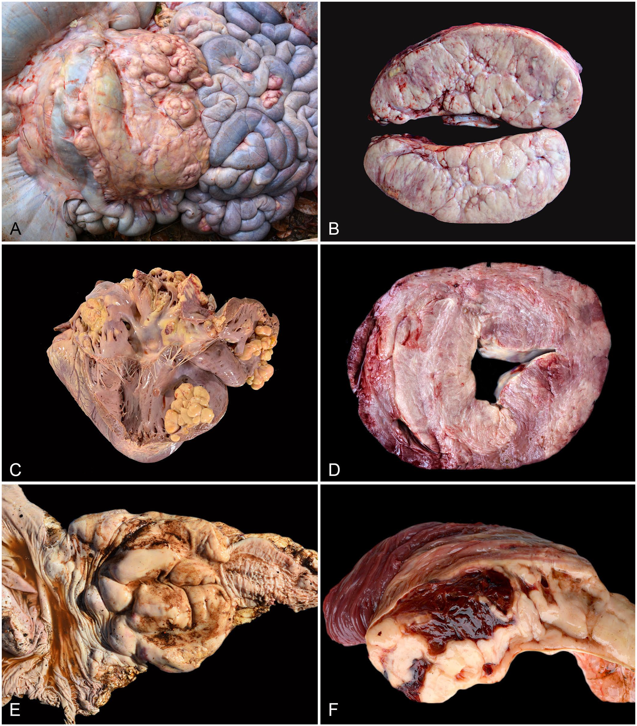
Bovine lymphoma.

Bovine lymphoma.
The existing literature on the anatomic distribution of lymphoma in cattle is conflicting. The consensus is that tumors most often occur in the heart, abomasum, uterus, lymph nodes, and epidural space of the spinal cord.3,6,18 However, the most frequently affected anatomic locations in our cases (lymph nodes, heart, abomasum, kidney, and intestinal wall) were slightly different than the classic 5 predilection sites cited.
The kidney was the fourth most frequently affected site in our study (32.7% of cases), followed by the intestinal wall (26.1% of cases) and forestomach (19.4% of cases). Uterus (18.8% of cases), epidural space, and retrobulbar area (7.9% of cases each) were less commonly involved. We initially considered the forestomach as a single anatomic region, but even when considering the rumen separately, we found that ruminal lymphoma was reported in 15 cases (9.1%), which was more frequent than spinal cord lymphoma (7.9%). If we consider only females (146) with a uterine lesion (31), the percentage changes to 21.2%, still behind kidney and intestinal wall.
Different studies have reported various frequencies in specific organs, including 18.6%, 17 32%, 1 and 44% 19 for lymphoma in kidney; 10.6% for tumors in the uterus; and 3.5% in the spinal cord. 17 We found a high prevalence of lymphoma in the intestine (26.1%); other studies indicate frequencies that ranged from 7.9–96.4% in this location.17,19 Similar discrepancies exist for diaphragmatic lymphoma, which was reported in 5.4% of our cases, contrasting with prevalences of 0.8–62.5% in other studies.17,19
In several of our cases, the neoplasm was observed to involve both the mesenteric lymph nodes and the intestinal wall, forming numerous nodules. In the abomasum, tumors were often associated with ulcers and pyloric outflow syndrome, a condition attributed to the proximity of the lymphoma to the pylorus, as documented in the literature. 5 Large neoplastic nodules, which were more frequent in the abdominal cavity, often had dark-red areas of hemorrhage and necrosis.
Splenic lymphoma is not often reported, but affected cattle may rapidly die after splenic rupture and abdominal hemorrhage.9,18 The prevalence of splenic lymphoma was higher at the TVDIL (22%) and AVDL (17.1%) compared to the LPV (10%). Splenic rupture was reported as the cause of death in 2 (TVDIL) and 4 (LPV) of the cases. Similarly, in one of our cases, death was attributed to hypovolemic shock secondary to hemoperitoneum resulting from tumor infiltration and rupture of the iliac artery. This is an uncommon finding, and the origin of the striking gross lesion must be differentiated from splenic rupture, which could also be caused by babesiosis. 9
In the heart, tumors were either infiltrative throughout the myocardium or formed nodules that bulged into the atrial and ventricular chambers; the latter has been described only rarely in the literature. 14 Reported in 4 cases was an uncommonly described epicardial lesion consisting of thickening of the epicardium with adhesions between the parietal and visceral pericardium 16 ; histologically, the lesion was papillary fibrovascular tissue diffusely infiltrated by large atypical lymphoblasts. The epicardial surface also had areas of hemorrhage and fibrin deposition with hydropericardium. These lesions need to be differentiated from incidental cardiac mesothelial hyperplasia, which lack neoplastic lymphocytes. 7
The distinct frequency of spinal cord involvement across the 3 diagnostic laboratories underscores the importance of comparing data among institutions. At the LPV, spinal cord was examined in all cases, and lymphoma was detected in only 4 (5%). Similar findings were described in an investigation reporting 113 bovine autopsies, in which spinal cord lymphoma was recorded in 3.5% of cases. 17 Spinal cord lymphoma was reported in 9 (25.7%) AVDL cases and in none of the TVDIL cases, resulting in an overall rate of 7.9% of spinal cord lymphoma in our dataset. These discrepancies might be explained by the higher prevalence of lymphoma in the paravertebral muscles in LPV cases (9 cases; 11.2%).
Metastatic neurolymphomatosis exhibits a predilection for specific nerves within the peripheral nervous system. In contrast, secondary spinal cord tumors display diverse types and patterns of local extension. Notably, in bovine, feline, and canine lymphoma, most spinal cord metastases originate from the vertebral bodies or paravertebral tissue and subsequently expand into the epidural space. 8 We hypothesize that cases described here most likely represent lymphoma originating in the lymphoid tissue near the vertebrae and subsequently infiltrate the surrounding skeletal muscles and spinal cord. But the possibility of metastatic lymphoma cannot be ruled out.
It is possible that the limited spinal cord involvement and extensive paravertebral muscle lesions observed were associated with a shorter clinical course. Neoplasia infiltrating the spinal canal through the spinal nerve roots, with a cream-colored soft tissue mass surrounding the lateral edges of the vertebral bodies, has been described.3,10 We speculate that in the 8.5% of cases with neoplasia confined to the paravertebral muscles, progression could lead to lymphoma involvement of the spinal cord within a few months. These animals frequently had clinical signs of ataxia. Another factor to consider is the proportion of dairy and beef cattle among the laboratories. The daily management of dairy cattle enables farmers to monitor their animals closely. We believe that, overall, dairy cattle are more likely to be brought to veterinary attention and autopsied in the early stages of the disease, when lymphoma is still confined to the paravertebral muscles. In fact, only 20.6% at the AVDL were dairy cattle, compared to 97.4% at the LPV.
One limitation of our work is that we did not perform PCR for retrovirus confirmation. Despite that, all of our cases were multicentric lymphoma in adult cattle, which is suggestive of BLV-AL. The age range of cattle in our cases (3–15-y-old) is characteristic of BLV-AL and beyond the typical age range for sporadic multicentric lymphoma (non-associated with virus).6,18
It is always difficult to compare autopsy data among diagnostic laboratories because the standard procedure on the necropsy floor can vary. One peculiarity of the distribution obtained at the LPV is that 93.1% of all cases were performed and/or diagnosed by the same pathologist, granting it high homogeneity. In fact, the statistical analysis showed no significant difference among laboratories in most of the data, except for the kidney, retrobulbar space, and spinal cord. Furthermore, these differences were mainly due to no cases of lymphoma in the retrobulbar space and spinal cord at the TVDIL. It should be noted that at the TVDIL, the spinal cord and retrobulbar space were examined only in cases with clinical signs of ataxia or exophthalmos, respectively. Despite that, the brain was analyzed in all cases, and no histologic lesion of lymphoma was observed. Additionally, the standardized procedures employed by these laboratories are similar and support a comprehensive examination of all organs, accompanied by a detailed macroscopic description of any lesions observed. Subsequently, thorough histologic analysis is mandated.
Footnotes
Acknowledgements
We thank the Instituto Federal Catarinense and the University of Georgia for supporting the veterinary pathology diagnostic laboratories, as well as the numerous veterinarians and farmers who facilitated and provided information regarding the reported cases.
ORCID iDs
Declaration of conflicting interests
The authors declared no potential conflicts of interest with respect to the research, authorship, and/or publication of this article.
Funding
We received financial support for our research from the Instituto Federal Catarinense and the Brazilian National Council for Scientific and Technological Development (CNPq), project 307086/2022-4.