
Abstract
Histiocytic sarcomas (HSs) are uncommon malignant neoplasms in cats, typically affecting older animals and not previously associated with feline leukemia virus (FeLV). Here, we describe 2 cases of disseminated HS in young FeLV-positive domestic shorthair cats that clinically and grossly mimicked lymphoma. Case 1, a 1-y-old female cat, was presented with hemiplegia and dysuria and was euthanized because of the clinical suspicion of spinal lymphoma. Case 2, a 4-y-old male cat, had hyporexia, ocular abnormalities, and bilateral renal enlargement and was euthanized because of the clinical suspicion of renal and ocular lymphoma. Both cases tested positive for feline leukemia virus (FeLV) infection with a SNAP test. At autopsy, case 1 had a white, irregular, intradural spinal cord mass at the C6–C7 segment, and case 2 had coalescing, unencapsulated, white nodules that effaced both renal cortices, along with uveal thickening in the left eye with hyphema. Both cases had highly pleomorphic neoplastic round-cell proliferation in neoplastic masses, with strong cytoplasmic labeling for vimentin and IBA1 on immunohistochemistry and no CD20 or CD3 labeling, confirming disseminated HS. The neoplastic cells also had strong cytoplasmic immunolabeling for FeLV, suggesting a possible oncogenic role of FeLV in feline HSs.
Histiocytic disorders in cats are uncommon, compared with dogs, and include pulmonary Langerhans cell histiocytosis, feline progressive histiocytosis, and the histiocytic sarcoma complex.18,19 Histiocytic sarcoma (HS) is a malignant neoplasm thought to originate from interstitial dendritic cells. 18 HSs can be either localized or disseminated. 19 In cats, the most common sites of HSs are the skin and subcutis, spleen, bone marrow, lymph nodes, liver, lungs, and articular and periarticular tissues. 18
Feline leukemia virus (FeLV; family Retroviridae, taxon species Gammaretrovirus felleu) causes one of the most common and important infectious diseases in cats, 15 and is associated with a higher frequency of lymphocyte-driven neoplasia in young cats, such as lymphoma and leukemia.6,15 On the other hand, HSs are mostly neoplasms of older cats 20 and are not directly associated with this retroviral infection. Here, we describe the clinical, gross, histologic, and immunohistochemical findings in 2 cases of HS in young FeLV-positive cats that clinically and grossly mimicked lymphoma. Aside from a single case of HS in a FeLV-positive cat detected by ELISA, 10 we retrieved no cases of HSs with FeLV immunolabeling within the neoplastic cells in a search of Google, PubMed, Web of Science, and Scopus, using the search terms “histiocytic sarcoma”, “malignant histiocytosis”, “cat”, “feline”, “FeLV”, and “feline leukemia virus”, suggesting that this possible association has not been reported previously in domestic cats.
We searched the online autopsy database of the Setor de Patologia Veterinária (College of Veterinary Medicine, Universidade Federal do Rio Grande do Sul, Porto Alegre, Rio Grande do Sul State, Brazil) between 2010 Jan and 2025 Jun for cases compatible with HS in FeLV-positive cats. Using the same search keywords, we retrieved 2 cases. Histologic sections stained with H&E were reviewed by 4 of the authors (G Hartmann, ÉC Lamego, SP Pavarini, L Sonne), and selected sections were submitted for immunohistochemistry (IHC). We used the following antibodies to immunophenotype the neoplasms: anti–pan-cytokeratin (AE1/AE3), anti-vimentin, anti-CD3, anti-CD20, anti–ionized calcium-binding adapter molecule 1 (IBA1), anti–E-cadherin, anti–FeLV gp70 antigen, anti–glial fibrillary acidic protein (GFAP), and anti-S100 ( Suppl. Table 1 ).
The 2 selected cases were both domestic shorthair cats, both positive for the FeLV p27 antigen and negative for feline immunodeficiency virus (FIV) antibodies via a rapid assay kit (SNAP FIV antibody/FeLV antigen combo test; Idexx). Case 1 was a 1-y-old spayed female, and case 2 was a 4-y-old castrated male. In case 1, the cat was presented with a 5-d history of right-sided hemiplegia and dysuria and was euthanized because of clinically suspected spinal cord lymphoma. Case 2 was presented with a 3-wk history of hyporexia, bilateral buphthalmos, anisocoria, and hyphema of the left eye. The cat also had marked bilateral renal enlargement and was euthanized because of clinically suspected renal lymphoma with ocular metastasis.
Gross findings in case 1 included a soft-to-firm, white, irregularly shaped intradural mass strongly adhered to the right side of the spinal cord at the C6–C7 segment, extending and encircling the adjacent nerve roots ( Fig. 1 ). The renal capsular surface was irregular, and white-to-pale-red, soft, 0.3–1.5-cm nodules were visible on cut surface. In the lungs, white, firm, up to 0.1-cm nodules were throughout all lobes. Additionally, the urinary bladder was markedly distended.
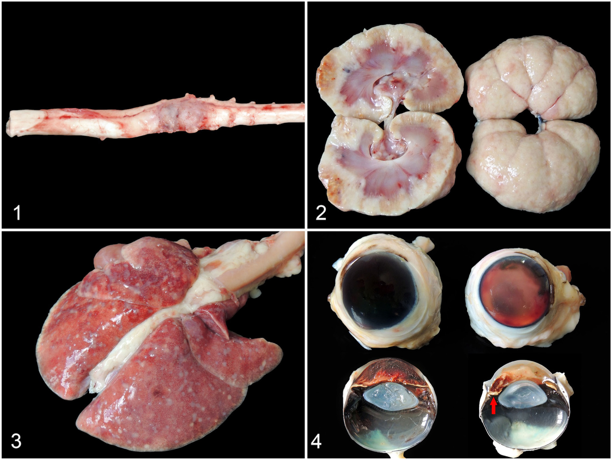
Histiocytic sarcoma in 2 FeLV-positive young cats.
In case 2, both kidneys were markedly enlarged, with irregularly shaped, pale, and firm capsular surfaces. On the cut surface, coalescing, white-to-yellow, soft, 0.1–1.5-cm nodules were noted ( Fig. 2 ). Marked hydronephrosis in the left kidney was caused by a similar 2.5-cm mass that blocked the ureteral lumen. In the lungs, coalescing, soft white, 0.1–0.5-cm plaques were present throughout all the lobes and visceral pleura ( Fig. 3 ). On the splenic cut surface, white, 0.3-cm nodules were found. The mesenteric lymph nodes were moderately enlarged. Additionally, the cat had moderate bilateral buphthalmos and hyphema. The left eye had moderate uveal thickening ( Fig. 4 ).
Microscopically, in case 1, an intradural and intraparenchymal highly cellular neoplastic proliferation of highly pleomorphic cells was arranged in sheets and bundles, supported by a delicate stroma ( Fig. 5 ). These cells expanded the white and gray matter and encircled and entrapped the spinal nerve roots. The neoplastic cells were round-to-fusiform, with moderate-to-abundant eosinophilic cytoplasm and distinct cell borders. The nuclei were round-to-reniform, slightly eccentric, with finely stippled chromatin and conspicuous nucleoli. Anisocytosis and anisokaryosis were marked, binucleate cells were frequent, multinucleate cells were rare, karyomegaly and odd-shaped nucleoli were noted, and 30 mitotic figures in 2.37 mm² (10 FN22/40x fields) were frequently atypical. Geographic necrosis was interspersed within the neoplasm, and a mild, multifocal infiltrate of neutrophils and foamy macrophages was associated with multifocal hemorrhage. In the adjacent white matter, frequent axonal degeneration with axonal spheroids was noted. The nodules observed grossly in the kidneys and lungs were also compatible with HS, and a neoplastic embolus was present within a urinary bladder blood vessel.

Histiocytic sarcoma in 2 FeLV-positive young cats.
In case 2, a neoplastic proliferation compatible with HS was observed in the kidneys ( Fig. 6 ), lungs, bone marrow, heart, thyroid gland, splenic red pulp, and skeletal muscle. However, only 3 mitotic figures were identified in 2.37 mm2, all within the neoplastic cells of the kidney. In both eyes, expansion of the anterior uvea with neoplastic lymphocytes ( Fig. 7 ) was observed, as well as moderate lymphoplasmacytic uveitis, pre-iridal fibrovascular membrane formation, and occlusion of the drainage angle. Furthermore, in the right eye, severe hyphema, significant fibrosis of the iris with reactive endothelium, and excavation of the optic disc were noted. The left eye had mild hyphema with fibrin deposition and mild atrophy of the retinal ganglion cell layer.
More than 90% of the neoplastic cells of the spinal cord, kidney, and lung (case 1), as well as the neoplastic cells of the kidney, lung, eye, bone marrow, heart, thyroid gland, spleen, and skeletal muscle (case 2), had strong cytoplasmic vimentin and FeLV immunolabeling, and strong membranous IBA1 immunolabeling ( Figs. 8–10 ). In both cases 1 and 2, no labeling for pancytokeratin, CD3, CD20, or E-cadherin was observed. Additionally, in case 1, no GFAP or S100 immunoreactivity was observed.
The diagnosis of disseminated HS in both FeLV-positive cats was based on the histologic and IHC findings. FeLV is an oncogenic retrovirus frequently associated with hematopoietic neoplasia, such as lymphoma and leukemia. 15 Rarely, other non-hematopoietic neoplastic processes are attributed to FeLV infection, such as osteochondromatosis 26 and multicentric fibrosarcoma induced by feline sarcoma virus (FeSV) infection. 12 FeSV is a hybrid virus that originated from the recombination of FeLV DNA provirus with cat proto-oncogenes. 12 All of these FeLV-driven neoplasms typically affect young cats up to 7-y-old.9,11 This age pattern is consistent with our findings; our cases were either juvenile or young adults. Additionally, in FeLV-related neoplasms, the neoplastic cells frequently have anti-FeLV cytoplasmic immunolabeling.6,9,11 In our search, no cases of HSs were attributed to FeLV infection. However, the IHC detection of FeLV gp70 antigen in the cytoplasm of neoplastic cells, combined with the young age of the affected cats, may suggest a role for this virus in the carcinogeneses in our cases. Further studies involving a larger number of animals and advanced molecular approaches, such as transcriptomic analyses of FeLV oncogenesis, might clarify this potential association.
In cats, HSs with CNS involvement are mostly reported affecting the brain,16,20 and, less frequently, the spinal cord.20,24 The most common spinal cord neoplasm in cats is lymphoma, 13 particularly in FeLV-positive cats 1–5-y-old.13,14,25 In cats with spinal neoplasms, paraparesis, and paraplegia are common clinical signs 13 and can be asymmetrical at initial presentation, 24 as we observed in case 1. Therefore, in FeLV-positive young cats with myelopathic signs, spinal lymphoma remains a major clinical differential diagnosis. 25 However, given their clinical and gross similarities, our case indicates that spinal cord HS should also be considered as a differential diagnosis in FeLV-positive cats.
Lymphomas are the most common primary renal tumors in cats, 17 and about half of the affected animals are FeLV-positive.11,17 Both lymphomas and HSs can be unilateral or bilateral masses. 3 In case 2, which involved enlargement of both kidneys by soft, white masses, the primary differential diagnosis—both clinically and grossly—was renal lymphoma. In cats, HSs with kidney involvement are quite uncommon and often part of a systemic disease, 5 as observed in case 2. Interestingly, similar findings with the same primary differential diagnosis were reported in a 5-y-old FeLV-negative cat with bilateral renal HS, including lung involvement. 27
As observed in case 2, ocular manifestations of HSs may be the initial presentation of systemic disease in cases with ocular involvement. 23 However, lymphoma was suspected first given that it is the most prevalent systemic neoplasm with ocular involvement in cats. 1 The uveal tract is the most common site of secondary intraocular neoplasia in cats, 1 and animals with uveal neoplasia frequently have intraocular hemorrhage, glaucoma, uveitis, or retinal detachment.1,7 Hyphema and pupil abnormalities, both observed in case 2, are reported in intraocular HSs within the uveal tract in cats. 23 Moreover, the hyphema, the buphthalmos, and the atrophy of the ganglion cell layer of the retina with excavation of the optic disc observed in case 2 are consistent with glaucoma, 7 which most likely was secondary to the uveal tract infiltration of neoplastic histiocytes and membrane formation with occlusion of the draining angle.
An IHC panel was conducted for case 1 given the highly pleomorphic nature of the neoplastic cells. The differential diagnoses of astrocytoma and spinal schwannoma were excluded based on the absence of GFAP and S100 immunoreactivity.21,22 Histologically, the neoplasm did not resemble lymphoma; however, the lack of CD20 and CD3 labeling ruled out a diagnosis of anaplastic variants, which can occur in the nervous system. 8 Despite this, the intense IBA1 cytoplasmic labeling in the neoplastic cells in both cases, which is typical of these neoplasms, 20 confirmed the diagnosis of disseminated HS. The absence of E-cadherin labeling confirmed the histiocytic origin by ruling out other histiocytic disturbances caused by Langerhans cells.18,20 Although IBA1 does not differentiate HS from hemophagocytic HS of macrophage origin, 20 no hemophagocytic activity was observed. Moreover, neither cat had gross lesions compatible with hemophagocytic syndrome, such as splenomegaly or jaundice. 19
In contrast to North America, where the prevalence of FeLV infection in cats is very low (3.1%), 4 in Brazil, the frequency of FeLV infection in domestic cats remains high (~22.3%). 2 This divergence is believed to be the result of the small number of vaccinated cats in Brazil. 2 The epidemiologic characteristics and dynamics of certain neoplasms, such as lymphomas, vary between countries in which FeLV remains a prevalent infection and those where the infection is under control. 11 This may apply to other putatively FeLV-related neoplasms, such as the HS cases that we describe here.
Supplemental Material
sj-pdf-1-vdi-10.1177_10406387261452652 – Supplemental material for Disseminated histiocytic sarcoma in 2 FeLV-positive cats in Brazil
Supplemental material, sj-pdf-1-vdi-10.1177_10406387261452652 for Disseminated histiocytic sarcoma in 2 FeLV-positive cats in Brazil by Gabriela Hartmann, Éryca C. Lamego, Yasmin Daoualibi, Joanna V. Z. Echenique, Maria Vitória G. Sanches, Marcele Bettim Bandinelli, Saulo Petinatti Pavarini and Luciana Sonne in Journal of Veterinary Diagnostic Investigation
Footnotes
Declaration of conflicting interests
The authors declared no potential conflicts of interest with respect to the research, authorship, and/or publication of this article.
Funding
Our work was supported by Coordenação de Aperfeiçoamento de Pessoal de Nível Superior (CAPES), Brazil–Finance code 001. We received partial financial support from Conselho Nacional de Desenvolvimento Científico e Tecnológico (CNPq grant 307277/2021-6).
Supplemental material
Supplemental material for this article is available online.
References
Supplementary Material
Please find the following supplemental material available below.
For Open Access articles published under a Creative Commons License, all supplemental material carries the same license as the article it is associated with.
For non-Open Access articles published, all supplemental material carries a non-exclusive license, and permission requests for re-use of supplemental material or any part of supplemental material shall be sent directly to the copyright owner as specified in the copyright notice associated with the article.