
Abstract
Upper digestive tract neoplasms associated with ingestion of Pteridium spp. (bracken fern) have been documented in cattle but not in buffalo. Here, we describe upper digestive tract lesions associated with the ingestion of P. esculentum subsp. arachnoideum in 3 water buffalo (Bubalus bubalis) in Brazil. Gross inspection revealed proliferative and ulcerative lesions in the mucosa of the base of the tongue, oropharynx, and esophagus in all 3 cases. Proliferative lesions were well-demarcated, white-to-brown, exophytic nodules on the lingual and oropharyngeal mucosa (papillomas, cases 1–3); a well-demarcated, white nodular fibroma in the distal esophageal submucosa (case 1); and locally infiltrative areas of mucosal thickening with ulceration and dark red-to-brown areas of hemorrhage in the base of the tongue and oropharynx (squamous cell carcinomas [SCCs], cases 1–3). Histologically, non-neoplastic lesions were irregular (cases 2, 3) or pseudocarcinomatous (case 1) mucosal hyperplasia with parakeratosis (cases 1–3) and dysplasia (cases 1, 2) in the base of the tongue, oropharynx, and esophagus. Neoplastic lesions were papillomas affecting the base of the tongue, oropharynx, and esophagus (cases 1–3); a submucosal distal esophageal fibroma (case 1); and SCCs affecting the base of the tongue and oropharynx (cases 1–3). Mucosal ulceration was associated with the SCCs in all cases. Inflammation was lymphoplasmacytic. Esophageal vascular myxomatous degeneration or proliferation with fibrosis were present. The diagnoses of Pteridium-associated disease were based on the epidemiologic and pathologic findings, which were identical to those observed in cattle.
Pteridium spp. (bracken ferns) have a cosmopolitan distribution and high carcinogenic potential for livestock because of their radiomimetic activity.
18
Ptaquiloside, a norsesquiterpene glycoside found in bracken ferns, is the toxic principle implicated in oncogenesis in cattle.5,18 This carcinogen leads to DNA alkylation and neoplastic transformation in the upper gastrointestinal tract and urinary bladder.
18
In cattle, consumption of Pteridium spp. may result in various clinical and pathologic conditions that depend on the dose and duration of ingestion.
18
Acute hemorrhagic diathesis typically results from daily ingestion of >10 g/kg of the plant for several weeks to a few months.
18
Two chronic conditions may occur individually or simultaneously—enzootic hematuria (
The occurrence of UDTNs associated with Pteridium spp. ingestion in cattle has been described in several countries around the world, 18 including Brazil,4,5,10 Bolivia, 9 Scotland, and England. 7 Although UDTNs have been associated historically with the carcinogenic effects of Pteridium spp.,4,5,10 bovine papillomavirus (BPV) has been proposed as a contributor to the pathogenesis or as the primary cause of these tumors. 7 UDTNs are progressive and lead to clinical signs that vary according to their location in the gastrointestinal tract. Stridor, dysphagia, and coughing are associated with lesions at the base of the tongue and pharynx; regurgitation of contents, sometimes through the nostrils, may occur in cases of esophageal tumors; and drooling, dysphagia, ruminal atony with tympany, diarrhea, emaciation, dyspnea, and death are reported in cases with lesions at the entrance of the rumen.4,10,11 Grossly, UDTNs are typically exophytic (papillomatous or polypoid) masses that are often ulcerated and infiltrate the surrounding mucosa and underlying tissues. Carcinomas may also be flat, endophytic, or infiltrative lesions that lead to mucosal thickening and ulceration rather than exophytic masses.4,5,10
Histologic changes in cattle typically fall within a spectrum of hyperplastic, dysplastic, and neoplastic mucosal lesions, with distinct degrees of local infiltration into the submucosa, muscle layers, and serosal surface. These changes may range from mucosal squamous hyperplasia, with or without dysplasia, to in situ squamous cell carcinomas (
The associations between chronic Pteridium spp. poisoning and UDTNs are described mainly in cattle.4,5,10 Although EH has been described in buffalo in Brazil, 16 Turkey, Formosa, Indonesia, and India, 15 and digestive tract viral papillomas are reported in buffalo in India, 8 a search of Google, PubMed, CAB Direct, Web of Science, and Scopus retrieved no UDTN cases associated with natural ingestion of Pteridium spp. in buffalo. Here, we describe UDTNs associated with the natural ingestion of P. esculentum subsp. arachnoideum by 3 water buffalo (Bubalus bubalis) in Brazil.
The natural outbreak occurred in August–September 2018 in a dairy cattle farm in São José do Barreiro (São Paulo, Brazil). The buffalo were used for milk and cheese production. Cows were being replaced by buffalo in the farm because buffalo were thought to be more resistant to the effects of Pteridium spp. During visits to the farm, three 6–8-y-old buffalo cows in good body condition and with a history of hematuria were evaluated before slaughter. All buffalo were raised under natural rangeland conditions, but some individuals were supplemented with citrus pulp and concentrate during the dry season (Jun–Sep). Pastures consisted of Urochloa spp. (Brachiaria spp.) and were heavily invaded (70–90% of the area) by P. esculentum subsp. arachnoideum (
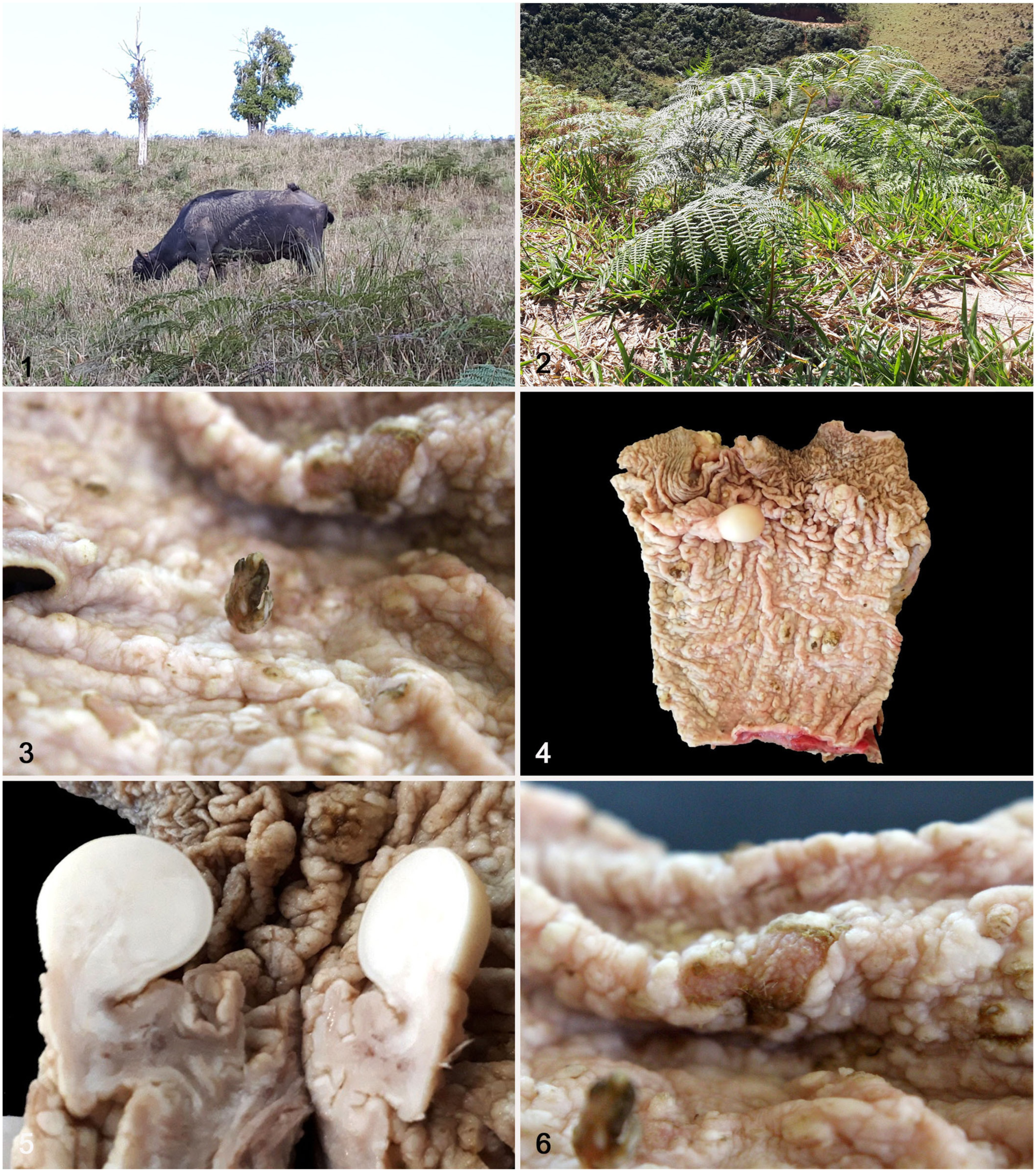
Grazing conditions and gross lesions associated with P. esculentum subsp. arachnoideum ingestion by 3 water buffalo.
Routine physical evaluation of the buffalo in the holding corrals at the slaughterhouse included behavioral observation (breathing, level of consciousness, posture and locomotion, coat, abdominal shape), body condition score (BCS) based on a scale of 1–5, and color of urine and feces. The 3 buffalo had normal levels of consciousness, posture, and locomotion. Their coats were clean and shiny with no visible ectoparasites. The BCSs were 3.5 (buffalo 1), 2.5 (buffalo 2), and 3 (buffalo 3). Macroscopic evaluation of the urine confirmed the presence of hematuria in all 3 buffalo; no alterations were observed in the feces. All 3 individuals were slaughtered using conventional methods (stunning by captive bolt).
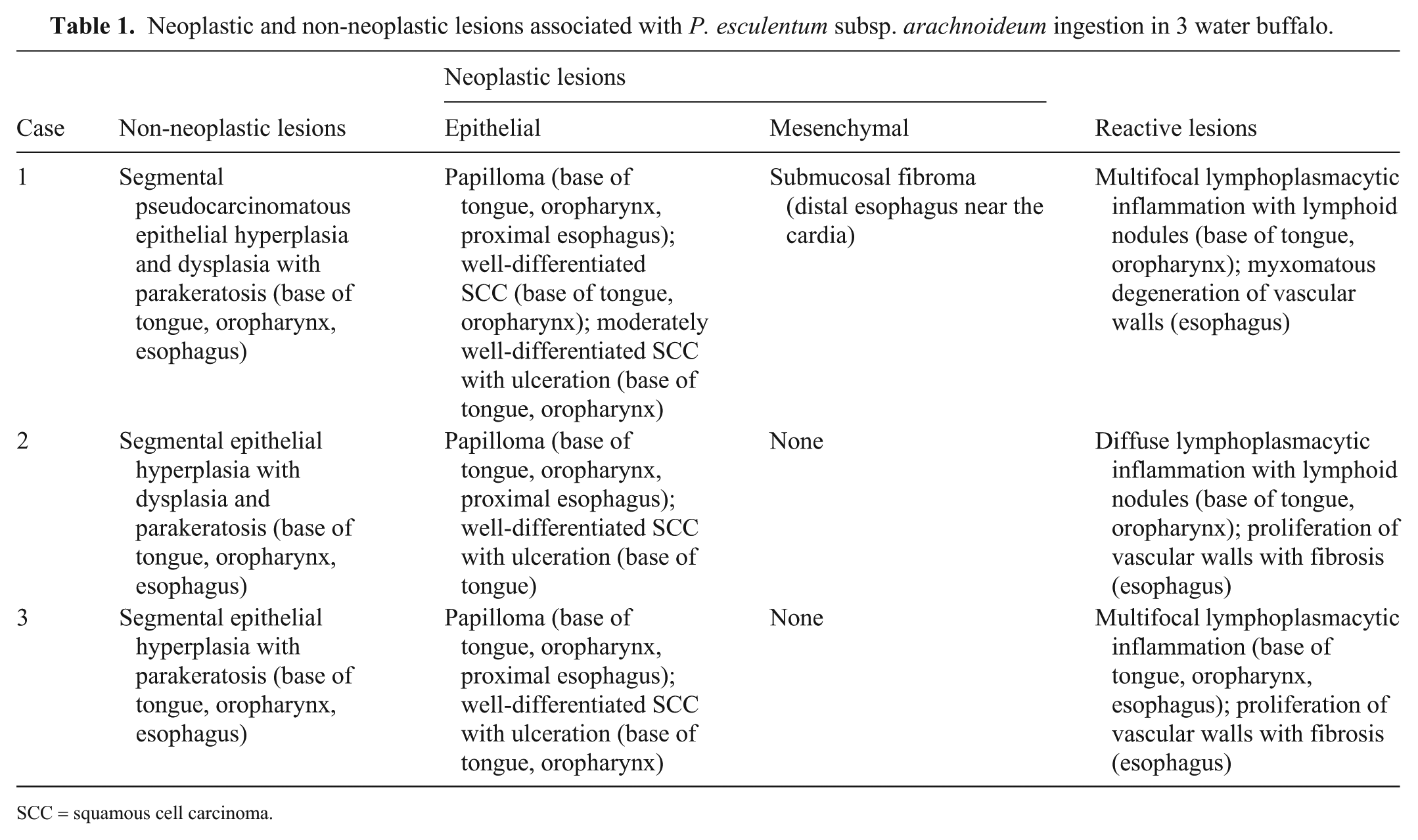
Gross examination of the carcass and multiple organs after slaughter revealed proliferative and ulcerative lesions in the mucosa of the base of the tongue, oropharynx, and/or esophagus in all cases. Papillomas (
Fig. 3
) consisted of well-demarcated, ~0.4–1-cm white-to-brown, exophytic, irregular nodules that occurred on the lingual and oropharyngeal mucosa (cases 1–3). The surrounding mucosa had circular, 0.4–0.8-cm white plaques with a depressed, dark-red center. The esophageal mucosa (case 1) had segmental areas of thickening with a well-demarcated, ~1-cm white smooth nodular fibroma near the cardia (
Histologically, all buffalo had a variety of non-neoplastic and neoplastic lesions affecting the base of tongue, oropharynx, and esophagus (
Table 1
). Non-neoplastic mucosal lesions were segmental areas of irregular (cases 2, 3) or pseudocarcinomatous (case 1) epithelial hyperplasia, with parakeratosis (cases 1–3) and dysplasia (cases 1, 2), affecting the base of the tongue, oropharynx, and esophagus in all cases (
Neoplastic and non-neoplastic lesions associated with P. esculentum subsp. arachnoideum ingestion in 3 water buffalo.
SCC = squamous cell carcinoma.

Upper digestive tract lesions associated with ingestion of Pteridium esculentum subsp. arachnoideum by 3 water buffalo. H&E.
Multifocal lymphoplasmacytic inflammation was associated with the mucosal lesions in the base of the tongue and oropharynx (case 1) and base of the tongue, oropharynx, and esophagus (case 3); lymphoplasmacytic inflammation was diffuse in the base of the tongue and oropharynx (case 2). Other lesions included intimal and medial myxomatous degeneration of small-caliber vessels (case 1) and vascular wall proliferation with fibrosis (cases 2, 3) in the esophagus.
A formalin-fixed, paraffin-embedded esophageal sample from each buffalo was tested via PCR assay for identification of a 480-bp fragment of the BPV genome. The samples were macerated with a mortar and pestle in 1 mL of PBS, pH 7.4, and centrifuged at 720 × g for 10 min. Total DNA was isolated from 100 µL of the sample using a phenol-chloroform–based protocol. 17 DNA quantity and quality were assessed by spectrophotometry (NanoDrop spectrophotometer; ThermoFisher). Although absorbance-based quantification may overestimate DNA concentration (because of potential co-purified contaminants), the measurements were used exclusively to standardize the amount of template added to PCR reactions within the range recommended by the manufacturer. DNA purity was evaluated using the A260/A280 ratio for suitable PCR amplification. Conventional PCR was performed using the primers FAP 59 (5′-TAACWGTIGGICAYCCWTATT-3′) and FAP 64 (5′-CCWATATCWVHCATITCICCATCATC-3′) to amplify a relatively conserved fragment of the L1 gene of all known papillomavirus types. 6 This approach may fail to detect novel papillomavirus genomes in papilloma lesions, given the lower base-pairing homology in the 3′ region of both primer binding sites. 3 The conditions for performing the PCR were as follows: 94°C for 5 min; 40 cycles of 94°C for 1 min; 55°C for 1 min and 72°C for 1 min; and 72°C for 5 min. The amplicons were electrophoresed in a 0.5% agarose gel and visualized under an ultraviolet light source. PCR results were negative for BPV.
Our findings are consistent with reports of UDTNs associated with the chronic consumption of Pteridium spp. in cattle in endemic areas.
11
Although the most frequently described clinical form of Pteridium spp. poisoning in cattle in Brazil is EH, cases in the south (Santa Catarina, Rio Grande do Sul) consist mainly of upper digestive tract (
A role for BPV in the development of UDTNs in cattle ingesting Pteridium spp. has been proposed but not confirmed. 5 It is thought that BPV-induced mucosal papillomas act as the starting point for the development of SCCs in cattle ingesting Pteridium spp. Although immunosuppression induced by the ingestion of the plant appears to facilitate BPV infection, and the presence of viral DNA is necessary for the induction of papillomas, BPV is likely not essential for their progression and transformation to SCCs. 1 This conclusion has been supported by the fact that BPV DNA has not been detected in bovine UDT papillomas by PCR on some occasions, weakening the link between BPV infection and SCCs in cattle ingesting Pteridium spp. 5 Accordingly, we did not detect BPV DNA by PCR testing in our cases. Based on our findings and other investigations, Pteridium spp. is the most likely cause of UDTNs in cattle and buffalo, as supported by epidemiologic evidence (consumption of the plant), the fact that Pteridium spp. ingestion alone appears to induce neoplastic transformation of the UDT mucosa not only in cattle but also in buffalo independent of the presence of BPV, and the failure to reproduce urinary bladder neoplasms in cattle after inoculation of BPV suspensions. 14 Finally, the experimental reproduction of SCCs at the base of the tongue, esophagus, and cardia following daily administration of 2.5 and 5 g/kg BW/d of P. esculentum subsp. arachnoideum to 6 cattle for 755–1,239 d solidified the role of Pteridium spp. in the development of UDTNs. 12
Similar to our affected buffalo, UDTNs in cattle are observed in individuals >4–5-y-old. 18 The lack of reports describing UDTNs in buffalo in Brazil is likely associated with the low population of buffalo in areas invaded by Pteridium spp. It is also possible, as stated by many producers in São José do Barreiro (São Paulo), that buffalo are more resistant to the effects of the plant, as reported for plants that cause sudden death. 13 The buffalo in our case were in good nutritional status and, with the exception of hematuria, had no clinical signs of UDTNs.
The gross and histologic findings in our buffalo are similar to those described in affected cattle.4,5,10,18 However, a mucosal fibroma was detected in the distal esophagus in one of our buffalo. Mesenchymal neoplasms (particularly hemangiomas and hemangiosarcomas but also fibromas) have been reported in cases of EH in cattle, 2 but no reports of mesenchymal UDTNs associated with the ingestion of Pteridium spp. have been documented in cattle. 18
Supplemental Material
sj-pdf-1-vdi-10.1177_10406387261452989 – Supplemental material for Neoplastic and non-neoplastic lesions in the upper digestive tract associated with the ingestion of Pteridium esculentum subsp. arachnoideum by 3 water buffalo in Brazil
Supplemental material, sj-pdf-1-vdi-10.1177_10406387261452989 for Neoplastic and non-neoplastic lesions in the upper digestive tract associated with the ingestion of Pteridium esculentum subsp. arachnoideum by 3 water buffalo in Brazil by Paulo V. Peixoto, Juliana F. Rocha, Bartolomeu B. N. Santos, Alexandre Galvão, Juliana C. Olegário, Claudio S. L. Barros, Daniel R. Rissi and Ticiana N. França in Journal of Veterinary Diagnostic Investigation
Footnotes
Declaration of conflicting interests
The authors declared no potential conflicts of interest with respect to the research, authorship, and/or publication of this article.
Funding
Our study was partially funded by the Coordenação de Aperfeiçoamento de Pessoal de Nível Superior, Brazil (financial code 001). Acknowledgements to the Graduate Program in Veterinary Medicine – Uinversidade Federal Rural do Rio de Janeiro.
ORCID iDs
Supplemental material
Supplemental material for this article is available online.
References
Supplementary Material
Please find the following supplemental material available below.
For Open Access articles published under a Creative Commons License, all supplemental material carries the same license as the article it is associated with.
For non-Open Access articles published, all supplemental material carries a non-exclusive license, and permission requests for re-use of supplemental material or any part of supplemental material shall be sent directly to the copyright owner as specified in the copyright notice associated with the article.