
Abstract
Background:
Cardiac biomarkers are recognized as potential indicators for brain function, emotion regulation, and psychiatric risk. While research in adults has suggested associations between cardiac variables of autonomic function and neural changes, little is known about these relationships in children and young adults with psychiatric conditions. This systematic review aimed to examine the evidence linking cardiac biomarkers with structural, functional, and connectivity-based brain changes in this population.
Methods:
A systematic review was conducted in accordance with Preferred Reporting Items for Systematic Reviews and Meta-Analyses guidelines. Multiple databases were searched for studies examining associations between any cardiac biomarker and neuroimaging findings in individuals under 21 years old with psychiatric diagnoses. After screening and full-text review, 11 eligible studies were included.
Results:
The included studies investigating a range of psychiatric conditions and cardiac biomarkers were associated with brain changes across three domains: structure, connectivity, and neural responses. Associations were most consistent between higher vagal tone and structural and functional integrity of emotion-regulating regions such as the prefrontal cortex, amygdala, and insula. However, findings were heterogeneous and potentially moderated by symptom severity or environmental stressors.
Conclusions:
This review supports the potential of cardiac biomarkers, particularly high-frequency heart rate variability and root mean square of the successive differences, as proxies of brain changes in youth with psychiatric disorders. While promising, current evidence is too limited and variable for clinical applications. Future research should prioritize large-scale, longitudinal studies using harmonized protocols and wearable technologies to validate these indices as translational tools in child and adolescent psychiatry.
Keywords
Introduction
The brain, as the central organ of the nervous system, regulates bodily functions through an extensive network of neural pathways. The central nervous system (CNS) coordinates sensory input, motor output, and autonomic processes essential for maintaining homeostasis. Among these functions, the CNS exerts significant influence over cardiovascular activity, highlighting a complex, bidirectional relationship between the brain and heart.
Neural regulation of heart rate (HR) occurs via the autonomic nervous system, particularly through the vagus nerve, which modulates HR by releasing neurotransmitters at the sinoatrial and atrioventricular nodes (Szczepanska-Sadowska, 2024). This relationship is bidirectional: changes in HR can influence brain activity, with elevated HR linked to increased brainwave amplitude, a phenomenon known as heart–brain synchronization (McCraty et al., 2009). Hemodynamically, the brain receives about 12% of cardiac output, reflecting its high metabolic needs. Disruptions in cardiovascular function can impair cerebral perfusion, affecting brain health through reduced blood flow, blood–brain barrier disruption, and microvascular damage (Moore and Jefferson, 2021; Testai et al., 2024). Conversely, neurological events, such as subarachnoid hemorrhage, can alter cardiac function (Moore and Jefferson, 2021). This dynamic interaction forms the basis of neurocardiology, the study of brain–heart connections (van der Wall and van Gilst, 2013).
One noninvasive marker used to explore this interaction is heart rate variability (HRV), an index of autonomic regulation derived from beat-to-beat (R–R) interval fluctuations. HRV reflects both the magnitude (time domain) and rhythmic patterns (frequency domain) of cardiac autonomic input (Stein et al., 1994) and is considered a putative proxy for autonomic activity, executive functions, decision-making, and emotional regulation (Arakaki et al., 2023). Abnormal HRV changes are associated with various neurological and psychiatric conditions (Arakaki et al., 2023). Individuals with greater emotion regulation ability have been shown to have higher levels of resting HRV (Appelhans and Luecken, 2006), while increased stress has been linked to lower HRV, indicating reduced parasympathetic activation. In psychiatric conditions such as depression, anxiety disorders, autism spectrum disorder (ASD), and bipolar disorder (BD), lower HRV reflects reduced stress reactivity and can distinguish affected individuals from healthy controls (Chalmers et al., 2014; Faurholt-Jepsen et al., 2017; Schiweck et al., 2019).
Commercial-grade and research digital platforms now make it feasible to collect cardiac and related physiological measures, including electrocardiogram, HR, HRV, respiratory rate, and body temperature at scale. These digital data are increasingly incorporated into biomarker development studies involving children and young adults with psychiatric conditions such as ASD, attention-deficit/hyperactivity disorder (ADHD), and other internalizing disorders (Welch et al., 2022). While parasympathetic measures (e.g., root mean square of the successive differences [RMSSD], high-frequency HRV [HF-HRV], respiratory sinus arrhythmia [RSA]) were of primary theoretical interest, this review also included other cardiac biomarkers (e.g., HR, blood pressure [BP], cardiorespiratory fitness [CRF]) because these measures may reflect related but distinct autonomic and cardiovascular processes that could also be associated with brain functioning and emotional regulation.
Accessible, scalable platforms capable of providing clinically meaningful biomarkers of neural functioning could advance both research and clinical care. However, there is currently limited research specifically examining the relationship between cardiac biomarkers and brain changes in children and young adults. Although there is currently limited research specifically examining the relationship between cardiac biomarkers and brain changes in children and young adults, this review focuses on individuals under 21 years of age with psychiatric disorders because these populations may show greater vulnerability to autonomic and neural dysregulation associated with psychopathology. Regulatory pathways for relevant devices also classify patients up to age 21 years of age as adolescents.
This review aims to summarize the current knowledge on associations between cardiac biomarkers and neural changes, as observed through neuroimaging, in individuals under 21 years of age with psychiatric disorders. Given the cost-effectiveness, noninvasive nature, and potential to identify brain changes earlier, scalable cardiac biomarkers could inform future protocols, enable timely interventions, and ultimately improve clinical outcomes.
Methods
A systematic review was performed following the guidelines of the Preferred Reporting Items for Systematic Reviews and Meta-Analyses (PRISMA). (Liberati et al., 2009; Fig. 1 and Supplementary Table S1) illustrates the detailed methodology. The protocol for systematic review was registered with Open Science Framework (OSF) (Sangster-Carrasco et al., 2024, October 23).
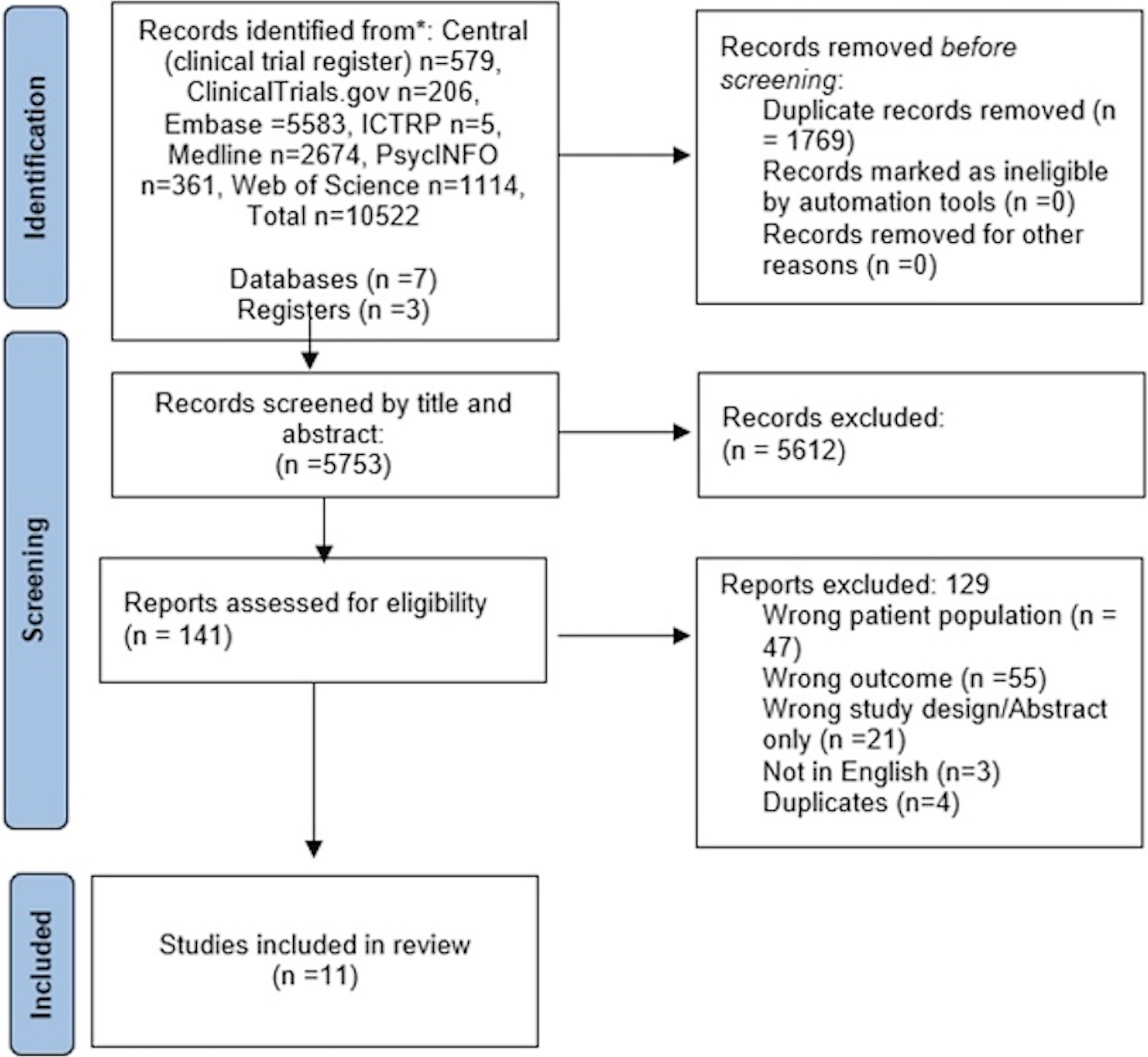
PRISMA flow diagram. PRISMA, Preferred Reporting Items for Systematic Reviews and Meta-Analyses.
Information sources and search strategies
A medical librarian assisted in the development of a search strategy and ensuing search focused on cardiac biomarkers, neuroimaging, and pediatric psychiatric disorders. Search strategies were designed with a combination of keywords and standardized index terms. Searches were run on October 4, 2024, in ClinicalTrials.gov (2000+), Ovid Cochrane Central Register of Controlled Trials (1991+), Ovid Embase (1974+), Ovid Medline (1946+ including epub ahead of print, in-process & other non-indexed citations), Ovid PsycINFO (1806+), Web of Science Core Collection (Science Citation Index Expanded 1975+ and Emerging Sources Citation Index 2015+), and the World Health Organization’s ICTRP clinical trial registry. After limiting results to the English language with conference abstracts and animal studies removed, a total of 10,522 citations were retrieved. Deduplication was automatically performed in Covidence, leaving 5189 citations for initial screening. The full search strategies are described in the supplementary materials (Supplementary Table S2).
The initial screening was conducted based on titles and abstracts by three authors (L.S.C., I.A., C.B.). Full texts that met the inclusion criteria were then assessed for eligibility by the same authors. Any disagreements during the process were resolved through consultation with another coauthor (P.E.C.). The last search was performed on January 8th, 2025.
Study selection and eligibility criteria
The initial study selection included those describing an association between any cardiac biomarker—such as fetal heart rate (FHR) reactivity, resting-state HR, HR, BP, inter-beat interval (IBI) deceleration and acceleration, HRV, HF-HRV and RMSSD, and heartbeat-evoked potential (HEP); RSA; and brain structural changes, activity, or connectivity in imaging studies of individuals under 21 years old diagnosed with a psychiatric disorder. Studies that did not meet these criteria were excluded. Articles were included only if the full text was available in English.
Presenting the findings
Data extraction from the studies meeting inclusion criteria was conducted using Covidence by L.S.C. and P.E.C. verified the data. Each study was carefully analyzed, summarizing the author, group/age, psychiatric disorder, cardiac parameter, brain structure/activity measurement methodology and parameter, as well as the correlation and whether it was positive or negative between brain structure/activity and the cardiac measure.
Results
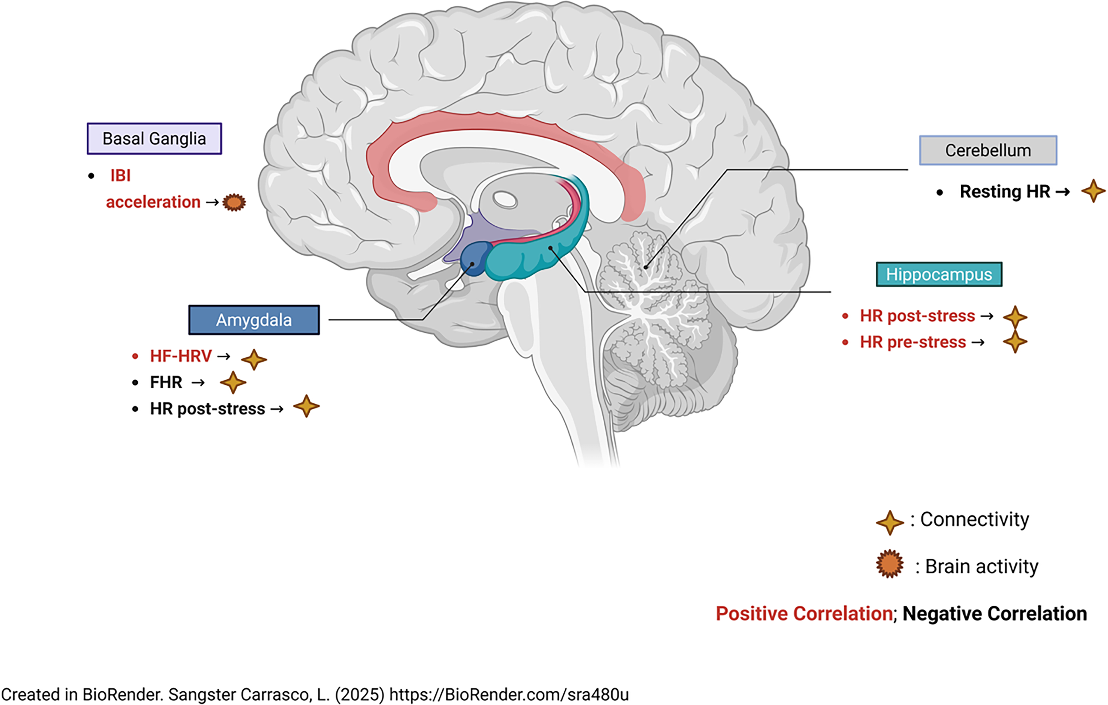
Findings are summarized in (Table 1) and visually illustrated in (Figs. 2 and 3), which depict regional correlations between brain measures and cardiac biomarkers. A total of 11 studies examined the association between cardiac biomarkers and brain structural changes or activity in children and young adults with psychiatric conditions. The biomarkers investigated included FHR reactivity, resting-state HR, HR, BP, IBI deceleration and acceleration, HRV, HF-HRV, RMSSD, HEP, and RSA. Overall, the literature suggested general patterns of association between autonomic functioning markers and brain structure, connectivity, and neural responses, although findings varied across cardiac and psychiatric conditions.
Summary of Cardiac Measures, Brain Structure Changes/Activity and Its Correlation
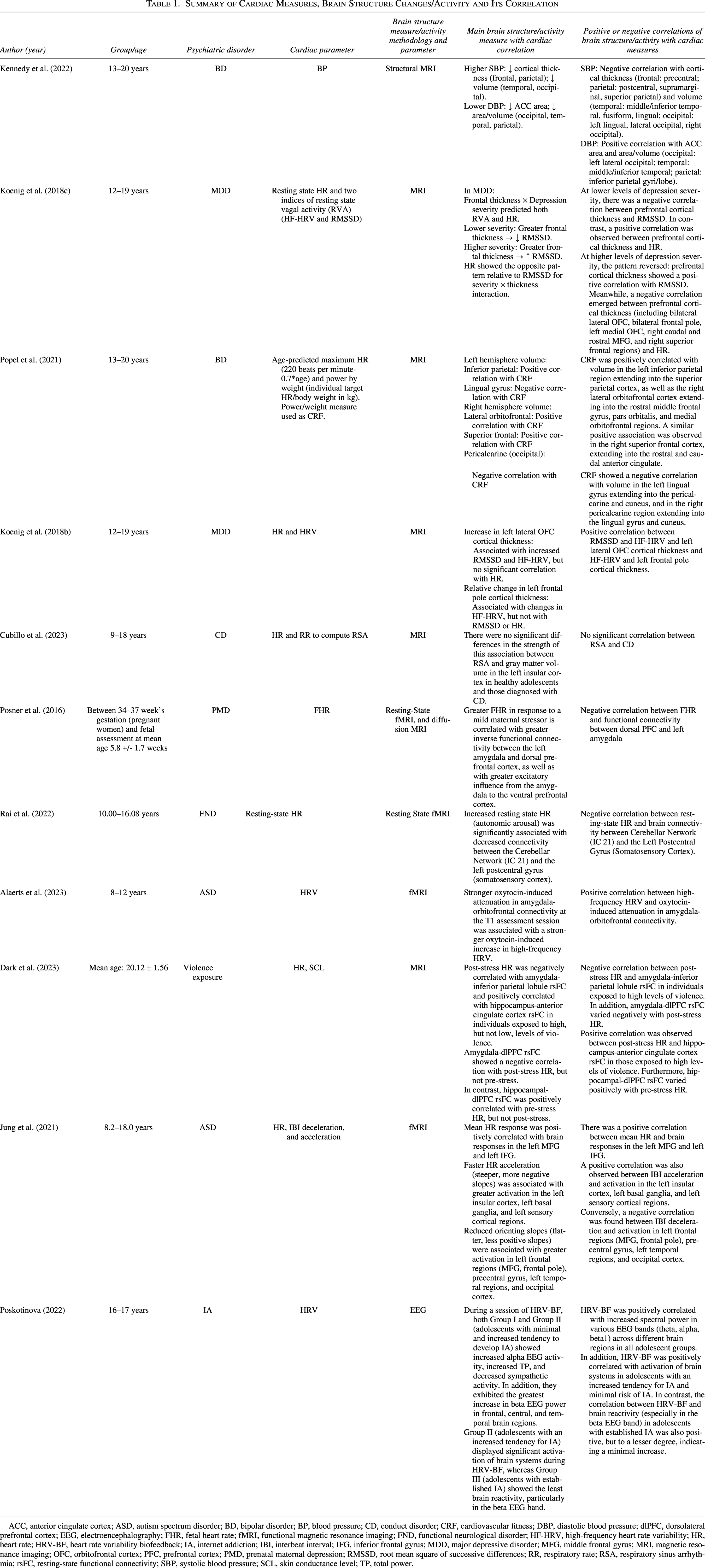
ACC, anterior cingulate cortex; ASD, autism spectrum disorder; BD, bipolar disorder; BP, blood pressure; CD, conduct disorder; CRF, cardiovascular fitness; DBP, diastolic blood pressure; dlPFC, dorsolateral prefrontal cortex; EEG, electroencephalography; FHR, fetal heart rate; fMRI, functional magnetic resonance imaging; FND, functional neurological disorder; HF-HRV, high-frequency heart rate variability; HR, heart rate; HRV-BF, heart rate variability biofeedback; IA, internet addiction; IBI, interbeat interval; IFG, inferior frontal gyrus; MDD, major depressive disorder; MFG, middle frontal gyrus; MRI, magnetic resonance imaging; OFC, orbitofrontal cortex; PFC, prefrontal cortex; PMD, prenatal maternal depression; RMSSD, root mean square of successive differences; RR, respiratory rate; RSA, respiratory sinus arrhythmia; rsFC, resting-state functional connectivity; SBP, systolic blood pressure; SCL, skin conductance level; TP, total power.
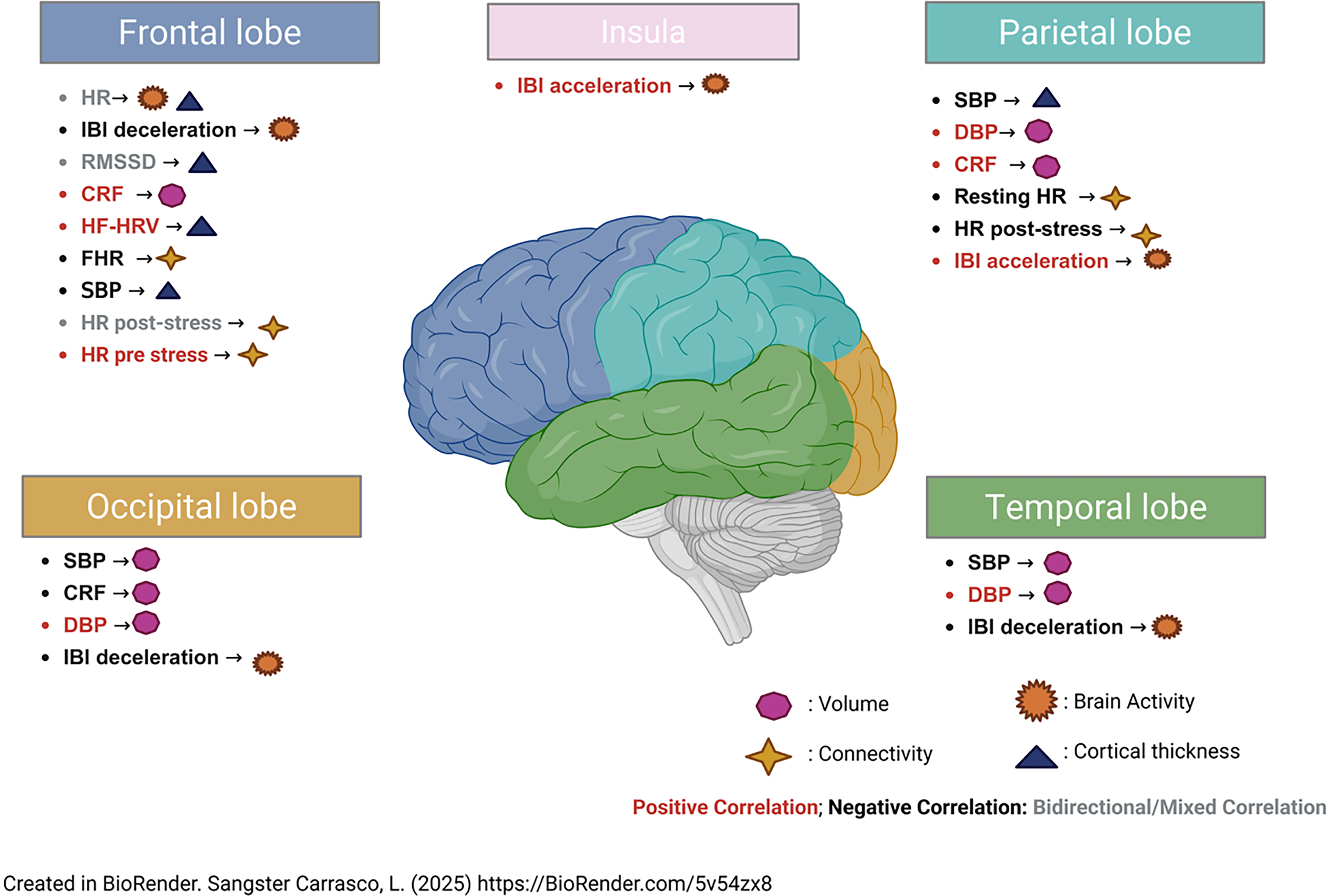
Associated brain changes.
Associated brain changes.
Brain structure and activity were assessed using magnetic resonance imaging (MRI), resting-state functional MRI (fMRI), structural MRI, and electroencephalogram (EEG). Among the studies, five examined the association between brain structural changes and cardiac biomarkers, four explored the connection between brain connectivity and cardiac biomarkers, and two investigated the relationship between brain responses and cardiac biomarkers. To improve synthesis, studies are presented below grouped by primary neuroimaging outcome and further organized by primary cardiac biomarker category when possible.
Correlations of cardiac biomarkers and brain structural changes
Regarding structural changes and cardiac biomarkers, Kennedy (Kennedy et al., 2022) examined 154 participants aged 13–20 with BD to examine the correlation between BP and structural brain changes using MRI. The study found that higher systolic blood pressure (SBP) was negatively correlated with cortical thickness in the precentral gyrus, postcentral gyrus, and supramarginal gyrus, as well as with volume in the middle temporal gyrus, lingual gyrus, fusiform gyrus, inferior temporal gyrus, left lingual gyrus, lateral occipital cortex, and right occipital cortex. Conversely, lower diastolic blood pressure (DBP) was positively correlated with surface area in the anterior cingulate cortex, as well as with both area and volume in the left lateral occipital cortex, middle temporal gyrus, inferior temporal gyrus, inferior parietal gyrus, and inferior parietal lobe. These findings suggest potential associations between vascular-related cardiovascular markers and cortical structural integrity.
Koenig et al. (2018b, 2018c) examined similar samples and therefore are likely derived from overlapping datasets; these studies are therefore presented together to avoid duplication of findings. A depressive symptom severity-dependent correlation was reported by Koenig (Koenig et al., 2018c) in a study of patients aged 12–19 years diagnosed with major depressive disorder (MDD). The study examined the relationship between resting-state HR, two indices of resting-state vagal activity—HF-HRV and RMSSD—and structural brain changes using MRI. Key findings showed that among individuals with lower depression symptom severity, greater prefrontal cortical thickness was associated with lower RMSSD but higher HR. In contrast, for those with higher depression severity, increased prefrontal cortical thickness was positively correlated with RMSSD and negatively correlated with HR. These results highlight potential moderating effects of clinical symptom severity on heart–brain associations.
Popel (Popel et al., 2021) also investigated young patients diagnosed with BD, examining the association between CRF, estimated using age-predicted maximum HR and power-to-weight ratio and brain hemisphere volumes using MRI. The study found a positive correlation between CRF and the volume of the left inferior parietal lobule, right lateral orbitofrontal cortex (OFC), and right superior frontal region. In contrast, CRF was negatively correlated with the volume of the left lingual gyrus and right pericalcarine region. Although CRF is not a direct autonomic marker, these findings suggest systemic cardiovascular fitness may be associated with structural brain differences during development.
Koenig (Koenig et al., 2018b) also explored the relationship between cortical thickness and HRV in patients aged 12–19 years with major depressive disorder. The study found a positive correlation between RMSSD and HF-HRV with cortical thickness in the left lateral OFC, as well as between HF-HRV and cortical thickness in the left frontal pole. These findings further support associations between parasympathetic functioning and prefrontal cortical structure.
The study conducted by Cubillo (Cubillo et al., 2023) examined children with conduct disorder (CD) with a mean age of 14 years. The study found no significant difference in the correlation between RSA and gray matter volume in the left insular cortex when comparing healthy adolescents with those with CD. This suggests that structural–autonomic associations may vary across psychiatric diagnoses.
Regarding brain connectivity and cardiac biomarkers, Posner (Posner et al., 2016) examined the relationship between FHR and functional connectivity in pregnant women between 34 and 37 weeks of gestation with prenatal maternal depression. Using fMRI and diffusion MRI, the study found a negative correlation between FHR and functional connectivity in the left amygdala and dorsal prefrontal cortex. This study was included because it examined fetal cardiac and neural measures during prenatal development.
Rai (Rai et al., 2022) reported a negative correlation between resting-state HR and connectivity between the cerebellar network and the left postcentral gyrus in patients with functional neurological disease compared with controls.
Alaerts (Alaerts et al., 2023) evaluated a population of children with ASD aged 8–12 years and found a positive correlation between HF-HRV and oxytocin-induced attenuation in amygdala-orbitofrontal connectivity in fMRI. These findings support potential interactions between parasympathetic functioning and social-emotional neural networks.
Dark (Dark et al., 2023) studied 297 young adults under the age of 21 who had been exposed to violence and found distinct correlations based on exposure levels. Overall, results suggested that stress and environmental exposure may moderate heart–brain connectivity relationships. In individuals with high levels of violence exposure, post-stress HR showed a negative correlation with amygdala–inferior parietal lobule resting-state functional connectivity (rsFC) and a positive correlation with hippocampus–anterior cingulate cortex rsFC. These correlations were not observed in those with low levels of violence exposure. Also, amygdala- dorsolateral prefrontal cortex (dlPFC) rsFC varied negatively with HR post-stress, and hippocampal-dlPFC rsFC varied positively with HR prestress.
Correlations of cardiac biomarkers and brain responses
Regarding brain responses and cardiac biomarkers, Jung (Jung et al., 2021) examined the relationship between HR, IBI deceleration and acceleration, and brain responses using fMRI in children with ASD. The study found a positive correlation between mean HR and brain activity in the left middle frontal gyrus (MFG) and left inferior frontal gyrus. In addition, IBI acceleration was positively correlated with activation in the left insular cortex, left basal ganglia, and left sensory cortical regions. In contrast, IBI deceleration showed a negative correlation with activation in the left frontal regions (MFG, frontal pole), precentral gyrus, left temporal regions, and occipital cortex. These findings suggest differential autonomic–neural response patterns across brain regions involved in cognitive and emotional processing.
Demin (Demin and Poskotinova, 2022) examined adolescents at varying risk levels for developing internet addiction (IA), including those with minimal risk, an increased tendency, and established IA, to explore the correlation between HRV and brain system activation. The study found a positive correlation between HRV biofeedback (HRV-BF) and increased spectral power in various EEG bands—alpha, theta, and beta1—across different brain regions in all adolescent groups. In addition, HRV-BF sessions were linked to greater activation of brain systems across all groups. However, differences emerged in beta1 activity depending on IA severity: adolescents with minimal risk and an increased tendency showed a more pronounced rise in beta1, whereas those with established IA exhibited only a slight increase. These findings further support the relationship between autonomic regulation and cortical electrophysiological activity.
Discussion
This review sought to examine existing literature related to cardiac and neural biomarkers correlates in children and young adults. This review demonstrates that variations in HRV and BP correlate with regional changes in cortical thickness and brain volume, especially in frontal, temporal, parietal, and occipital lobes. These correlations appear to vary depending on clinical severity. However, symptom severity was not directly tested as a formal statistical moderator in the included studies; rather, these interpretations are based on descriptive patterns across studies reporting clinical symptom associations with cardiac–neural relationships. CRF was also linked to greater volume in cognitive, emotional, and sensory process brain areas and reduced volume in regions involved in visual processing. The lower HR or FHR correlated with weaker functional connectivity between emotion-regulatory regions like the amygdala and prefrontal cortex and sensory and motor areas.
The HF-HRV was associated with enhanced social and emotional connectivity, especially in children with ASD and stress-related HR fluctuations influenced connectivity patterns differently based on environmental adversity. Although environmental adversity was not uniformly analyzed as a formal moderator variable across studies, several studies demonstrated context-dependent autonomic–neural associations influenced by stress exposure and psychosocial risk factors. In ASD, higher HR and IBI acceleration correlated with increased activation in frontal and insular areas, involved in social cognition and interoception. HRV-BF increased EEG spectral power, especially in youth at lower risk of IA. Overall, across structural, functional, and connectivity measures, cardiac biomarkers—especially HRV—consistently related to brain areas involved in emotion regulation, executive function, and sensory integration.
Rather than focusing solely on specific physiological indices or diagnoses, this review also contributes to the literature by synthesizing evidence across psychiatric disorders, supporting a cross-diagnostic perspective on heart–brain interactions. Cross-diagnostic approaches to autonomic–neural functioning have been previously proposed in theoretical work (Beauchaine, 2015; Beauchaine and Cicchetti, 2019; Beauchaine and Thayer, 2015), which emphasize transdiagnostic mechanisms underlying psychopathology.
These associations differ by psychiatric diagnosis, symptom severity, and environmental stressors, suggesting their potential as noninvasive indicators of brain health and developmental risk.
Previous reviews have explored the cardiac–brain relationship in adults or focused narrowly on specific physiological indices or diagnoses (e.g., HRV in anxiety/depression). Consistent with cross-diagnostic neurodevelopmental models, this review expands prior work by examining cardiac–neural relationships across multiple psychiatric conditions in youth. Our review adds by emphasizing symptom severity and environmental adversity as moderators of heart–brain interactions during a critical developmental window.
Two theoretical frameworks, the Neurovisceral Integration Model (NIM) (Thayer and Lane, 2000) and the Polyvagal Theory (PT) (Porges, 1997), provide a useful framework for interpreting these findings. The NIM suggests that the prefrontal cortex, through its connections with subcortical and brainstem regions, plays a key role in regulating autonomic function, linking HRV with executive and emotional processes. On the other hand, the PT highlights the role of vagal pathways in supporting social engagement, emotional regulation, and adaptive responses to stress. These frameworks are primarily applicable to HRV-related cardiac measures, as HRV reflects vagal autonomic regulation. Although both frameworks are considered, a clearer alignment of the current findings with the predicted circuits of each model is needed. For instance, the observed links between HRV and prefrontal–amygdala connectivity, as well as between HF-HRV and social-emotional network function, are consistent with PT predictions. In contrast, correlations between HRV and executive function–related cortical regions lend support to the NIM. Future research should purposefully integrate these models with empirical data to provide a clearer understanding of the underlying mechanisms.
A key strength of the present review is a multimodal scope, incorporating MRI, fMRI, and EEG data across diverse psychiatric conditions. Limitations include the small number of studies, methodological heterogeneity, and lack of longitudinal data. Future studies should aim for harmonized protocols and larger, developmentally stratified cohorts.
From a translational standpoint, the observed links between cardiac function, brain structure, and cognitive-emotional processes suggest that interventions aimed at modulating autonomic activity could be tested for their potential to influence neurodevelopmental trajectories in psychiatric populations. In addition, integrating physiological cardiac measures with biochemical and immune markers could help identify systemic pathways mediating heart–brain interactions. These approaches could also help clarify the role of vagal nerve health as a mediator between cardiac and neural outcomes.
The neurovisceral integration model (Thayer and Lane, 2000) and Polyvagal theory (Porges, 1997) offer frameworks linking autonomic function to emotion regulation. A metanalysis (Beissner et al., 2013) identified brain regions—including the anterior cingulate cortex (ACC), insula, vmPFC, thalamus, amygdala, hippocampus, and hypothalamus—as key in autonomic control.
Based on these findings, we propose that cardiac biomarkers may serve as proxies for brain structural changes in children and young adults with psychiatric disorders. For example, Koenig (Koenig et al., 2018a) showed that HRV is inversely related to cortical thickness in healthy adolescents, suggesting that higher HRV may support cortical development and thinning during adolescence. Other studies reported a positive correlation between HRV and left cortical thickness (Koenig et al., 2018b), and higher depression severity was associated with greater insula and frontal cortical thickness as well as increased resting-state vagal activity, a key HRV component, in adolescents with major depressive disorder (Koenig et al., 2018c). Together, these findings suggest that higher HRV may relate to cortical thinning in healthy adolescents but to increased cortical thickness in those with depression.
Psychiatric conditions such as BD have been linked to cardiovascular comorbidities (Shah et al., 2024) such as high BP (Naiberg et al., 2016), and higher body mass index has been previously reported as associated with brain structural changes in adolescents with BP. Kennedy (Kennedy et al., 2022) showed that higher SBP was associated with smaller brain structures, while conversely to what was hypothesized, lower DPB unexpectedly showed similar effects. Popel (Popel et al., 2021) found mixed correlations between CRF and brain volume in BD youth. More recent studies examining endothelial function (Kennedy et al., 2023), a marker of early atherosclerosis in BD youth, found that better endothelial function is associated with smaller brain structures and lower cerebral blood flow (CBF) (Sultan et al., 2024), despite previous findings of increased CBF in certain brain regions in BD youth (MacIntosh et al., 2017). These inconsistencies could be related to potential blood flow-oxygen mismatches among these patients (Karthikeyan et al., 2019). Findings in frontal asymmetry thickness remain mixed: A significant positive association was reported between HRV and thickness of the right but not left ACC (MacIntosh et al., 2017), while other studies (Yoo et al., 2018) reported the opposite. In contrast, studies in adolescents with MDD (Koenig et al., 2018b; 2018c) reported a positive correlation between HRV and cortical thickness in prefrontal areas bilaterally. These findings could support the previous evidence that both hemispheres are under vagal control at some level (Winkelmann et al., 2017; Yoo et al., 2018). Autonomic function may reflect or even influence neurodevelopmental patterns in psychiatric conditions, and this is reflected in this review, where the prefrontal cortex, crucial for emotional regulation and executive function, shows sensitivity to HRV and BP changes. Three studies demonstrated a negative correlation between autonomic arousal and brain connectivity, especially in the amygdala and prefrontal cortex (Alaerts et al., 2023; Dark et al., 2023; Posner et al., 2016) in children and adolescents with different psychiatric conditions. Adolescents with underlying psychiatric disorders often exhibit amygdala overactivity (Hafeman et al., 2017), leading to increased autonomic arousal. On the other hand, if PFC regulation of the amygdala is impaired (Navarro-Nolasco et al., 2025), this can result in heightened autonomic responses such as elevated HR and reduced HRV. Impaired vagal control (Thayer and Lane, 2009), hypothalamic-pituitary-adrenal axis dysregulation, and chronic stress may exacerbate this cycle, contributing to emotional dysregulation (Philippi et al., 2024). This highlights the importance of studying the bidirectional dynamic between the heart and brain, where dysfunction in either system could contribute to pathology in the other, reinforcing cycles across different psychiatric conditions.
Autonomic activity indexed by HRV appears to modulate functional brain connections underlying emotion regulation and stress response, and these effects may be diagnosis-specific and environmentally sensitive. The only study that reported a positive correlation between post-stress HR and brain connectivity was by Dark (Dark et al., 2023), specifically in the hippocampus–ACC region, which is crucial for emotion regulation (Bush et al., 2000), cognitive control, executive functions (Botvinick et al., 2004), and processing and response to pain (Vogt, 2005), while hippocampus is involved in memory formation (Squire and Zola-Morgan, 1991) and stress regulation (McEwen, 2007). This positive correlation could suggest the significant role of the hippocampus–ACC interaction when exposed to stress.
Artificial intelligence is an emerging tool in psychiatry for diagnosis, prediction, and treatment through the use of computational techniques and algorithms (Fakhoury, 2019). Wearable technologies with cardio-fitness chest straps or wrist-worn biosensors in watches collect passive data such as HR, HRV, RMSSD, and RSA and have shown promising results in diagnosis and prediction in children with different psychiatric disorders like ADHD, ASD, BD, and others (Welch et al., 2022). A feasibility study involving children with disruptive behavior disorders found that after 7 days of continuous smartwatch use, physiological biomarkers were successfully collected, and a machine learning approach was able to identify predictive markers of impending disruptive behaviors (Romanowicz et al., 2023). These findings could suggest that data from wearable devices could be used to explore correlations between physiological cardiac biomarkers and brain changes in morphology or connectivity, potentially aiding in the early detection of psychiatric disorders in children and young adults.
Findings from this review suggest that cardiac biomarkers show promise as indicators of neural functioning and psychiatric risk in children and young adults, particularly given their associations with brain regions involved in emotion regulation, executive function, and stress processing. However, the current literature is too heterogeneous and preliminary for routine clinical use. While some studies consistently link HRV to prefrontal cortical structure and amygdala connectivity, others show condition-specific or context-dependent patterns influenced by symptom severity, developmental stage, and environmental adversity. This variability highlights both the potential and the challenges of using cardiac metrics in child and adolescent psychiatry. Although these findings support theoretical models of bidirectional heart–brain communication, including NIM and PT, clinical translation will require larger, longitudinal, and methodologically rigorous studies to clarify causal pathways and determine practical applications. Future research should also incorporate immune and systemic biomarkers, HRV monitoring, and multimodal imaging to capture the complexity of heart–brain interactions. For now, cardiac biomarkers should be seen as complementary research tools to deepen our understanding of brain–body interactions during critical neurodevelopmental periods. In addition, the method of HRV measurement remains a key limitation; many studies rely on lab-based assessments, which may not reflect HRV dynamics in naturalistic settings. Few have examined HRV over extended periods or real-world contexts, where heart–brain interactions may differ. Future research incorporating long-term, naturalistic HRV assessments could improve ecological validity and enhance the clinical utility of cardiac biomarkers.
Conclusions
In summary, our study identified several cardiac biomarkers that correlated with structural, functional, and connectivity-based brain changes in children and young adults with psychiatric conditions. According to the studies, patterns consistently pointed to associations between vagal tone and brain regions involved in emotion, with some modulation by clinical severity and environmental factors. Among the indices studied, HRV, especially HF-HRV, and RMSSD, emerged as the most consistently associated with brain structure and connectivity and demonstrated sensitivity to both symptom variation and external stressors. Resting-state HR and BP also showed important associations, though findings were more condition-specific. In contrast, IBI and RSA were less frequently studied and yielded more variable results. In addition to using wearable devices to collect these biomarkers, their implementation may help explore the relationship between physiological cardiac signals and brain alterations in morphology or connectivity.
Taken together, these findings suggest that HF-HRV and RMSSD may be the most promising and reliable cardiac indices for future research exploring heart–brain interactions in youth mental health. However, broader, longitudinal studies are still needed to validate these measures as clinical tools.
Authors’ Contributions
L.S.-C. and P.E.C.: Conceptualization, methodology, investigation, formal analysis, data curation, writing—original draft, writing—review and editing, visualization, supervision, and project administration. I.A., C.B., K.D., D.G., M.R., and A.P.A.: Formal analysis, data curation, writing—review and editing, visualization, validation, supervision, and project administration.
Footnotes
Author Disclosure Statement
Dr. Croarkin has received research support from the Agency for Healthcare Research and Quality (AHRQ), National Institutes of Health (NIH), National Science Foundation (NSF), Brain and Behavior Research Foundation and the Mayo Clinic Foundation. Dr. Croarkin has received research support from Pfizer, Inc. He has received grant-in-kind equipment support from Neuronetics, Inc. and MagVenture, Inc. for investigator-initiated studies. He received grant-in-kind supplies and genotyping from Assurex Health, Inc. for an investigator-initiated study. He served as the principal investigator for a multicenter study funded by Neuronetics Inc., a site principal investigator for a study funded by NeoSync, Inc., and site principal investigator for a study funded by Innosphere. Dr. Croarkin served as a paid consultant for Engrail Therapeutics, Meta Platforms, Inc, MindMed, Myriad Neuroscience, Procter & Gamble Company, and Sunovion. Dr. Croarkin is employed by Mayo Clinic. He receives compensation as the Editor-in-Chief for the Journal of Child and Adolescent Psychopharmacology. Dr. Athreya receives funding from National Science Foundation (Award IIS-2041339), National Institutes of Health (R01NR020362), and Physician Foundation. The other authors have no disclosures.
Supplemental Material
Supplemental Material
References
Supplementary Material
Please find the following supplemental material available below.
For Open Access articles published under a Creative Commons License, all supplemental material carries the same license as the article it is associated with.
For non-Open Access articles published, all supplemental material carries a non-exclusive license, and permission requests for re-use of supplemental material or any part of supplemental material shall be sent directly to the copyright owner as specified in the copyright notice associated with the article.