
Abstract
Background:
Microplastics (MPs) have been identified in multiple human tissues and are increasingly implicated in systemic health risks. Their presence in the thyroid gland, however, remains unexamined. Autoimmune thyroiditis (AIT) is the most frequent autoimmune thyroid disorder and the leading cause of hypothyroidism. This study aims to detect the presence of MPs in the thyroid and their potential relevance to AIT.
Methods:
In this case–control study, thyroid tissues were obtained from 29 patients with histologically confirmed AIT and 29 age- and sex-matched non-AIT controls who underwent thyroidectomy due to thyroid nodules. MP burden was quantified by pyrolysis-gas chromatography–mass spectrometry (Py-GC/MS). Particle-level polymer identity and particle characteristics, including size, shape, and color, were assessed using micro-Raman spectroscopy, whereas scanning electron microscopy (SEM) was employed for morphological observation.
Results:
MPs were detected in thyroid tissues from both groups. Py-GC/MS revealed significantly higher total MP concentrations in the AIT group compared to controls (median: 19.9 vs. 1.9 μg/g; p=0.012). This elevation was primarily driven by polyvinyl chloride (PVC), which was significantly higher in AIT patients. Micro-Raman spectroscopy identified particles ranging from 33.9 to 1467 µm. The AIT group contained significantly increased MPs abundance compared with the non-AIT control group (172 vs. 50.2 items/g, p=0.037). Morphological profiling revealed no significant differences in the size, shape and color of MPs between groups.
Conclusion:
An increased MPs burden with the particular enrichment of PVC was observed in patients with AIT, suggesting a potential association between environmental MPs exposure and thyroid autoimmunity. Further mechanistic and epidemiological studies to clarify the impacts of chronic MPs exposure are needed.
Summary of the Research Problem
Microplastics (MPs) are synthetic polymer particles smaller than 5 mm in size. These particles primarily originate from the degradation of plastic packaging, textiles, and personal care products. Human exposure occurs mainly through ingestion and inhalation, with minimal absorption through the skin. 1 Studies have confirmed the presence of MPs in various human organs and suggest that chronic exposure to MPs, even at low doses, may be associated with adverse cardiovascular and metabolic outcomes.2–4 A recent postmortem study detected MPs in the thyroid. 5 However, their quantitative burden and potential health effects remain unclear. This study aims to assess the presence of MPs in human thyroids and their association with autoimmune thyroiditis (AIT), which is the most frequent autoimmune thyroid disorder and the leading cause of hypothyroidism.
Methods
This case-control study was approved by the Medical Ethics Committee of Nanfang Hospital, Southern Medical University (NFEC-2025-462). The research was completed in accordance with the 2024 version of the Declaration of Helsinki. Thyroid tissues were obtained from 29 patients with pathologically confirmed AIT and 29 age- and sex-matched non-AIT controls. All participants underwent thyroidectomy for thyroid nodules between March and December 2025. Written informed consent was obtained from every participant. This consent authorized the use of residual specimens for scientific research after the completion of pathological diagnosis. During surgical procedures and specimen collection, strict precautions were implemented to avoid plastic contamination. Tissue samples were handled exclusively with metal surgical instruments and were transferred into glass containers.
MP Identification and Characterization
MPs in thyroid tissue were characterized and identified using integrated pyrolysis–gas chromatography–mass spectrometry (Py-GC/MS), micro-Raman spectroscopy, and scanning electron microscopy (SEM). Bulk polymer quantification (Py-GC/MS) and particle-level analyses (micro-Raman spectroscopy and SEM) were performed on parallel aliquots processed using method-specific preparation procedures.
Py-GC/MS was performed to quantify 12 common MP polymers, including polystyrene (PS), polyethylene (PE), polymethyl methacrylate (PMMA), polypropylene (PP), polyvinyl chloride (PVC), polyethylene terephthalate (PET), polycarbonate (PC), polyamide 6 (PA6), polyamide 66 (PA66), acrylonitrile–butadiene–styrene copolymer (ABS), styrene–butadiene rubber, and polyurethane. Samples were digested, extracted, and analyzed using a GCMS-QP2020NX system (Shimadzu, Japan) coupled to a PY-3030D pyrolyzer. A procedural blank and a glass surrogate were included to monitor background contamination and analytical performance.
For particle-level analyses, thyroid tissues were subjected to chemical digestion to remove organic matter, followed by membrane filtration. Particles retained on polytetrafluoroethylene (PTFE) membranes were identified using a micro-Raman imaging system (DXR3xi, Thermo Scientific). To ensure representative detection, each sample membrane was systematically scanned across multiple predefined regions. Polymer types were determined by spectral matching against a custom spectral database and the Raman Sample Library. Particle size, morphology, and color were quantified using Nano Measurer software (v1.2). SEM was performed to examine the morphology of MP particles. Target particles were precisely marked on the membrane, sputter-coated with gold, and imaged using SEM.
Detailed descriptions of instrumental parameters, analytical procedures, and quality assurance/quality control methods are provided in the Supplementary Data.
Statistics
Continuous variables that did not follow a normal distribution were assessed using the Mann–Whitney U test. Comparisons of categorical variables were performed using either the chi-square test or Fisher’s exact test, as appropriate. Statistical analyses were carried out in SPSS version 26 (IBM, USA), and a p value of less than 0.05 was considered statistically significant. Graphics were created using Prism version 10.0 (GraphPad Software, USA). The figures were created using BioRender (BioRender.com).
Results
This study included 58 participants (29 patients with AIT and 29 matched non-AIT controls). The mean (standard deviation [SD]) age of the overall cohort was 41.4 (12.1) years, and 83% of the participants were women (48 of 58). There were no statistically significant differences in age and body mass index (BMI) between the two groups (Table 1). Bulk polymer profiling by Py-GC/MS identified 9 polymer types, including PVC, PMMA, PA66, PP, PA6, PC, PET, ABS, and PS. Total MP concentrations were significantly elevated in the AIT group compared with non-AIT controls (median 19.9 vs. 1.9 μg/g; p = 0.012). This difference was primarily attributable to PVC, which was significantly elevated in AIT patients (Table 1, Fig. 1A). The PVC burden in the AIT group ranged from 5.1 to a maximum of 370 µg/g. Stratification by PVC detection status revealed no significant differences in baseline demographics (age, sex, and BMI) or smoking history, whereas AIT prevalence was significantly elevated in the PVC-positive group (Supplementary Table S1).
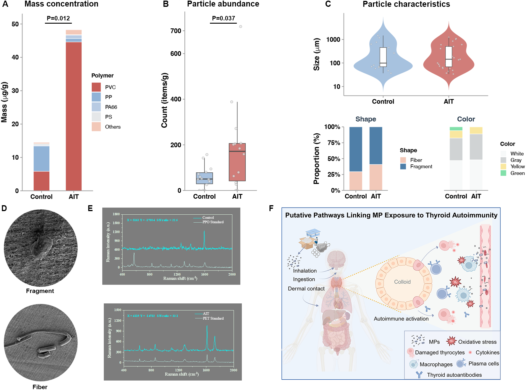
Detection, characterization, and potential mechanism of microplastics (MPs) in human thyroid tissue.
Baseline Characteristics of the Participants
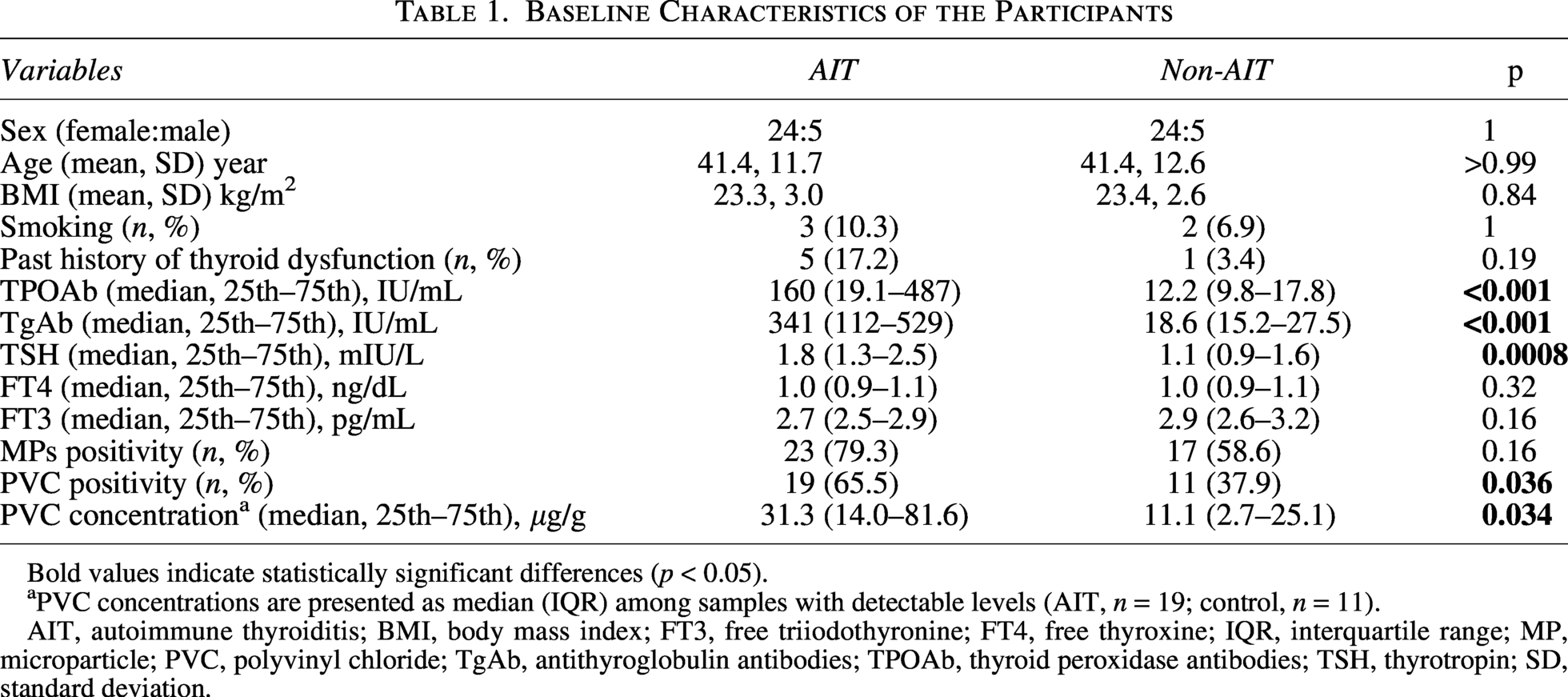
Bold values indicate statistically significant differences (p < 0.05).
PVC concentrations are presented as median (IQR) among samples with detectable levels (AIT, n = 19; control, n = 11).
AIT, autoimmune thyroiditis; BMI, body mass index; FT3, free triiodothyronine; FT4, free thyroxine; IQR, interquartile range; MP, microparticle; PVC, polyvinyl chloride; TgAb, antithyroglobulin antibodies; TPOAb, thyroid peroxidase antibodies; TSH, thyrotropin; SD, standard deviation.
Micro-Raman spectroscopy was subsequently applied as a complementary, particle-level approach to further characterize MPs in tissues from AIT patients and non-AIT controls (n = 14 for each group). This analysis confirmed the presence of major polymers identified by Py-GC/MS, including PET, PP, and PS, and additionally revealed polyphenylene oxide (PPO), PE and poly(butylene adipate-co-terephthalate) (Supplementary Figs. S1 and S2). The AIT group contained a significantly increased MP abundance compared with the non-AIT group (172 vs. 50.2 items/g, p = 0.037; Fig. 1B). We identified 44 particles with sizes ranging from 33.9 to 1,467 µm. Particles larger than 150 μm were predominantly fibers (80%, 16/20) with a median transverse diameter of 15.9 μm, suggesting a size dimension compatible with tissue translocation. There was no significant difference in particle size between the AIT group and the control group (p = 0.75). Regarding morphology, the AIT group contained 27 particles (40.7% fibers and 59.3% fragments), while the non-AIT control group contained 17 particles (29.4% fibers, 70.6% fragments; Fig. 1C–D). However, this difference in shape distribution was not statistically significant (p = 0.45). Particle color, predominantly white and gray, did not differ significantly between groups (p = 0.99; Fig. 1C–D).
Discussion
To our knowledge, this is the first study to integrate mass-based and particle-level approaches to demonstrate an increased burden of MPs in human thyroid tissue and its association with AIT. The detection of MPs in thyroid tissue, along with the significantly higher burden observed in AIT patients, particularly of PVC, raises important questions regarding their potential biological and clinical relevance.
The thyroid gland is highly vascularized, with each follicle surrounded by fenestrated capillaries, a unique anatomical feature that may facilitate the deposition and retention of circulating MPs. Our findings suggest the possibility that MPs can traverse epithelial barriers and disseminate via the bloodstream to remote organs. 1 Although this cross-sectional study cannot establish causality, it provides initial human evidence of an association between MP accumulation and AIT. Notably, animal studies suggest that dietary exposure to PP, PET, and PS, as well as inhalational exposure to PS, can induce thyroid dysfunction,6,7 supporting biological plausibility. While PP, PET, and PS were detected in thyroid tissue by both Py-GC/MS and Raman spectroscopy, their similar levels between groups likely reflect ubiquitous background exposure, with group differences driven instead by overall MP burden and polymer composition (Fig. 1F). In this context, the selective predominance of PVC warrants particular attention, as its higher density may favor retention following extravasation and its associated additives, such as phthalates, have been implicated in oxidative stress and immune dysregulation. 8 Together, these observations underscore the need for further mechanistic and longitudinal studies.
Py-GC/MS analysis revealed a wide range of MP burdens in thyroid tissues. In AIT patients, PVC concentrations ranged from 5.1 to 370 µg/g, with a median value of 31.3 µg/g. This level of variability is in line with recent mass-based quantifications in other highly vascularized organs. Previous studies have reported median total plastic burdens of approximately 300–430 µg/g in highly perfused organs such as the testis, liver, and kidney.9,10 PVC concentrations in liver tissue were generally in the range of several tens of micrograms per gram, comparable to the median level observed in thyroid tissue in this study, whereas median PVC concentrations in brain tissue were higher, reaching several hundred micrograms per gram. In contrast, MP burdens in human lung tissue were substantially lower, with a median concentration of approximately 2.19 particles/g. 11 These findings highlight organ-specific MP accumulation and suggest that intrinsic structural and functional features may contribute to MP retention in thyroid tissue.
Physically, Raman analysis revealed that the AIT group exhibited a distinct accumulation of fibrous MPs. This morphological pattern mirrors recent observations in human colorectal cancer, where fibers constitute up to 98.8% of detected particles. 12 High–aspect ratio particles may more readily penetrate tissue barriers and become mechanically entrapped within the thyroid. Moreover, the persistence of fibrous synthetic polymers, which are resistant to enzymatic degradation, may promote a foreign body response and fibrous encapsulation, thereby limiting physiological clearance and facilitating long-term retention. 13
We observed a methodological divergence between the two detection platforms regarding PVC identification. Py-GC/MS identified PVC as a dominant contributor to total MP burden, whereas it was not detected by micro-Raman spectroscopy. This discrepancy likely reflects Raman-specific limitations, including fluorescence interference from PVC additives and region-based particle sampling, which may miss heterogeneously distributed polymers. Additives in PVC frequently generate strong background fluorescence, which can effectively mask characteristic spectral peaks and render the polymer invisible to Raman imaging. 14 Consistent with our findings, PVC has been identified in thyroid tissue using optical photothermal infrared microscopy, a technique less susceptible to fluorescence interference and therefore complementary for polymer identification. 5 Accordingly, the integration of Py-GC/MS and Raman spectroscopy enabled a more comprehensive characterization of thyroid MPs contamination. Importantly, despite platform-specific differences, both approaches consistently demonstrated MP accumulation in human thyroid tissue and significantly elevated burdens in patients with AIT.
This study has several limitations. First, the small sample size and cross-sectional design preclude causal inference. We cannot determine whether PVC accumulation triggers pathogenesis or if inflammatory pathology facilitates secondary retention. Consequently, these exploratory findings require validation in larger prospective cohorts. Second, the absence of an individual’s environmental data prevents assessing personal exposure–response relationships. Third, our restricted polymer panel may underestimate the total MP burden. Finally, the use of tumor-adjacent tissues introduces the tumor microenvironment as a potential confounder. Although this factor was consistent across groups, it may limit generalizability. Despite these constraints, the presence of MPs in the thyroid raises critical questions about environmental determinants of autoimmunity. Future studies are warranted to clarify whether MPs act as initiators or inert bystanders in these pathological processes.
Authors’ Contributions
S.-t.Y.: Investigation (lead), resources (lead), methodology (supporting), conceptualization (supporting), and writing—review and editing (supporting). J.F.: Writing—original draft (lead), funding acquisition (lead), formal analysis (lead), and visualization (lead). Z.F.: Data curation (supporting), resources (supporting), and data interpretation (supporting). Q.Y.: Data curation (supporting), resources (supporting), and visualization (supporting). S.Z.: Conceptualization (lead), methodology (lead), investigation (lead), formal analysis (lead), visualization (lead), data curation (lead), and writing—original draft (supporting). H.G.: Conceptualization (lead), data interpretation (lead), supervision (lead), funding acquisition (lead), and writing-review and editing (lead).
Ethical Considerations
This case-control study was approved by the Medical Ethics Committee of Nanfang Hospital, Southern Medical University (NFEC-2025-462).
Data Availability
Data for analysis are available upon reasonable request.
Footnotes
Author Disclosure Statement
H.G. is Associate Editor of Thyroid, but she had no role in the review of this article and was blinded to the review process. S.-t.Y., J.F., Z.F., Q.Y., and S.Z. have nothing to disclose.
Funding Information
J.F. received the funds from the Foundation of Guangzhou Basic and Applied Basic Research Scheme (2025A04J4737) and the National Natural Science Foundation of China (82400916). H.G. received the funds from the National Key R&D Program of China (2024YFA1802700) and the National Natural Science Foundation of China (82372600). The funders were not involved in the design of the study; the collection, analysis, and interpretation of data; and writing of the report and did not impose any restrictions regarding the publication of the report. S.-t.Y., Z.F., Q.Y., and S.Z. have no funding to report.
Supplemental Material
Supplemental Material
Supplemental Material
Supplemental Material
References
Supplementary Material
Please find the following supplemental material available below.
For Open Access articles published under a Creative Commons License, all supplemental material carries the same license as the article it is associated with.
For non-Open Access articles published, all supplemental material carries a non-exclusive license, and permission requests for re-use of supplemental material or any part of supplemental material shall be sent directly to the copyright owner as specified in the copyright notice associated with the article.