






Abstract
Crouzon syndrome with acanthosis nigricans (CAN) is caused by a mutation in the fibroblast growth factor receptor (FGFR) 3 gene that presents clinically as Crouzonoid craniofacial features in association with other anomalies such as acanthosis nigricans and benign odontogenic tumors. Diagnosis through the use of genetic mutational analysis is critical, as it alerts the surgeon to the need for careful screening for jaw tumors so that timely treatment in the form of curettage or segmental resection can be provided.
Introduction
Crouzon syndrome (CS), first described by Octave Crouzon in 1912, is characterized by craniosynostosis, shallow orbits, a beaked nasal deformity, midface hypoplasia, a V-shaped palate, mandibular prognathism, and an absence of extremity findings (Hennekam et al., 2010). It is the most common form of FGFR-related syndromic craniosynostosis, occurring in 1:60 000 live births (Hennekam et al., 2010). In contrast, Crouzon with acanthosis nigricans (CAN) is a rare condition affecting 1 of every 1 000 000 patients that is clinically and genetically distinct from classic CS (National Library of Medicine, 2016). Acanthosis nigricans, a dermatologic condition characterized by hyperpigmented velvety plaques that predominantly occur in intertriginous areas, most commonly occurs in conjunction with endocrine disorders such as obesity or diabetes mellitus. The neck and axilla are the most common sites of involvement, and the plaques are usually asymptomatic. The association between Crouzonoid facial features and acanthosis nigricans was first described by Curth in 1968 (Curth, 1968). Characteristics of CAN include craniosynostosis with Crouzonoid appearance, acanthosis nigricans, choanal stenosis, hydrocephalus, Chiari malformation, short stature, spinal stenosis, scoliosis, and jaw tumors (Arnaud-López et al., 2007; Di Rocco et al., 2011; Herman et al., 2014; Mir et al., 2013; Nagase et al., 2000). Genetically, CAN is caused by a single mutation (p.Ala391Glu) in the fibroblast growth factor receptor (FGFR) 3 gene on chromosome 4 as opposed to CS, which is typically caused by mutations in FGFR2 on chromosome 10 (Robin et al., 2011). We report 2 cases of CAN that presented to the Children’s Hospital of Philadelphia between 2010 and 2015 and were confirmed by FGFR3 mutational analysis in an effort to increase awareness of this distinct syndrome. Of particular interest to the craniofacial surgeon is the need for careful screening for these odontogenic tumors, as they are a distinct cause of maxillomandibular discrepancy that may be mistaken for the typical midface hypoplasia associated with CS.
Case Description: Patient 1
Patient 1 presented at age 2.5 months with increased work of breathing secondary to laryngomalacia and choanal stenosis. He was a former term male infant with a history of gastroesophageal reflux, respiratory distress while feeding, and Crouzonoid features. Three-dimensional head computed tomography (CT) revealed shallow orbits and midface hypoplasia without evidence of craniosynostosis, and molecular studies demonstrated a normal FGFR2 mutational analysis, normal Muenke (FGFR3 Pro250Arg) mutational analysis, and normal TWIST sequencing, deletion, and duplication analysis. At 5 months of age, he developed hydrocephalus requiring a ventriculoperitoneal shunt, and his bilateral coronal sutures closed shortly thereafter (Figure 1). He subsequently underwent 2 cranial expansion procedures, first posterior vault distraction osteogenesis at 7 months of age and then fronto-orbital advancement at 14 months of age. He was followed closely in our Craniofacial Anomalies team where he was routinely examined by the craniofacial surgeon, neurosurgeon, ophthalmologist, ENT, geneticist, audiologist, orthodontist, and pediatric dentist. As he progressed through infancy, his obstructive apnea worsened, and he was placed on continuous positive airway pressure (CPAP) ventilation. Over the subsequent 2 years, his midfacial growth appeared to lag behind his mandibular growth, and this was felt to be associated with his syndromic midfacial hypoplasia in combination with the CPAP mask. At age 4 years, he was noted to have thick scarring and pigmentary changes involving the axilla, neck, inguinal folds, and perioral region consistent with acanthosis nigricans (Figure 2). These skin changes along with difficulties with chewing due to malocclusion alerted the team to the fact that his maxillomandibular discrepancy had worsened, this time due to a combination of maxillary hypoplasia and mandibular prognathism due to a large mandibular symphyseal tumor. CT revealed additional smaller maxillary tumors, and biopsy confirmed the diagnosis of cemento-ossifying fibroma (COF) (Figure 3); these were treated with segmental resection of the mandible with fibula free flap reconstruction and curettage of the maxillary tumors (Figure 4). Additionally, the head CT that had been obtained to evaluate the mandible revealed the presence of an asymptomatic Chiari 1 malformation that radiographically progressed over the course of 7 months and eventually led to Chiari decompression at age 5 years. Subsequent mutational analysis revealed the Ala391Glu mutation in the FGFR3 gene consistent with CAN. Additional comorbidities include exotropia, seizures, developmental delay, and obstructive sleep apnea.

Patient 1 at age 6 months with classic Crouzonoid features prior to craniofacial surgical interventions.
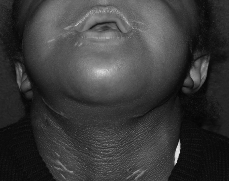
Acanthosis nigricans and scar hypopigmentation on the neck and in the perioral region.

Patient 1 presents to the craniofacial clinic at age 4 years with worsening midface hypoplasia and mandibular prognathism (A, B). Three-dimensional (3D) CT scans demonstrate small maxillary tumors and a large mandibular tumor histopathologically proven to be cemento-ossifying fibromas (C, D). Postoperative photos (E, F) and 3D CT scans (G, H) following fibular bone graft to maxilla and free fibular osteocutaneous flap for mandibular reconstruction. Computer-aided surgical planning was utilized to plan the segmental tumor resection and free fibula osteocutaneous flap reconstruction (I, J).

Intraoperative photographs. The mandibular tumor measuring 8 × 6 cm is shown in situ (A) and following completion of segmental resection (B). A fibula osteocutaneous free flap was harvested (C) and utilized to reconstruct the mandible (D).
Case Description: Patient 2
Patient 2 presented with respiratory and feeding difficulty, a bulging fontanelle, unicoronal craniosynostosis, shallow orbits, mild proptosis, and bilateral choanal stenosis at 9 days of age. Genetic testing consisting of a genome wide SNP microarray and a comprehensive craniosynostosis panel (FGFR1, FGFR2, FGFR3, TWIST) revealed the Ala391Glu mutation. She underwent posterior vault distraction osteogenesis and fronto-orbital advancement at age 3 (Figure 5) and 17 months, respectively, both times because of preoperative papilledema that resolved after each cranial expansion. She has a mild Chiari malformation that is being monitored. She is currently 26 months of age, tracheostomy dependent, and will likely require significant midfacial advancement and choanal stenosis repair prior to tracheostomy decannulation. She has not yet manifested evidence of acanthosis nigricans or tumors of the maxilla or mandible.

Patient 2 at age 3 months with midface hypoplasia, shallow orbits, and right unicoronal craniosynostosis.
Discussion
CAN is an extremely rare craniosynostosis syndrome caused by a single mutation between the immunoglobulin-like III domain and the tyrosine kinase domain (c.1172C>A; p.Ala391Glu) of the FGFR3 gene (Meyers et al., 1995). Most cases result from de novo mutations but some may be inherited in an autosomal dominant manner (Robin et al., 2011). Although CAN and CS share features such as craniosynostosis, shallow orbits, and midface hypoplasia, CAN is more frequently associated with multiorgan system involvement (Di Rocco et al., 2011). For example, patients with CAN may present with choanal atresia, hydrocephalus, Chiari malformation, dermatologic findings, skeletal abnormalities, or renal dysfunction (Arnaud-López et al., 2007). When present with Crouzonoid features, choanal atresia is suggestive of CAN and may lead to upper airway obstruction and respiratory distress, as was seen in both of our patients (Schweitzer et al., 2001). Hydrocephalus, Chiari malformation, and ventriculomegaly are also common with CAN, which is highly consistent in our 2 cases. Interestingly, Arnaud-Lopez et al. noted that although up to 75% of children with classic CS may have Chiari malformations, only 8 of 35 (23%) patients with CAN that they reviewed in the literature had evidence of Chiari malformations, making this particular manifestation less common in CAN compared to classic CS (Arnaud-López et al., 2007). Both of our patients manifested Chiari malformations, and it will be interesting to see whether our second patient develops symptoms requiring decompression. Other conditions resulting from FGFR3 mutations include skeletal disorders such as hypochondroplasia, achondroplasia, severe achondroplasia with developmental delay and acanthosis nigricans (SADDAN), and thanatophoric dysplasia (Meyers et al., 1995); accordingly, skeletal findings like spinal stenosis, short vertebral bodies, and scoliosis have been reported in patients with CAN (Arnaud-López et al., 2007). Compared to classic CS, CAN may also have increased frequency of renal involvement, ranging from dysplastic kidney to membranous glomerulonephritis resulting in end-stage renal disease; because of this risk, screening for renal pathology should be performed in children with CAN (Arnaud-López et al., 2007).
The most common dermatologic finding in CAN is widespread AN, manifesting as brown-black velvety skin hyperpigmentation that occurs in areas such as the neck, axillae, groin, and breasts but may extend beyond the folds to include perioral and periorbital involvement (Di Rocco et al., 2011). It is usually early-onset, with 80% of patients presenting before age 10 and almost all patients presenting prior to puberty (Arnaud-López et al., 2007). Unlike with other causes of acanthosis nigricans in the general population, which are typically endocrine in origin, the acanthosis nigricans seen in CAN is likely due to FGFR3 activation and has no effective treatment (Mir et al., 2013). Mir et al. described using topical alpha-hydroxyl acids, retinoids, and minocycline hydrochloride, which were temporarily effective but provided no lasting benefit (Mir et al., 2013). Other dermatologic abnormalities like prominent scars, multiple melanocytic nevi, and warty acanthomas may also be present (Di Rocco et al., 2011). Specifically, craniofacial surgeons should be aware of incision placement because incisions in AN-prone areas, such as the neck or eyelid, may cause prominent scarring (Breitbart et al., 1989).
Importantly, odontogenic and osteogenic tumors can occur, and thorough examination for maxillary or mandibular growths may enable early detection. In general, cemento-ossifying fibromas (COFs) occur in young adults, are asymptomatic until aesthetics or jaw function are affected, and can be treated successfully with curettage or enucleation; however, the juvenile variants often require wider resection due to their growth patterns and higher recurrence rates of up to 60% (Abramovitch and Rice, 2016). Although mandibular tumors have been described to occur in association with CAN, they are a less common manifestation of the syndrome, occurring in only 2 of 32 CAN patients in a recent literature review (Arnaud-López et al., 2007; Superti-Furga et al., 1996; Suslak et al., 1985). Suslak et al. described the case of a young woman with CAN who, while undergoing orthodontic preparation for a subcranial LeFort III midface advancement, was incidentally found to have multiple odontogenic tumors that were thought to be odontomas; her main symptom was malocclusion (Suslak et al., 1985). Superti-Furga et al. described the case of a 13-year-old boy with Crouzonoid features, choanal atresia, and acanthosis nigricans who also had psammous desmo-osteoblastomas arising at multiple sites in the maxilla and mandible (Superti-Furga et al., 1996). Diagnosis of a mandibular odontogenic tumor may be difficult because of the relative prognathism that results from maxillary retrusion, which may mask the presence of true prognathism caused by benign bone tumor growth. In the case of Patient 1, routine physical examination during a craniofacial team visit revealed severe midface retrusion and mandibular prognathism with greater than 20 mm of maxillary-mandibular discrepancy, which led to further evaluation via a 3D CT scan prior to segmental resection with fibula osteocutaneous free flap reconstruction. Overall, although odontogenic maxillary and mandibular tumors are relatively rare within CAN syndrome, they were present in 1 of our 2 cases and early diagnosis and treatment may result in reduced surgical morbidity and is therefore of great clinical importance.
Genetic testing for CAN involves detection of the heterozygous missense Ala393Glu (C to A) mutation at nucleotide 1172 of the FGFR3 gene. While the presence of clinical signs and symptoms such as Crouzonoid features with choanal atresia may suggest a diagnosis of CAN, the gold standard of genetic testing is essential for differentiating CAN from other similar syndromes such as Beare Stevenson syndrome, which can also present as craniosynostosis in association with acanthosis nigricans (Di Rocco et al., 2011). Because of the clinical implications of early diagnosis as well as the relatively straightforward genotype-phenotype correlation seen with CAN, we recommend performing a mutational analysis including the FGFR3 (p.Ala391Glu) mutation either initially as part of a comprehensive screen or following normal FRFR2 results.
In conclusion, because of its potential for multisystem involvement, the diagnosis of CAN is an important condition for craniofacial surgeons, geneticists, and the general healthcare team to consider when a patient presents with Crouzonoid features. Although patients with CAN may demonstrate similar craniofacial features to patients with classic CS, they tend to have a more dramatic phenotype, requiring multiple surgical procedures by plastic surgeons, neurosurgeons, and otolaryngologists. Additionally, awareness of the risk for odontogenic tumors and knowledge of how to manage these pathologies is essential in caring for this patient population. Therefore, management-guiding genetic testing for the (p.Ala391Glu) mutation in the FGFR3 gene should be considered early in the treatment course of any child with Crouzonoid features, which may aid in earlier diagnosis and improved health care delivery for children with CS with acanthosis nigricans.
Footnotes
Author Note
This manuscript was presented orally in September 2015 at the 16th Biennial Congress of the International Society of Craniofacial Surgery (ISCFS) in Tokyo, Japan.
Declaration of Conflicting Interests
The author(s) declared no potential conflicts of interest with respect to the research, authorship, and/or publication of this article.
Funding
The author(s) disclosed receipt of the following financial support for the research, authorship, and/or publication of this article: Donna McDonald-McGinn has presented lectures on 22q11.2 deletion syndrome for Natera.