






Abstract
Background:
Anatomical variation and deficits of velocardiofacial syndrome patients are related to unsatisfactory treatment results in surgical correction of speech abnormalities. The main purpose of the article is to investigate the clinical significance of thinned levator veli palatini muscle in VCFS patients.
Methods:
The authors reviewed medical records of all children with velocardiofacial syndrome who received pharyngeal flap surgery between March 2007 and September 2015. Data including thickness of levator veli palatini in magnetic resonance examination; preoperative velopharyngeal gap size from nasoendoscopy; and preoperative and postoperative speech outcomes were collected.
Results:
Total of 36 velocardiofacial syndrome patients with preoperative objective data and postoperative speech outcomes were identified. Preoperative velopharyngeal gap showed significant correlation with thickness of levator veli palatini (correlation coefficient: 0.297/0.397, P = .02/.03) and gap size showed correlation with postoperative speech improvement (0.347/0.413, P = .04/.02). However, muscle thickness showed no correlation with speech outcomes (0.046/0.037, P = .77/.86).
Conclusion:
Thinned levator veli palatini muscle in velocardiofacial syndrome patients are related to widened velopharyngeal gap and production of hypernasal speech, and can give negative impact on postoperative surgical outcome of pharyngeal flap surgery.
Introduction
Velocardiofacial syndrome (VCFS) is a congenital disorder caused by genetic microdeletion on band q11.2 location of chromosome 22. It is considered one of the most frequent genetic anomaly syndromes, for its frequency is observed to be 1/2000 to 1/7000 (Shprintzen, 2008). Disease characteristics include more than 180 anatomical and behavioral clinical features, and speech and language disorders with hypernasal speech occur in 75% of VCFS patients (Brandao et al., 2011; Goorhuis-Brouwer et al., 2003). Severe articulation impairments (D’Antonio et al., 2001; Gibbon et al., 2008) are also reported. Accurate diagnosis and careful surgical intervention are important for correcting hypernasal speech, and the reported treatments are highly effective (Brandao et al., 2011; Losken et al., 2006; Wang et al., 2009; Witt et al., 1999).
Hypernasal speech, or inordinate nasal resonance, is produced when velopharyngeal closure is not intact during production of wide variety of vowels, oral plosives, and fricatives (Gray et al., 1996). Appropriate retraction of the velum by the levator veli palatini (LVP) muscle and coordination of the posterior and lateral pharyngeal wall motion lead to effective closure of the velopharyngeal port (Coston et al., 1986; Kuehn et al., 1993). As patients with VCFS have combined anatomical and physiological variation and abnormality, more factors should be considered in the management of velopharyngeal insufficiency.
In a previous prospective study (Park et al., 2015), we reported that muscle thickness of the levator veli palatini is significantly less in VCFS population compared to nonsyndromic patients when seen on magnetic resonance imaging (MRI). As a further step, in the present study, we aimed to analyze the clinical significance of the thin levator muscle in the VCFS population. In this study, which included more patients with muscle thickness data, the relationship between muscle thickness and preoperative velopharyngeal gap and follow-up postoperative speech after pharyngeal flap surgery were analyzed.
Patients and Methods
In our previous study, 17 VCFS patients and nine nonsyndromic submucous cleft palate patients were enrolled for a prospective study on levator veli palatini muscle thickness with MRI before undergoing a pharyngeal flap operation. In this study, under approval of the institutional review board of Seoul National University Bundang Hospital, 19 more VCFS patients were included, who also underwent preoperative MRI and received pharyngeal flap surgery. Among these 36 patients, patients younger than 7 and older than 14 were excluded for homogeneity of data and to prevent bias related to growth. In addition, patients without postoperative speech analysis results were excluded. A total of 27 patients met the inclusion criteria and were enrolled in the study, and preoperative velopharyngeal gap, MRI data, and postoperative speech outcome were retrospectively analyzed by chart review. Written consent was obtained from all patients after the physician explained the study’s purpose to them.
MRI and Evaluation of LVP Thickness
MRI examination before pharyngeal flap surgery was primarily intended for preoperative assessment of medial deviation of the internal carotid artery. Using a Philips Achieva 3.0-tesla scanner (Philips Healthcare, Best, The Netherlands), we obtained sagittal, axial, coronal, and oblique coronal images of the nasopharynx. The MRI examination was done the day before the surgery. The age limit of patients, 7 to 14, was the same as in our previous study in order to minimize growth-related bias. The patients were sedated with chloral hydrate during the examination to prevent motion artifact, and were monitored by pulse oximetry until 1 hour after the examination was finished. The MRI protocol parameters, including selection of oblique coronal images of the correct plane of the LVP, were followed from the previous study (Park et al., 2015), as well as the measurement method of muscle thickness (Figure 1). To maintain the consistency of the data, all measurements were performed by the first author (M.Y.J.). We used the mean value of the LVP thickness of 6 different locations for statistical analysis.
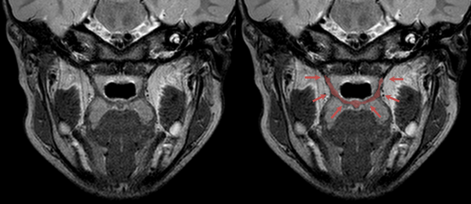
Levator veli palatine muscle thickness measurement from a tilted coronal magnetic resonance image. Left: The section with the most obvious slinglike image of the muscle was chosen for measurement of muscle thickness. Right: A total of 6 reference points were chosen, and the mean value was calculated.
Velopharyngeal Gap
For preoperative preparation, velopharyngeal closure patterns and gap sizes were measured at an outpatient clinic using nasopharyngoscopy before the surgery within 1 week. The velopharyngeal gap was measured during repetitive pronunciation of high to low oral and nasal pressure-loaded words and sentences, following the instructions of a speech pathologist. We used the Golding-Kushner (Golding-Kushner et al., 1990) and D’antonio (D’Antonio et al., 1989) method for reporting and assessing the velopharyngeal gap via nasopharyngoscopy. Based on the checkpoints of previous studies, velopharyngeal gap sizes of patients were evaluated in 6 grades. Grade 0 was designated as complete closure, 1 was pinhole closure, 2 was a resting gap closure over 80%, 3 was a resting gap closure of 50% to 80%, 4 was gap closure less than 50%, and grade 5 was complete hypodynamic movement of the velopharynx (Figure 2).

Images of nasopharyngoscopy examinations. Upper row, left: The resting gap of a patient with grade 5 hypodynamic velopharynx. Upper row, right: The gap shows almost no closure during production of bilabial plosive consonants. Second row, left: The resting gap of a grade 3 patient. Second row, right: Incomplete closure is observed during high nasal pressure-loaded speech. Third row, left: The resting gap of a grade 1 patient. Third row, right: The gap shows almost complete closure. Bottom row, left: Postoperative velopharyngeal resting gap 6 months after pharyngeal flap surgery in a patient who previously had grade 5. An intact flap on the midline of the gap is observed. Bottom row, right: The gap shows complete closure with no nasal escape during high-pressure loaded speech production.
Operative Technique
After the patient was placed in the supine position with their neck extended, intraoral irrigation was performed with normal saline and betadine solution. The size of the pharyngeal flap was estimated according to the velopharyngeal gap size of preoperative nasoendoscopy and lateral pharyngeal wall motion, by the surgeon (senior author: R.M.B.). Midline incision was performed in the soft palate to access the posterior pharynx, and bilaterally 2 soft palatal flaps were raised in the soft palate to establish contact between the raw surfaces of the raised pharyngeal flap and soft palatal flaps. After design of the flap on the posterior pharyngeal wall, 1:200 000 diluted epinephrine solution was injected for vasoconstriction. The flap was elevated above the muscular fascia, and the defect of the flap donor was closed primarily. The caudal edge of the pharyngeal flap was sutured to 2 soft palatal flaps to synchronize the size and direction. Closure of the oral lining of the soft palate was performed, and no raw surface was left opened.
Speech Analysis
Pre- and postoperative speech analysis was performed in all patients who received pharyngeal flap surgery. A single speech language pathologist performed all speech evaluations, to achieve reliability and homogeneity of results. Preoperative and postoperative results were derived from perceptual speech evaluation, and a statistical comparison between 2 examinations was performed. All preoperative speech evaluations were performed within 1 week before surgery, and postoperative speech evaluation results were collected only from examinations performed at least 3 months after the surgery, to ensure stabilization and adaptation of the pharyngeal flap and soft palate tissue after the operation (range, 6-49 months; average, 23.6 months).
A total of 5 variables—hypernasality, hyponasality, nasal emission, articulation error, and intelligibility—were collected in the speech evaluation. Because there have been no standard evaluation indicators for evaluating velopharyngeal insufficiency in the Korean-language population, the speech language pathologist revised methods and suggestions from previous studies based on hypernasality and nasal emission. According to Swanson et al. (2011), John et al. (2006), and a Pittsburgh study, weighted values for speech symptoms are associated with velopharyngeal incompetence (McWilliams and Philips, 1979). Four categories (hypernasality, nasal emission, intraoral pressure, and intelligibility) were analyzed, and the degree of improvement after the surgery was evaluated using the point scale. Weighted values were given to the hypernasality score and combined nasal emission, intraoral pressure, and speech intelligibility scores. The speech language pathologist provided 5-point evaluation results: 1 point was the least improvement and 5 points represented the most improvement after the surgery.
Points based on percentages of nasal pressure flow through the nasopharyngeal gap were monitored using a nasometer (Nasometer II 6400, Kay Elemetrics/KayPENTAX, Lincoln Park, NJ), with the device connected to a computer with an external sound card (Audigy Model SB0300, Creative Labs, Singapore). Test words were repeated bilabial plosive consonants, “papa” and “pipi,” and pressure measurements of nasal escape were recorded. The difference between preoperative and postoperative points and mean nasalance score of the normal Korean population was assessed (Park et al., 2014), and the number of changes after the surgery was recorded and analyzed.
Statistical Analysis
To investigate the correlation between preoperative velopharyngeal gap in the 6-point scale and muscle thickness seen on MRI, Kendall rank correlation method was used, and statistical analysis was done using SPSS (version 21.0, IBM Corp, Armonk, NY). For analysis of the correlation between preoperative velopharyngeal gap, muscle thickness with 5-point scale of speech improvement after the operation, and difference of nasometry outcomes, Kendall rank correlation method was also used, along with the Spearman rank correlation coefficient method. Independent variables were velopharyngeal gap size scale and muscle thickness, and dependent variables were speech analysis improvement scale and difference in preoperative and postoperative nasometric points. Additional regression analysis was done with simple logistic regression between measured continuous variables, muscle thickness, and nasometry score difference. Statistical significance was set at P <.05.
Results
The preoperative velopharyngeal gap showed a significant correlation with the thickness of the levator veli palatini measured by MRI (correlation coefficient: Kendall 0.297, Spearman 0.397, P = .02 and P = .03, respectively) (Table 1).
Statistical Analysis of the Correlation Between Preoperative Velopharyngeal Gap and Levator Muscle Thickness and Postoperative Speech Analysis.

Note: Bold numbers indicate the results with statistical significance.
a*Statistically significant correlation, P < .05.
In both Kendall tau-b correlation and Spearman correlation tests, the preoperative velopharyngeal gap showed a statistically significant correlation with the amount of speech improvement (correlation coefficient 0.347 and 0.413, P = .04 and P = .02, respectively). On the other hand, muscle thickness showed no correlation with speech analysis results (correlation coefficient 0.046 and 0.037, P = .77 and P = .86) (Table 1).
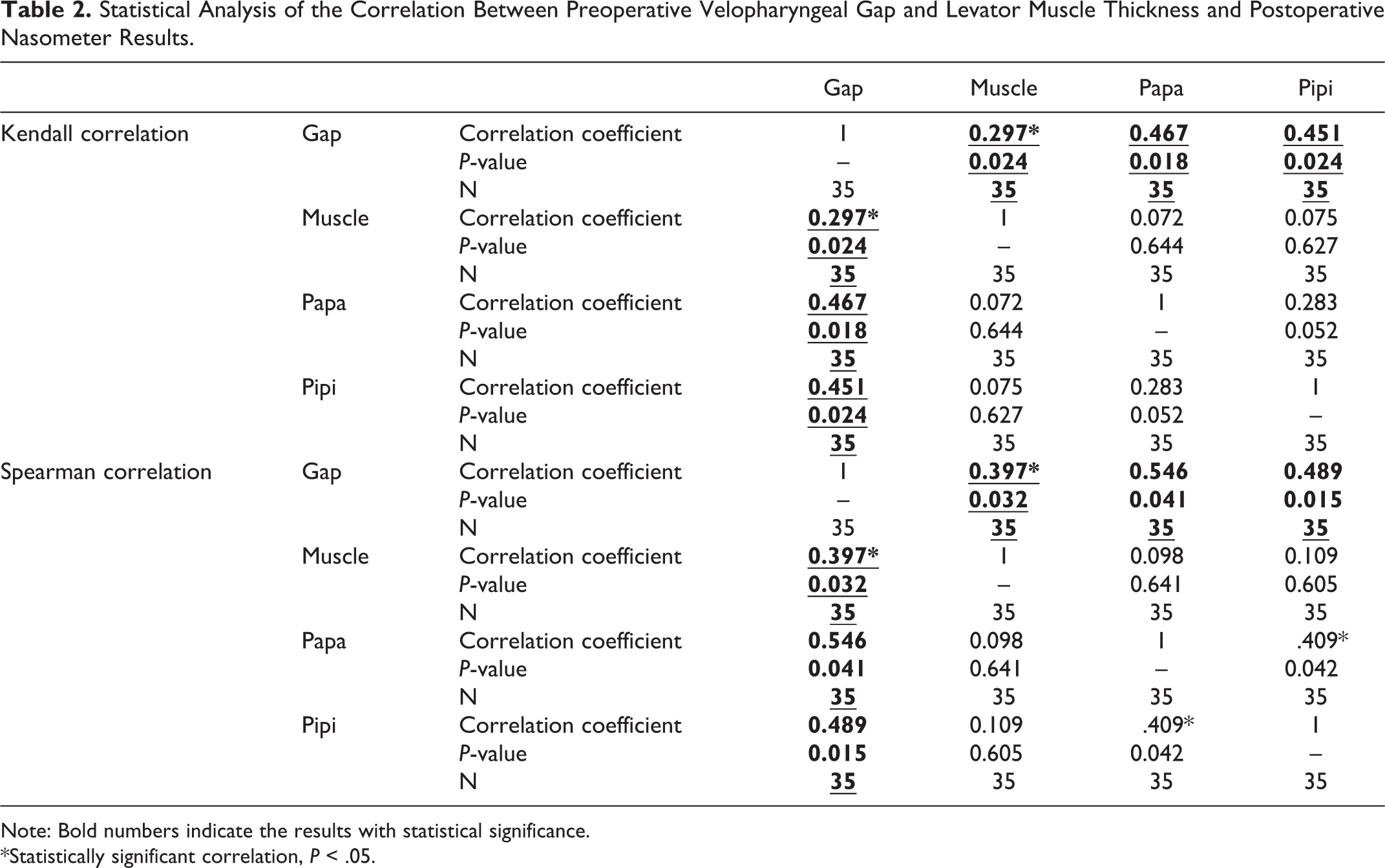
In the correlation analysis between velopharyngeal gap and nasometry point changes, the preoperative gap showed a statistically significant correlation with nasometry score improvement (“papa”: correlation coefficient 0.467 and 0.546, P = .018 and P = .041, respectively; “pipi”: correlation coefficient 0.451 and 0.489, P = .024 and P = .015, respectively). However, there was no significant correlation between muscle thickness and nasometry scores (“papa”: correlation coefficient 0.072 and 0.098, P = .644 and P = .641; “pipi”: correlation coefficient 0.075 and 0.109, P = .627 and P = .605, respectively) (Table 2).
Statistical Analysis of the Correlation Between Preoperative Velopharyngeal Gap and Levator Muscle Thickness and Postoperative Nasometer Results.
Note: Bold numbers indicate the results with statistical significance.
*Statistically significant correlation, P < .05.
Finally, from regression analysis between muscle thickness and nasometry score changes, there was no significant relationship in pronouncing “pipi” (correlation coefficient 0.238, constant 45.310, P = .251) and “papa” (correlation coefficient 0.252, constant 20.213, P = .144).
Discussion
In a previous prospective study (Park et al., 2015) with MRI, we observed that LVP muscle thickness is significantly decreased compared to that in the normal population in VCFS patients, and also that there was significant asymmetry of thickness between the right and left side of the LVP. To investigate the clinical significance of our observation, in the current study, we added more patients and collected the results of speech analysis of VCFS patients who received pharyngeal flap surgery for correction of velopharyngeal insufficiency. We also analyzed the relationship between postoperative results and the preoperative thickness of the LVP, as well as velopharyngeal gap size.
With LVP thickness in VCFS patients that was measured with methods used in the previous study, and preoperative velopharyngeal gap size from nasoendoscopy before pharyngeal flap surgery, analysis between speech outcome after the operation and preoperative data was performed, and the correlation between the variables was investigated. Postoperative speech outcome and amount of improvement showed a significant correlation with preoperative velopharyngeal gap and did not appear to correlate with preoperative levator muscle thickness. Regarding the previous study (Park et al., 2015) and other studies observing levator muscle thickness in nonsyndromic and normal populations (Ettema et al., 2002; Ha et al., 2007; Perry et al., 2013), it can be deduced that in VCFS patients with an already hypodynamic and weakened LVP, the LVP has little or no effect on surgical outcome, including speech analysis, especially after pharyngeal flap surgery that does not involve an additional levator muscle rearrangement or strengthening procedure. On the contrary, a smaller preoperative velopharyngeal gap lead to better surgical outcomes of pharyngeal flap surgery, as the primary goal of pharyngeal flap surgery is to effectively obstruct the gap size in patients with velopharyngeal insufficiency.
Moreover, by analyzing the relationship of levator muscle thickness and preoperative velopharyngeal gap, there was statistically significant correlation between thickness and gap size, as increased muscle thickness is related to more complete closure of the velopharyngeal gap. The results have shown that functional capacity and power of the LVP play a certain role in velopharyngeal closure that can eventually lead to improved speech of patients and better surgical outcomes after pharyngeal flap surgery, although there was no observed significance between muscle thickness and operative outcome. Moreover, the findings could lead to determining the outcomes on treatment of velopharyngeal insufficiency patients related to VCFS, who received other surgical methods such as sphincter pharyngoplasty and Furlow palatoplasty.
Air flow pressure during repeated production of bilabial plosive consonants measured by nasometer is related to velopharyngeal closure (Dalston et al., 1991; Fukushiro et al., 2015; Keuning et al., 2004). The degree of hypernasality and nasal emission increases with severe nasal escape, and the amount of nasal escape can objectively measure velopharyngeal function. The speech evaluation and the difference of nasometry scores showed a statistical correlation with the preoperative gap, with a smaller gap leading to bigger improvement. However, muscle thickness did not affect changes of postoperative nasal escape, both in correlation tests and regression analysis. Although the LVP muscle affects lateral pharyngeal wall motion, in speech improvement after pharyngeal flap surgery, the role of the LVP muscle is limited. We can assume that the predisposing factors of the outcome of pharyngeal flap surgery is very different from other surgeries for the treatment of velopharyngeal insufficiency, such as sphincter pharyngoplasty (Losken et al., 2003; Witt et al., 1999) or palatal advancement with Z-plasty (Lin et al., 1999). Also, the results can give background for analysis of previous reports of pharyngeal flap surgery in patients with velocardiofacial syndrome and velopharyngeal insufficiency (Swanson et al., 2011; Wang et al., 2009).
This study was based on an MRI study in which we performed pharyngeal flap surgery before analyzing the thickness of the LVP muscle, and the study reports the largest number of velocardiofacial syndrome patients to this date. The study aimed to investigate the clinical significance of a relatively thin and insufficient LVP muscle in patients with velocardiofacial syndrome compared to the nonsyndromic population. We performed an analysis of the thickness of the LVP, preoperative velopharyngeal gap, postoperative speech outcome, and improvement of nasometric scores. The findings of this study will help to understand the pathophysiology of VCFS-associated velopharyngeal insufficiency, although the results are not clinically applicable.
Our study has some limitations. Because we only studied the thickness of the LVP, electromyography that directly evaluates muscular function or direct visualization with devices such as videonasopharyngoscopy could have strengthened our evaluation of LVP function. Also, another shortcoming is that different factors with a possible influence on postoperative speech analysis, such as patients’ mental status or cognitive function, were not included in the present study. Further studies should be performed on other factors. Nevertheless, as a step further from a previous study, this study reported the effect of anatomical abnormality of VCFS patients on clinical outcome, and the results can act as a theoretical background in the treatment of language and speech abnormality in VCFS patients.
Footnotes
Declaration of Conflicting Interests
The author(s) declared no potential conflicts of interest with respect to the research, authorship, and/or publication of this article.
Funding
The author(s) received no financial support for the research, authorship, and/or publication of this article.