
Abstract
Introduction
Vomer flap is a technique to close cleft lip and palate. This technique is a simple procedure that has many benefits. However, the vomer flap's application together with primary lip closure is still questionable.
Objective
To find out whether the vomer flap's application in primary cleft lip repair can provide significant benefits
Design
A systematic review was conducted using the PRISMA methodology has been licensed in PROSPERO databases (CRD42023399487).
Setting
A comprehensive search was set out, utilizing eight data sources up to March 2023.
Participants
Both cohort studies and randomized control trials regarding the use of vomer flaps performed concurrently with cleft lip repair in children up to six months old.
Results
This article involved 8 studies involving 542 patients who met the inclusion criteria, consisting of 6 retrospective cohort studies, 1 RCT study, and 1 prospective cohort study. Vomer flaps provide a reduction in palatal cleft distance of 3–5 mm, a relatively small number of fistulas (0–4%), improvement of velopharyngeal function (nasal tone and nasal emission), maximal development of the maxilla although it is still controversial.
Conclusion
The vomer flap's application in primary cleft lip repair provides many advantages, such as reduced palatal and alveolar clefts, decreased risk of oronasal fistula, increased velopharyngeal function, and increased maxillary growth. It is reliable for the management of cleft lip and palate.
Introduction
Cleft lip and palate (CLP) is a congenital dysmorphology commonly found within the craniofacial region.1,2 According to WHO, the prevalence of CLP is around 1: 700 live births, the highest being of Asian descent with 14: 10000 live births.3,4 Indonesia is recorded to have a prevalence of CLP of around 0.2%. 5 There have been many procedures introduced to treat CLP, all with the same goal in mind: an aesthetically pleasing appearance, adequate speech outcome and minimal impact on craniofacial growth. Several complications most commonly found are surgical wound dehiscence, partial or total necrosis of the flap, floating palate, and oronasal fistula, in which the latest studies reported variation in oronasal fistula incidence between 0% and 77.8%, making it the most frequent complication in palatal repair.6–8 The most common signs of oronasal fistula are nasal regurgitation and hypernasality of speech. In terms of surgery techniques and experiences, the major reason is thought to be the restoration of cleft palate under stress, while vascular injuries, poor hygiene, and infection may also constitute contributing factors. However, certain cleft palates are somewhat large, and having enough tissue to rebuild the palate appears insufficient. 9
Pichler, in 1934, explained a palatal closure method that involves tucking a single cranial-based flap of the vomerine's mucoperiosteum and the nasal septum's mucoperichondrium under the raised margin of oral mucoperiosteum on the opposite side of the cleft, exposing the raw surface of the nasal septum exposed and allowing it to heal with epithelialization.10,11
This technique uses a soft tissue flap from the vomer or nasal septum to close a defect in the hard palate region. The tissue is delicate but highly vascular, and if properly manipulated, will develop as the free-floating flap. The procedure is relatively simple but with satisfying results. 12 Several studies showed no reports of maxillary growth stunts. The dental arch grew as it should, providing sufficient space for dental accommodation. The facial growth was reported to be adequate with no sub malar depression and no disorders of the maxillofacial growth. Therefore, this procedure was frequently employed, albeit with modest modifications to meet each circumstance.13,14
However, there are investigations reporting contradictory results, demonstrating that the vomer's flap application causes scarring at the vomero-premaxillary suture, which means slowing anterior midface development. 15 Despite patients having final palatoplasty using the “push back” method, Friede and Johanson's cohort study discovered poor craniofacial growth in complete bilateral and unilateral vomer flaps performed between 1964 and 1970. 16
Deshpande et al. initiated a retrospective analysis on 101 non-syndromic CLP, calling into doubt the efficacy of simultaneous hard palate and cleft lip repair employing a vomer flap. The findings found that almost 100% flap successes were seen in about half of the cases, whereas 50% flap successes occurred in nearly a quarter of the cases. Vomer flap's failure is linked with a greater probability of fistula and wound dehiscence even after palatal cleft closure.17,18 Even though vomer flaps succeeded, the palate failed to be closed without releasing the incision, limiting subsequent palatal repair.14,18,19
Considering the multiple contradictory reports on the efficacy and complications of early vomer flaps, this research examines the utilization of vomer flaps performed concurrently during labioplasty up to the age of six months.
Method
This research has been licensed in PROSPERO databases (CRD42023399487) and was completed in line with the Preferred Reporting Items for Systematic Review and Meta-Analyses (PRISMA) statement. 20
Eligibility Criteria
For our study's inclusion criteria, we specifically considered published papers that met certain parameters. These parameters included both cohort studies and randomized control trials that showed data on any outcome related to palate repair using a vomer flap along with cleft lip repair in children up to six months old. Additionally, we included controlled studies focused solely on cleft lip repair in children up to six months old. To ensure consistency, we limited our analysis to articles available in full text, written in English, and conducted on human subjects.
Conversely, we excluded articles featuring samples that underwent surgery either with or without vomer flaps, whether performed simultaneously with cleft lip repair or not, but at an age exceeding six months. This decision was based on our study's goal of evaluating early vomer flap repair, which was performed at the initial stage of CLP surgery, namely cleft lip repair, when children were up to six months old. By implementing these inclusion and exclusion criteria, we aimed to ensure the relevance and validity of the data included in our study.
Search Strategy and Selection Studies
To identify studies related to the use of vomer flaps in cleft palate and cleft lip closure, we systematically searched the PubMed, Scopus, Science Direct, Cochrane, EBSCO, Web of Science, Cilinicaltrial.gov, and Proquest databases that were published through March 2023, apart from that we also looked for gray literature using Pre-print and Medrxiv. The term the search is as follows: vomer flap, early vomer flap, cleft lip repair, early cleft lip repair, primary cleft lip repair, labioplasty, cheilorrhaphy, early hard palate repair, early palatoplasty, and early palatorrhaphy. This term is used in combination with “AND” or “OR”, depending on the search engine utilized (see the Appendix). Two investigators conducted the literature review autonomously, with a third reviewer settling any disagreements as required.
Quality Assesment
Two authors autonomously selected necessary data, including author, year of publication, article title, country of origin, type of study, and variable measured, then determined the size of the sample, sex characteristics, and average age when treated in the study. Then we will extract the results of the variables measured in each study found.
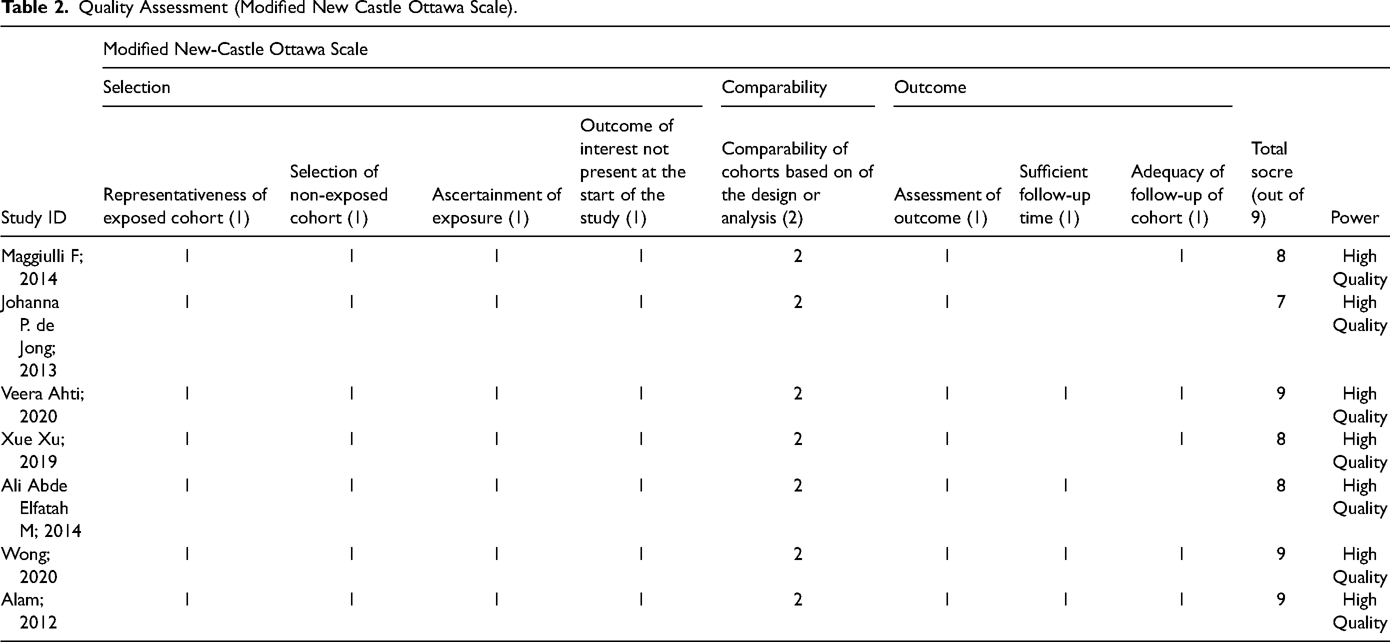
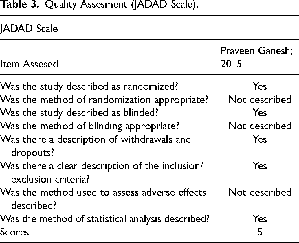
To assess the quality of the included studies, we employed specific evaluation tools. We used a quality assessment tool for the cohort study using a modified New Castle Ottawa (NOS) scale, 21 with maximum score of nine points. The NOS assesses three domains: study group selection (four points), group comparability (two points); and ascertainment of exposure and outcome (three points). In accordance with the overall score, we sorted the studies as having a low risk of bias (≥7 points), a moderate risk of bias (5–6 points), or a high risk of bias (≤4 points). Meanwhile, for the randomized controlled trial study, we used the JADAD score consisting of three items: randomization (0–2 points), blinding (0–2 points), and dropout and withdrawal (0–1 points). 22 The response to each item is “Yes” (1 point) or “No” (0 points). Final points range from 0 to 5 points, with higher points signifying better-quality reporting. JADAD scores of 2 points or less indicate low quality, whereas JADAD scores of 3 points or above indicate excellent quality. All authors discussed and settled any disputes.
Statistical Analysis
Due to significant variations in the comparisons made among research articles and the diversity of outcome measures, conducting a meta-analysis was not feasible for our study. Instead, we will employ a narrative synthesis approach to analyze each piece of evidence individually. This process will involve dividing the outcomes into relevant subsections using appropriate subtitles. Subsequently, we will compare the treatment group to the control group in a descriptive manner, providing a comprehensive narrative analysis of the available evidence. This method will allow us to synthesize the findings effectively, despite the inability to perform a meta-analysis due to the aforementioned differences in study designs and outcome measures.
Result
Study Selection
Based on screening strategies, 2568 pertinent investigations were retrieved from sources. We used the Mendeley Reference Manager to remove 558 duplicate studies, meta-analyses, systematic reviews, case reports, case series, and reviews. Subsequently, 986 pertinent investigations were omitted based on the title and abstract, and 328 papers were eliminated after reviewing the whole article. In the gray literature we found 6 related studies, but then we excluded all studies. Moreover, this systematic review comprised a total of eight studies. Figure 1 depicts the PRISMA flowcharts of the included investigations. 20
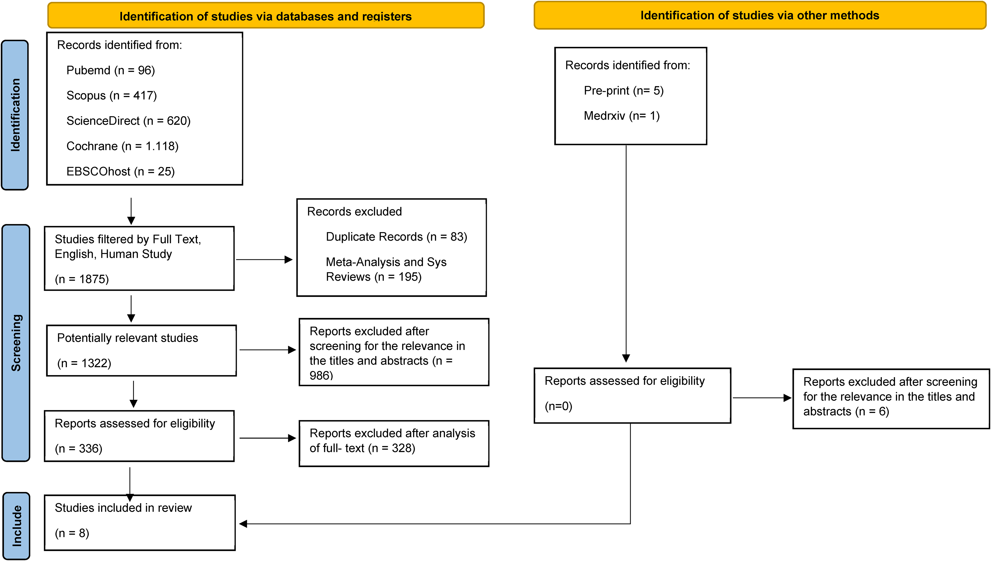
PRISMA flowchart.
General Characteristic
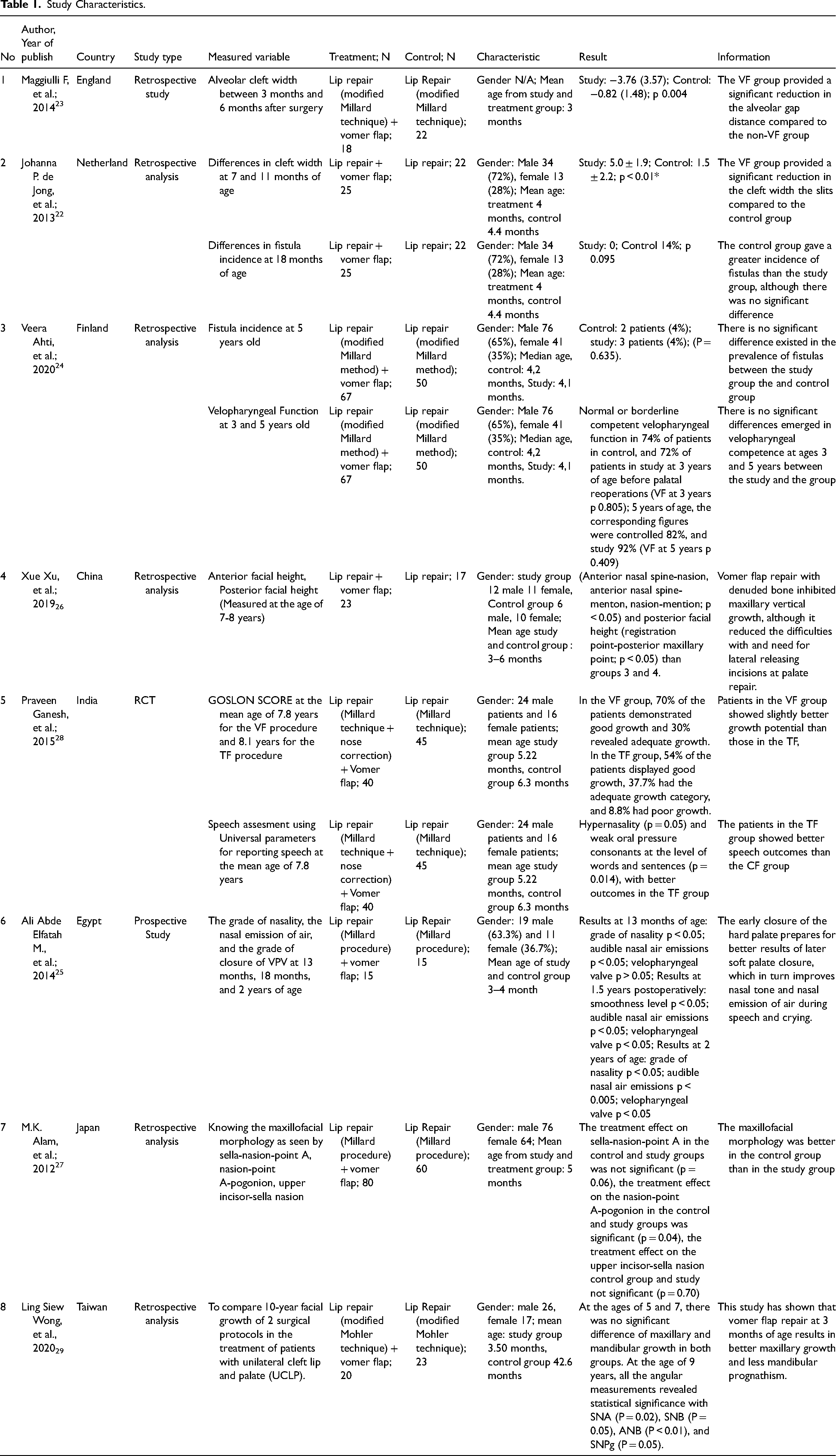
Three studies were from Continental Europe (England, Netherlands, Finland), four studies were from Continent Asia (China, Taiwan, Japan, and India), and 1 study was from the African continent (Egypt). The total number of samples from the eight articles that we used was 542, and each article had a sample size ranging from 40 to 140. The number of male patients ranged from 12 to 76, while the number of female patients ranged from 11 to 41. The average age of patients undergoing surgery in the vomer flap and lip repair treatment group was 4.1 months, while the mean age of patients undergoing surgery in the control group was 4.2 months. Seven articles on lip repair use a modified Millard technique, while 1 article uses a modified Mohler technique. Six studies were retrospective cohort studies, 1 prospective cohort study, and 1 RCT study. Table 1 shows the overall features of the eight selected studies.
Study Characteristics.
Quality Assessment
The study assessment used the New Castle Ottawa score for the cohort study, where in 7 studies scores ranged from 7–9 which were included in high-quality studies, while the RCT study assessment used the JADAD score and obtained a score of 5, which was included in good research. The detailed results of the study quality assessments are listed in Table 2 and Table 3.
Quality Assessment (Modified New Castle Ottawa Scale).
Quality Assesment (JADAD Scale).
Gap Reduction
The use of a vomer flap provides an advantage in terms of reducing the gap between the alveolar gap and the gap itself. Two published research articles showed that the vomer flap's application resulting a substantial reduction in gap distance (3–5 mm) compared to the control group using vomer flaps (0.8-.15 mm) (p < 0.005).23,24 See Table 1.
Incidence of Fistula
Fistula is one of the complications that often occur in flap closure. Closing the cleft palate using the vomer flap method provides advantages by reducing the incidence of fistulas. Two studies have shown that the incidence of fistulas in patients who use vomer flaps is only around 0–4% compared to patients who do not receive vomer flaps 4–14%. This study also shows an evaluation of the occurrence of fistulas at the age of 18 months, where at the age of 18 months a definitive cleft palatal closure is usually done, so that the use of this vomer flap can also facilitate the second stage of surgery.23,25 See Table 1.
Velopharyngeal Function
The use of vomer flaps also provides benefits in terms of velopharyngeal function, a study shows that vomer flaps provide good output on nasal tone and nasal emission so that patients can provide better sound output. Two subsequent studies showed that the vomer flap group provided better outcomes in terms of velopharyngeal function than the non-vomer flap group, although two did not provide statistically significant differences.25,26 See Table 1.
Maxillary Growth
The 4 studies presented in this article present the pros and cons of using the Vomer Flap, respectively. Xu et al. and Alam et al. noticed that the Vomer flap had adverse consequences for maxillary growth. However, Xu et al. conclude that the vomer flap's application is favorable in terms of offering efficiency for palatal closure in the next steps. In contrast to the two previous studies, Ganesh et al. and Wong et al. observed that the vomer flap had a positive effect on the growth of the facial bones, particularly the maxillary bone.27–30 See Table 1.
Discussion
Treatment for unilateral CLP has evolved significantly over the previous few decades, and surgical procedures are continually advancing. To protect the nasal lining during palatal closure, the Vomer flap employed mucoperichondrial and mucoperiosteal flaps, which are still questioned in terms of risks and benefits. 31 However, the study analysis presented in this publication reveals that the vomer flap's application together with primary lip repair up to 6 months of age can be exceptionally trustworthy, although risks can still arise. 24
In numerous investigations, the ages of patients with CLP employing the vomer flap ranged from 4 months to 9 years. 32 It is predicted that by sealing the hard palate, the space left in the soft palate would close spontaneously as well. However, there is limited evidence that using a vomer flap has any significant advantage in reducing the cleft, particularly in children under the age of six months. 24 Therefore, we gathered some literature that can offer data on the usage of vomer flaps in children under the age of six months.
In several articles, it has been shown that the use of vomer flaps can reduce the distance between clefts, the distance between palatal slits, the distance between alveolar slits, and the distance between clefts of the uvula. 24 De Jong et al. 23 showed a significant reduction in the distance (p < 0.05), so it was hoped that narrowing the clefts would be of benefit in reducing complications such as fistula occurrence and facilitating surgery for definitive closure of the hard palate. This reduction in cleft spacing may be due solely to the new tissue creation from the vomer flap used for anterior palate closure during labioplasty. In addition, many studies also state that the greater the reduction in the distance between the gaps, the lower the number of fistulas.
There are differences between 4 studies regarding early vomer flap and maxillary growth. In this maxillary development, the average study provides follow-up after 7–10 years. Ganesh et al. 29 and Wong et al. 30 concluded that vomer flap technique is favorable in terms of maxillary growth. Meanwhile, Xu et al. 27 and Alam et al. 28 concluded that vomer flap technique is unfavorable in terms of maxillary growth. Alam et al. believed that scarring slows maxillary growth, a later age for hard palate closure will result in less growth disruption and better maxilla final growth. However, the number of patients in the study conducted by Ganesh et al. 29 and Wong et al. 30 was greater than the study conducted by Xu et al. 27 and Alam et al., 28 so this could provide insight that the vomer flaps technique is favorable in terms of maxillary growth. Moreover, the research conducted by Ganesh et al. 29 is a randomized controlled trial, whereas the other studies are retrospective analyses. Xu et al. 27 later emphasized the importance of deferring the final evaluation until the facial bone is completely mature. This is also consistent with Ganesh et al. 29 and Alam et al., 28 suggesting further follow-up once maxillary growth is complete at puberty. Genetic and/or environmental factors influence the growth of the maxilla and mandible.
Fistula incidence is an important factor to be assessed since it has a negative impact on articulation.17,19 Numerous investigations have shown that hard palate closure with a vomer flap correlates with a reduced number of fistulas. Which may be the consequence of significant cleft narrowing. However, a study from Ahti et al. 25 found that labioplasty and hard palate closure with a vomer flap at 4 months of age followed by soft palate closure at 10 months of age versus labioplasty at 4 months of age followed by complete palate closure at 12 months of age did not differ (4% versus 4%). Another study by de Jong et al. 23 also revealed no significant fistula incidence between patients receiving lip repair and vomer flap and patients receiving only lip repair on their first surgery. Though it is important to note, that smaller residual clefts make way for a simpler follow-up repair regardless of the fistula incidence.
The patient's speech quality frequently serves as a barometer for the success or failure of cleft surgery results. However, it is not a simple measurement due to the fact that it is subjective, depending on the examiner. Several reports have stated different outcomes in the quality of speech of patients. In 2015, Ganesh et al. 29 employed a universal metric for reporting speech and discovered that the Two Flap approach is superior to the Vomer Flap in terms of hypernasality and weak oral pressure in patients aged 4 to 6 years. Another study, Ahti et al. 25 found no statistically significant variance between the vomer flap and non-vomer flap groups at 3 and 5 years of age. However, Ali et al. 26 demonstrated conflicting results, in which they documented the audio recordings of the patients and had three judges score them, as well as tracked the velum's movement and lateral pharyngeal walls using video nasoendoscopy. At 13 months, 18 months, and 24 months, they reported substantial changes in favor of the vomer flap group. Among these studies, the study by Ali et al. 26 seems to be more advantageous since they include an objective measure from the nasoendoscopy. Another difficulty in speech assessment is determining when the right time is for its evaluation. One study argues speech assessment right after surgery, or at least one month after surgery, has a risk of being misinterpreted as edema secondary to surgery. 26 Another study stated that evaluations are warranted after speech development is completed and highlighted the importance of assessments done at 3 years of age. 25
This article provides the strength that the use of vomer flaps can be one of the techniques used in repairing palatal clefts, but we are still aware of several limitations in writing this article, including the relatively small number of studies, small number of samples in each study, single-center study, and the assessment parameters are not uniform. We hope that future studies will provide uniform assessment parameters with a larger sample size in a multicenter study.
Conclusion
The vomer flap's application in primary cleft lip repair provides many benefits, including reduction of palatal and alveolar clefts, reduced risk of oronasal fistula, improved velopharyngeal function, and improved maxillary growth, although this is still controversial. Overall, this is a reliable technique for CLP.
Footnotes
Acknowledgment
We thank the authors of articles included in this systematic review for the advancement of science.
Authors Contributions
Conceptualization, ILP, CDK; Data Collection, ESW, SL, SR; Methodology, ILP, CDK; Writing-Original Draft Preparation, ESW, SL, SR; Writing-Review & Editing, ILP, CDK. All authors have critically reviewed and approved the final draft and are responsible for the content and similarity index of the manuscript.
Declaration of Conflicting Interests
The authors declared no potential conflicts of interest with respect to the research, authorship, and/or publication of this article.
Ethical Approval
No ethical approval was required since the study is a systematic review.
Funding
The authors received no financial support for the research, authorship, and/or publication of this article.