
Abstract
Lateral synechiae syndrome is a rare congenital malformation characterized by cleft palate and lateral intraoral adhesions from the free borders of the palate to the floor of the mouth. Synechiae can be isolated or occur more frequently in association with other congenital anomalies such as cleft lip and/or palate. These synechiae can cause functional deficits, especially in the respiratory and feeding difficulties, as well as facial abnormalities at later stages. Surgical excision of the adhesions as soon as possible is the well-adapted protocol at most centers. We report a case of a 15-day-old male infant reported to the outpatient department for feeding difficulties with a cleft palate associated with bilateral lateral intraoral synechiae.
Keywords
Introduction
Congenital lateral palatal synechiae are defined as adhesions or bands connecting the sides of the cleft palate to the lateral parts of the tongue or the floor of the mouth. 1 It is a rare condition and very few cases have been reported worldwide (Fuhrmann et al 2 ). 3 These connections can be of fibromucosal tissue or fibrous bands defined as synechiae, or they may be a bony connection, which is defined as synostosis, leading to fusion of maxilla and mandible. Epithelial fusions are more common than bony fusions. 4 Lateral synechiae syndrome is defined as a cleft palate along with unilateral or bilateral palatal synechiae. 4 These adhesions may lead to respiratory or feeding difficulties in infants due to restricted mouth opening in early stages and later facial growth discrepancies at later stages. The aim of treatment is to attain an adequate mouth opening, facilitating both the airway and feeding, and to allow oral and facial development as well as surgical treatment of the cleft. Hereby, we report a case of congenital bilateral lateral palatal synechia associated with a cleft palate and its management.
Case Report
A 15-day-old male infant reported to our outpatient department with an intraoral fibrous band present bilaterally, along with a cleft palate. The infant was the first son of the couple. Parents were healthy and had a nonconsanguineous union with no family history of orofacial clefts or other congenital malformations. He was born at 38 weeks’ gestation via normal vaginal delivery with a birth weight of 3 kg and did not need any resuscitation. No symptoms of respiratory distress had been observed since birth. Informed consent was taken from parents before the procedure.
Intraoral examination was difficult due to restricted mouth opening. There were bilateral membranous adhesions noted between the free margin of the complete cleft palate and the floor of the mouth lateral to the tongue on the left side (Figures 1 and 2). There was no associated syndrome with no hearing abnormality. Digits and extremities showed normal appearance. No cardiac abnormality was noted. The congenital oral synechia appeared as a thin membrane with a broad attachment at the floor of the mouth and narrowing at the palatal margin. The infant faced feeding problems due to the restricted mouth opening, and therefore an immediate surgical excision of the synechia was planned.

Bilateral Palatal Synechia.
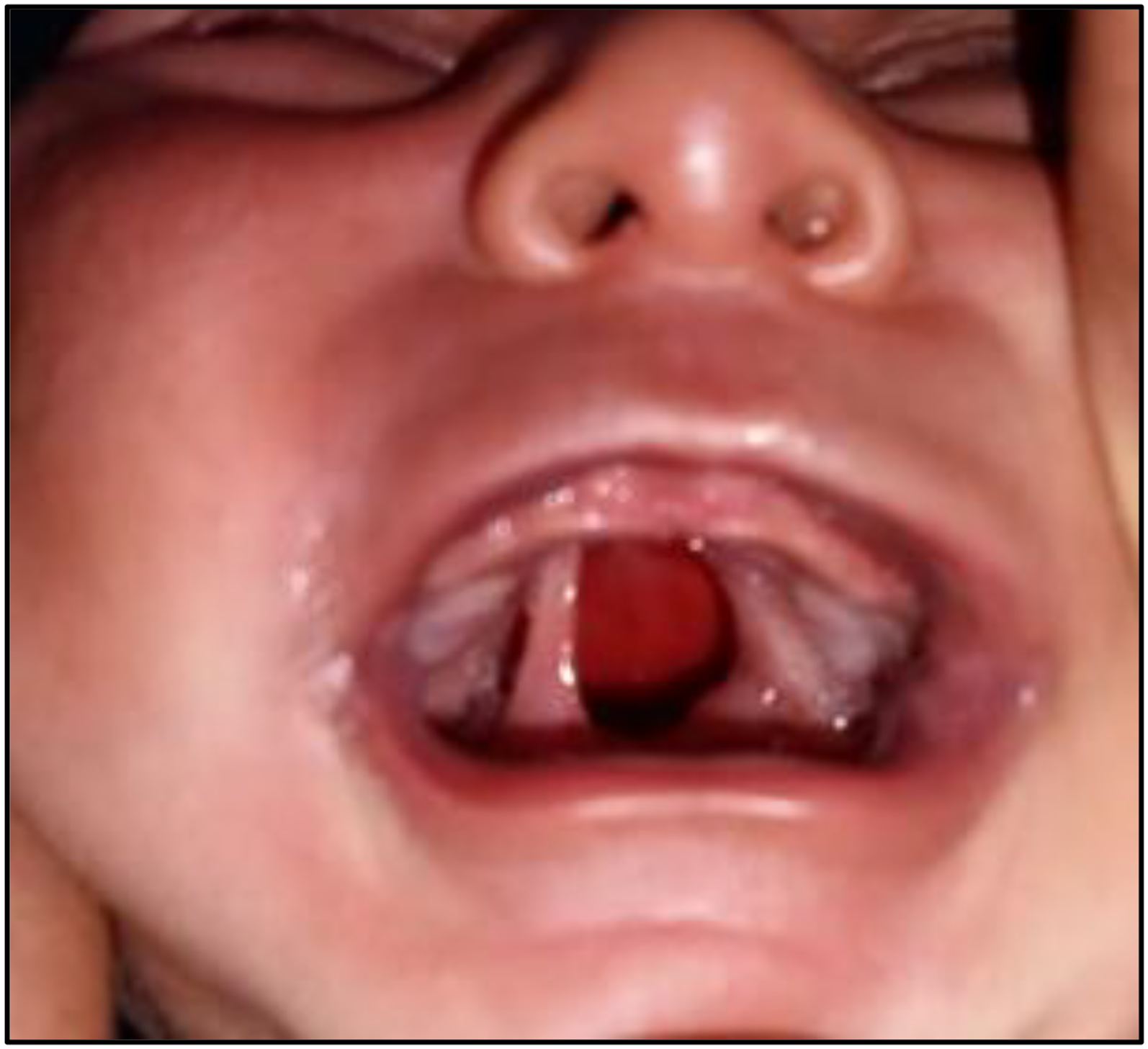
Bilateral Palatal Synechia.
As intubation was not feasible in the infant, excision of fibrous bands was planned under intravenous sedation, restoring adequate mouth opening (Figures 3 and 4). Palatoplasty of the complete cleft palate was planned for a later date. After complete excision of the band, complete visualization of the oral cavity was done, and hemostasis was achieved. The infant did fairly well in the recovery period, and normal feeding was restored.
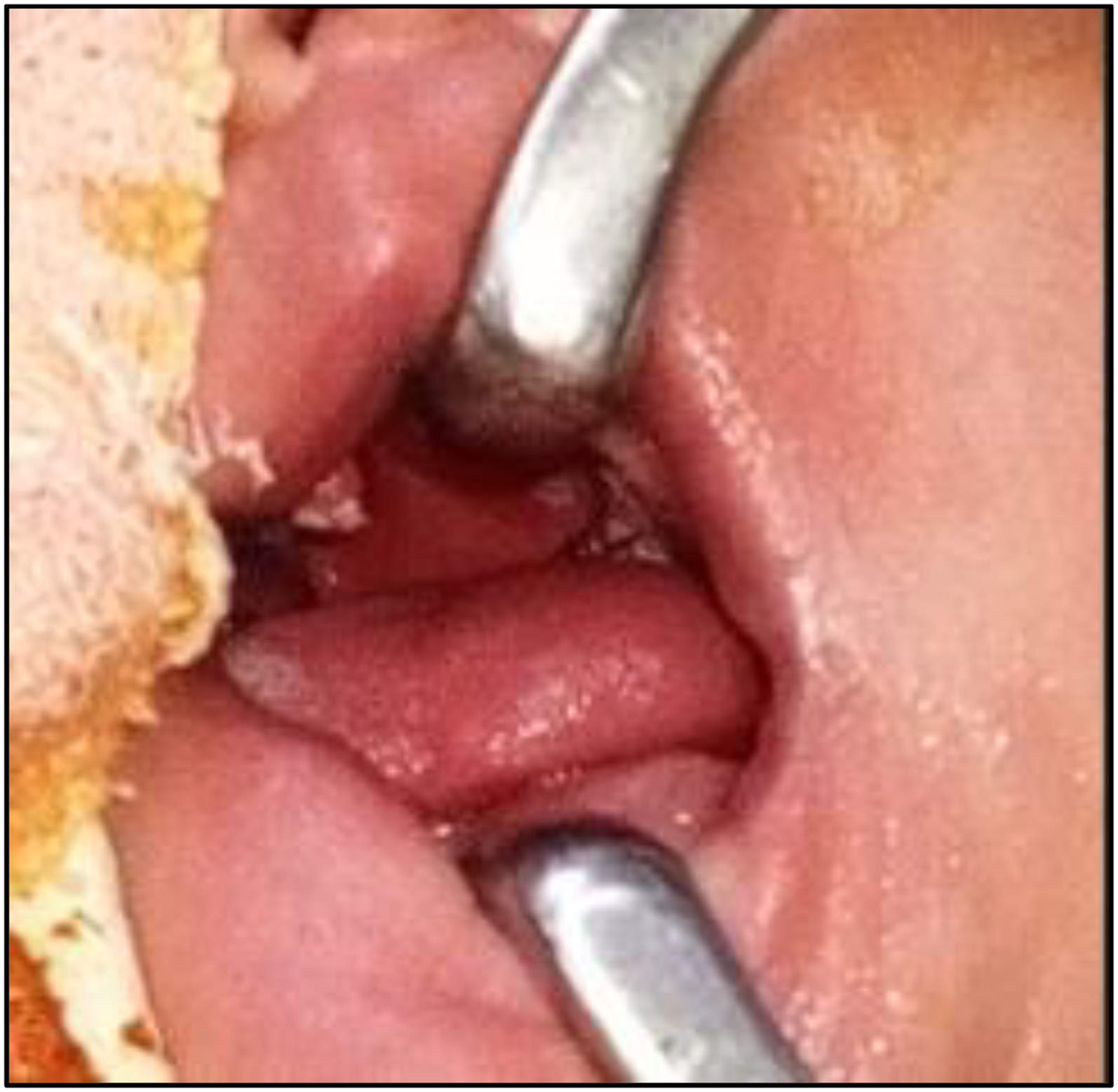
Excision of Palatal Synech.
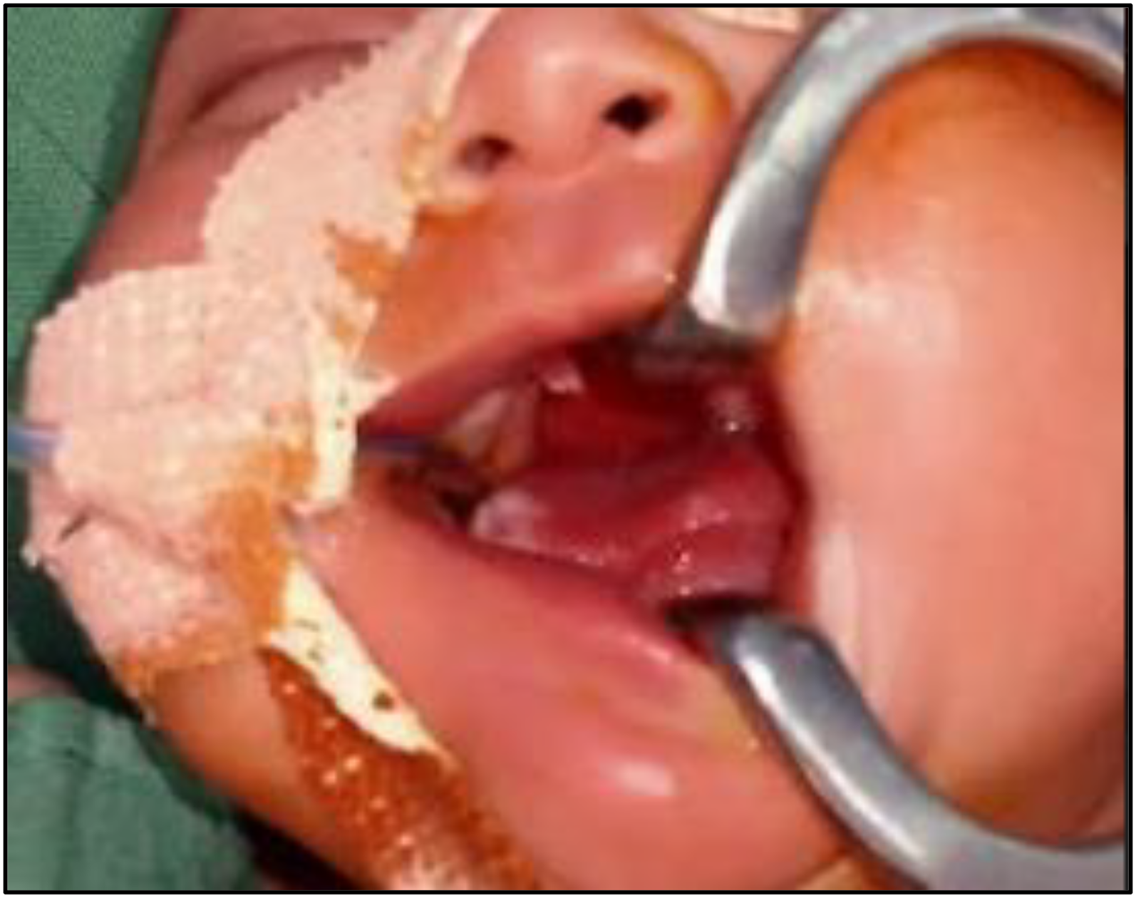
Excision of Palatal Synech.
Discussion
Palatal synchaie are a rare entity with approximately 60 cases reported worldwide. 5 The first case of oral synechia was reported by Illera in 1875 6 and the first documented case of a lateral synechia was by Hayward and Every in 1957. 7 Out of almost 60 cases reported, 52 are lateral synechia and 8 were of the median variety.
Pathogenesis of these palatal synchaie relates to the abnormalities occurring during embryological development. It has been postulated that during normal oral development, the tongue moves downward and forward in the 7th week to allow for fusion of the palatal shelf. Protrusion out of the oral cavity prevents fusion between the oral mucosae. Mathis, in 1962, gave the most widely accepted theory, reporting that the formation of oral synechiae is they are the remnants of the buccopharyngeal membrane. 8 The presence of a cleft palate in some children with synechiae can be further correlated as a sequence in that the presence of an abnormal membrane restricts normal movement of the tongue in development and therefore prevents the fusion of the palate. 9 According to Longacre, oral synechia may be due to the persisting buccopharyngeal membrane and, therefore, associated with micrognathia and cleft palate. 10
Ogino et al
11
described 5 varieties of palatal synchaie:
Adhesion of the alveolar mucosa to one or both sides of the upper and lower jaw (alveolar synechiae). Membranous adhesion on the hard palate and floor of the mouth, excluding the back of the tongue (lateral synechiae). Bands partially involving the hard palate and tongue. Soft palate and tongue are involved. Membranous adhesion between the hard palate and lower lip.
Fuhrmann et al, 2 in their study reported on 5 family members with cleft palates and synechia, 1 having a cleft palate without synechia, and 1 transmitted the gene but did not express. According to various studies reported on the treatment of cleft palate, lateral synechia syndrome is surgical excision of the synechia as early as possible so that the infant can attain adequate mouth opening and resolve breathing as well as feeding concerns. Palatal closure can be planned at a later date. Donepudi et al, 12 in their study have reported that the synechia can provide additional tissue during palatoplasty so that palatal flaps can be closed under less tension. Dalal et al, 13 reported a study of intra-alveolar synechia in 2 siblings, one of whom had a natural resolution of the adhesion and the other required surgical excision.
Acquiring a safe and sound airway for the release of the bands may be challenging in such patients. The customary oro-tracheal intubation or a laryngeal mask is usually not feasible due to the presence of the synechia. Fiberoptic nasotracheal intubation is also not possible due to the difficulties of getting a bronchoscope for such infants. 14 Therefore, many centers choose to perform it under local anaesthetic and sedation as it was done in our patient.
Thorough examination of the oral cavity should be done after excision to ensure proper hemostasis, as there might risk of bleeding complications and aspiration.
Conclusion
Congenital palatal synchaie are rarely reported in the literature, and early intervention is often required, especially in infants with respiratory and feeding difficulty. They can occur in an isolated way or more frequently with cleft palate, as in our case. An early and adequate treatment eases the mouth opening and correction of the associated malformations.
Footnotes
Funding
The authors received no financial support for the research, authorship, and/or publication of this article.
Declaration of Conflicting Interests
The authors declared no potential conflicts of interest with respect to the research, authorship, and/or publication of this article.