
Abstract

This column includes a short case presentation and differential diagnosis. It is followed by a discussion of the disease or condition and how the school nurse should handle it.
History
R.Q. is a 13-year-old who has been waiting in your office. She stopped earlier in the day and asked if she could talk to you. She tells you that she is embarrassed to talk about her problem. You notice that she has her hands closed and hidden in her clothes. You wait while she tells you that her hands are ugly, and no one will hold or touch her hands.
You ask R.Q. to show you her hands. She has many large dome-shaped, grey-brown hyperkeratotic growths on her fingers, around and under her nails, and on the back of her hands. You examine her skin for any other growths and find that the lesions are confined to her hands. She tells you that she has had them for several years, they are getting larger, and she is getting more of them. She also tells you that she does not have any other illnesses, and no one else in the house has any of these growths. Her vaccines are up-to-date, and you do not see any other problems on her health record.
You ask R.Q. about treatments. She tells you that she went to her primary care provider and some of the growths were treated, but they did not completely go away. She has also used an over-the-counter product, but it did not help. When questioned further, she tells you she put the product on for only a “week or two.” The lesions do not hurt, itch, or have any other symptoms.
Physical Findings
R.Q. appears to be a healthy, well-developed 13-year-old Hispanic female. Despite the fact that she has told you she is ashamed of her lesions, she looks you in the eyes and does not appear depressed. Her vital signs are normal. She has eight lesions on her right hand, including lesions over the cuticle on her middle, ring, and first finger. She has a shallow raw-looking lesion on her little finger (Figure 1). She tells you she “picked and bit” that lesion on the day before. She has four lesions on her left hand, including a large blackened lesion on her middle finger (Figure 2). This is the lesion she has been treating with the over-the-counter product. A careful examination of the rest of her skin, including around her mouth, does not reveal any other findings.
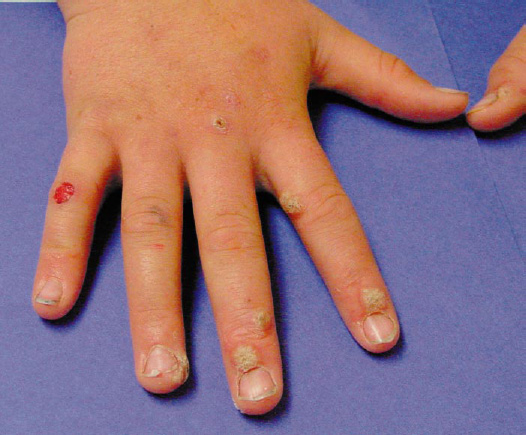
Eight Lesions Including Lesions Over the Cuticle of the Middle, Ring, and First Finger
Differential Diagnosis
Molluscum contagiosum.
Verrucae vulgaris (common warts).
Verrucae plana (flat warts).
Condyloma acuminata.
Discussion
This child has verrucae vulgaris or common warts. Warts are caused by human papilloma viruses (HPV). There are as many as 100 different types of HPV that infect epithelial cells of the skin, mouth, esophagus, larynx, trachea, and conjunctiva (Habif, 2004). Warts are one of the most common skin disorders of childhood (Hurwitz, 1993). Warts are benign epidermal neoplasms, or tumors, that occur on the very top layer of the skin. Warts are unrecognized by the body as foreign because they do not have roots that penetrate into the dermis. Therefore, they do not trigger an immune response so the body can rid itself of the virus.
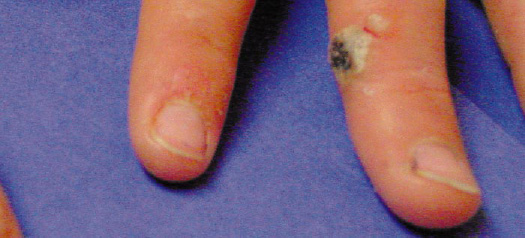
Four Lesions Including a Large Blackened Lesion on the Middle Finger
Common warts occur most often in children and young adults, but they can occur at any age with the highest incidence occurring between 10 and 19 years of age (Hurwitz, 1993). Warts are commonly found on the fingers, hands, and feet of children. These viral lesions are spread by touch; therefore, it is not uncommon to see two lesions side-by-side or “kissing” on adjacent fingers or toes. Warts are often spread to the lips or cuticles of nails when children attempt to bite or pick off the warts. They can also spread to the eyelid border if the child rubs the eyes with fingers that have warts on them. Warts also appear at sites of trauma, so scratching may spread the warts in a line in the direction where the child has been scratching.
There are several different kinds of common warts. Some form cylindrical projections that look like fingers, called digitate warts, which are common on the face or other thin skin. Common warts start on thicker skin and have cylindrical projections that become fused together to form a domed-shaped grey-brown hyperkeratotic lesion with a mosaic pattern on the surface (Habif, 2004). Black dots often seen in warts are thrombosed vessels. Warts that are black (Figure 2) are often in the process of spontaneous resolution. Warts can be discrete lesions or can be so numerous that they become confluent and cover large areas of skin. Ver-rucae plana are flat warts. They occur commonly on the face, neck, arms, and legs. “They are usually seen as smooth, flesh-colored to slightly tan or brown, slightly elevated papules, 2 to 5 mm in diameter, with a round or polygonal base. They vary from two to several hundred in any given individual” (Hurwitz, 1993, p. 330). In teenagers, they may occur in a line where they have been spread by shaving the face or legs.
Plantar warts are warts on the soles of the feet or toes. They can be acquired from walking barefoot on moist surfaces such as around swimming pools, showers, and dressing and locker rooms (Cohen, 1999). They usually appear at the place where maximum pressure occurs on the foot or toe, such as the heels. Plantar warts can be very painful because a thick callus forms from the pressure. This often causes the child to complain of foot, leg, or back pain from distortion of the posture in response to the pain. Plantar warts often do not look very big on the surface, but are often deep lesions that can be confused with corns, calluses, or scars. To help with the diagnosis, when the top surface of a wart is removed by filing, shaving, or paring, the wart will have black dots that will bleed with additional paring or shaving. Also lateral pressure on a plantar wart will cause pain while a corn will be painless.
Warts are unpredictable and can be highly resistant to treatment. Warts are usually self-limiting, but may become a serious management problem in children who are immu-nosuppressed. The incubation period for warts varies from 1 to 6 months or more, and the duration varies from a few months to 5 years or more. Approximately 25% of warts will spontaneously disappear within 3 to 6 months and 65% will disappear spontaneously within 2 years (Hurwitz, 1993). Treatment should be limited to children who have spreading, enlarging, subject to trauma, or cosmetically objectionable lesions. The choice of therapy is different in children than adults. The first rule is that the treatment not be harmful and should depend on the age of the child and the location, number, and size of the lesions. No treatment should be given that might cause scarring. Yet, to be effective, the entire wart and all viruses must be eradicated or the wart will return.
The most benign treatments include use of topical irritants, including salicylic acid and lactic acid in an occlusive vehicle or under tape occlusion. These include over-the-counter products such as Duofilm, Occlusal, Duoplant, Compound W, and Trans-Ver-Sal. These products usually recommend that the wart have the hard surface pared or filed off before the product applied. This needs to be done daily with improvement seen in 2 to 4 weeks, but usually requiring 6 to 12 weeks of continual application. The most common cause of failure is termination of treatment before the wart is completely gone. Care must be taken to treat only the wart and not the surrounding skin, or irritation may occur. Liquid nitrogen, light electrocautery, or blunt dissection of resistant or large lesions should be done by the primary care provider in the office. Freezing can be painful, and one side effect of cryosurgery can be the spreading of the wart to the blister edge.
Suggestive therapy generally works in children through the age of 10. It is unclear how or why this works, but almost anything applied to the skin (banana peel, penny, potato eye, and holy water) for a period up to 2 weeks has resulted in spontaneous cures. The effectiveness of suggestive therapy can be explained by the natural course of the wart.
Duct tape can be applied in a way that it completely covers the wart and left in place for 6 days. It is removed at home and then reapplied in a similar manner 12 hours later and remains in place for an additional 6 days. This procedure is repeated for up to 2 months. This can be used for warts that are under fingernails, but the entire tip of the finger and nail must be covered to prevent oxygen from reaching the lesion. These treatments are harmless, nontraumatic, and when they work, can save the child and parent from a more painful therapeutic approach. These treatments can be suggested to the parents. All other options for treatment need referral to a primary care provider or dermatologist.
Key Points for School Nurses
Warts are viruses that are usually self-limiting. Children should be told that warts will eventually go away on their own, even if not treated.
Warts are spread by touch, and children should be reminded not to bite, pick, or scratch at their warts.
Teenagers should be warned that shaving can spread warts that are located on the face or legs.
Plantar warts may spread in locker room showers, pool decks, or gym, if barefoot.
Check for the plantar warts when children come to the office complaining of foot, leg, or back pain.
Refer all children for treatment if conservative methods are not working and the child is concerned about the cosmetic appearance of warts.