






Abstract
The Bethesda system (BS) for reporting thyroid fine-needle aspiration (FNA), which classifies nodules as nondiagnostic (ND), benign (B), atypia/follicular lesion of undetermined significance (AUS/FLUS), suspicious for follicular neoplasm (SFN/FN), suspicious for malignancy (SFM), or malignant (M), uses clinically valuable management guidelines. The authors employed a similar in-house classification system (IS) for thyroid FNAs, using the categories of ND, B, suspicious follicular cells (SFC), follicular lesion/neoplasm (FL/FN), SFM, and M. The authors compared IS and BS, and assessed the utility of BS in clinical practice. A total of 581 nodules with cytological/histological follow-up were examined and indeterminate lesions by BS were reclassified. The sensitivity and specificity for malignancy using IS were similar to that of BS (77% vs 99%). However, when SFN/FN and SFM were both considered positive, the results for IS and BS were as follows: sensitivity, 85% versus 85%; specificity, 87% versus 94%; and diagnostic accuracy, 86% versus 90%, respectively. Discrepancies between cytological and histological data were evident in 35 cases among all categories of BS except AUS/FLUS. The rate of surgery for nonmalignant nodules was lesser (20% vs 9%) by BS. Among 34 AUS/FLUS cases with follow-up data, hypocellularity was the case in 11 (46%) nonneoplastic and 10 (100%) neoplastic nodules. The use of BS results in a lower rate of surgery for nonmalignant nodules even though patients with borderline cytopathologic features are still encountered. AUS/FLUS category can be separated into subgroups according to the factors causing difficulties in the interpretation. There is a need of accumulation of AUS/FLUS cases to do further evaluations and studies.
Introduction
Thyroid fine-needle aspiration (FNA) is a well-established procedure used in diagnosis of thyroid nodules. FNA allows classification of nodules as benign or malignant, and patients with malignant nodules are scheduled for surgery.1,2 Various organizations have provided diagnostic guidelines for reporting of thyroid FNA results, but communication difficulties between pathologists and clinicians continue to exist.3-5 For example, Redman et al 6 reported that pathologists use various diagnostic categories and that clinicians differ in their perceptions of pathology reports. Thus, there is a need for a classification system that considers communication and collaboration between pathologists and clinicians; the method must employ evidence-based clinical management.
At the National Cancer Institute (NCI) Thyroid FNA State-of-the-Science Conference (October 2007), various aspects of thyroid FNA were discussed by 6 committees. 7 Committee IV was responsible for diagnostic terminology and for establishing the morphological criteria of thyroid FNA. 8 The proposed 6-tier system for classification of thyroid FNA included the following diagnostic categories: nondiagnostic (ND), benign (B), atypia of undetermined significance or a follicular lesion of undetermined significance (AUS/FLUS), suspicious for follicular neoplasm/follicular neoplasm (SFN/FN; including Hurthle cell lesions), suspicious for malignancy (SFM), and malignant. The criteria and explanatory notes for each category were subsequently published in an atlas. 9
The most problematic lesions in thyroid cytopathology are those with focal cellular atypia, a high degree of cellularity, scant colloid, and a predominance of microfollicles.10-13 The similarity of morphologic features between benign and malignant follicular lesions causes discrepancies among cytological and histological data when follicular patterned lesions are examined. The Bethesda reporting system classifies thyroid follicular lesions showing microfollicle predominance and lack of colloid into the suspicious for follicular neoplasm category. This system allows patients showing focal atypia to undergo repeat aspiration opposed to surgery. However, patients with repeated AUS/FLUS diagnoses will usually go for surgery given that the rate of malignancy approaches 20%. 9
Our institution used a similar 6-tier system before adopting the 2007 proposal of the NCI, although there were some differences in both terminology and diagnostic criteria (described in the Materials and Methods section). In the present study, we compare our in-house reporting system with the new Bethesda classification. Our specific questions were as follows:
Does the new classification offer any advantages (compared to our existing system) in reporting thyroid FNA results and facilitating clinical management in routine practice?
Do the new detailed diagnostic morphologic criteria resolve problems with the classification of indeterminate lesions?
Is the new classification better than our in-house system when used to triage patients with thyroid nodules?
We reviewed all thyroid FNAs with histological follow-up that were diagnosed in our department between 2004 and 2007. We also performed a detailed analysis of instances where cytology and histology results were discordant, to identify possible sources of error in classification.
Materials and Methods
Our data were acquired from a retrospective computerized search of data on all thyroid FNA patients (n = 3444) interpreted at the Department of Pathology, Istanbul Faculty of Medicine, Istanbul University, between January 2004 and December 2007. A total of 581 nodules from 515 patients, who had received either histologic or cytologic follow-up, were included. In patients with multiple nodules, data on each aspirated nodule were included. Patient age, gender, nodule size, and nodule location (lobe or isthmus of the thyroid) were recorded when possible. In patients who had experienced multiple aspirations of the same nodule, the FNA with higher diagnostic category and histological follow-up data were considered.
On-site evaluation is routinely performed on most thyroid aspiration samples taken at our institution. FNA was performed by an endocrinologist or radiologist under ultrasound guidance using a 25-gauge needle. One to two slides for each nodule were stained with hematoxylin–eosin for the evaluation of specimen adequacy. All other slides were subjected to Papanicolau staining after 95% (v/v) alcohol fixation and Romanowsky staining after air drying. The needle was washed with 50% (v/v) alcohol prior to cell block preparation.
In our department, a classification scheme similar to that developed by the NCI Thyroid Fine-Needle Aspiration State-of-the-Science Conference (Bethesda) was in use for thyroid FNA specimens until the 2007 Bethesda classification was promulgated. Our categories were as follows: 1, nondiagnostic; 2, benign cytology; 3, suspicious follicular cells; 4, follicular lesion or neoplasia; 5, suspicious for malignancy; and 6, malignant. All patients were classified by the Bethesda system, and definite diagnoses were used whenever possible in the present report.
Our detailed criteria were as follows:
Nondiagnostic (ND): The adequacy criteria used were 6 to 10 clusters of well-preserved follicular cells with 10 to 20 cells per group, on 2 different slides. 14 Patients who did not fulfill these criteria, and who did not show cytologic atypia, were classified as nondiagnostic.
Benign cytology (B): Nodules categorized as benign had abundant colloid, mixed follicular and Hurthle cells in mostly macrofollicular patterns, and no cytologic or architectural atypia. Aspirations with accompanying abundant lymphocytic infiltration, a finding compatible with diagnosis of chronic lymphocytic thyroiditis (CLT), were included in this category.
Suspicious follicular cells (SFC): This category was used for patients showing focal cellular atypia with chromatin clearing and/or crowding, and/or nuclear overlapping, that were not sufficient for a diagnosis of papillary thyroid carcinoma (PTC). Those showing cellular groups with macrofollicular patterns, enlarged nuclei, prominent nucleoli, vesicular chromatin with/without nuclear irregularities, atypical spindle cells with a suspicion of a cystic cavity, and reactive changes, were included in this category. Colloid was described as scant, compact, or dark-stained. Patients with low cellular material with mild cellular atypia, or a moderate-to-high cellular material, with 3-dimensional grouping and/or microfollicles, were included in this category.
Follicular lesion or neoplasia (FL/FN): Patients showing cellular material with crowding and overlapping without or with scant colloid, including those with Hurthle cell differentiation, were placed in this category. Patients with more microfollicular than macrofollicular cells, and with the presence of remarkable discohesive cells, were also included.
Suspicious for malignancy (SFM): This category included patients showing evidence of malignancy but lacking in some features of any specific type of malignant thyroid tumor, usually PTC. Some patients showing a predominant monotonous microfollicular pattern were included in this category, with suspicion of a follicular carcinoma.
Malignant: Patients showing typical features of any type of thyroid malignancy were placed in this grouping. The diagnoses included PTC, follicular thyroid cancer (FC), medullary thyroid cancer (MC), and other thyroid cancers.
Discrepancies in the classification of indeterminate follicular lesions are evident in the literature, and we therefore reevaluated patients placed in categories 3 (SFC), 4 (FL/FN), and 5 (SFM), and reclassified these patients according to the Bethesda method, which features well-defined cytologic criteria for each category within a 6-tiered system. 8 The reevaluation of the slides was made by 2 experienced cytopathologists (DY, YO) who were blind to histologic data and previous cytologic category. Discordant cases were discussed and a mutual decision was made for each discordant case. The Bethesda categories are nondiagnostic, benign, atypia of undetermined significance/follicular lesion of undetermined significance (AUS/FLUS), suspicious for follicular neoplasm/follicular neoplasm (SFN/FN), suspicious for malignancy (SFM), and malignant.
Cytologic or histologic follow-up was used as the gold standard for evaluation of cytologic diagnosis. Rates of malignancy (positive predictive values [PPVs]) were calculated for each diagnostic category, as were sensitivity and specificity. Data on patients showing discrepancies between cytologic and histologic diagnoses were reanalyzed to determine the possible reasons for error.
All findings were entered into a computerized database. χ2 analysis and Student’s t test were performed, as appropriate, to identify significant differences between diagnostic and clinical findings.
Results
Demographic Data
We examined 581 nodules from 515 patients, all of whom had cytological or histological follow-up (Table 1). Among the 581 nodules, histological follow-up data were available for 442. The other 139 biopsies, featuring reaspirations from 122 nodules, were examined by cytological follow-up only. In all, 94 patients (18%) were male and 421 (82%) were female. Nodule diameters were calculated from ultrasound data in the pathology reports. The mean nodule diameter was 20.8 ± 13.03 mm (median = 18 mm; range = 3.5-90 mm; n = 410). Of all nodules, 218 (51%) were in the right lobe, 179 (42%) in the left lobe, and 27 (7%) in the isthmus.
Demographic Data on 581 Nodules From 515 Patients
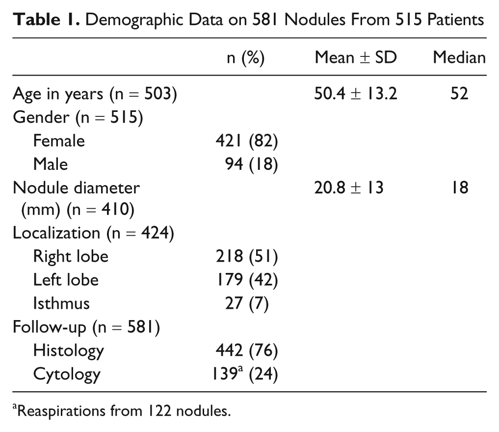
Reaspirations from 122 nodules.
Cytologic Diagnoses According to the In-House and Bethesda Reporting Systems
The overall distribution of cytologic diagnoses using our in-house system was as follows: 67 (11.5%) nondiagnostic, 284 (48.9%) benign, 57 (9.8%) SFC, 55 (9.4%) FL/FN, 38 (6.5%) SFM, and 80 (13.8%) malignant.
We reclassified patients with follicular patterned lesions, and who were in the suspicious categories (SFC, FL/FN, and SFM), using the Bethesda system. Of the 57 diagnoses of SFC, Bethesda diagnosis classified 20 as benign, 25 as AUS/FLUS, 2 as SFN, and 10 as SFM (Figure 1). For the 55 diagnoses of FL/FN, the Bethesda system classified 13 as benign, 6 as AUS/FLUS, 33 as SFN/FN, and 3 as SFM. For the 38 diagnoses of SFM, the Bethesda method classified 2 as benign, 3 as AUS/FLUS, 2 as SFN/FN, 30 as SFM, and 1 as malignant. Thus, the overall distribution of cytologic diagnoses based on the Bethesda classification was as follows: 36 (8.1%) nondiagnostic, 223 (50.5%) benign, 25 (5.7%) AUS/FLUS, 35 (7.9%) SFN, 42 (9.5%) SFM, and 81 (18.3%) malignant (Table 2). Two cytomorphologic examples of cases that displayed changes in diagnostic category for two reporting systems are shown in Figure 2.
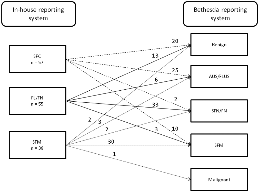
Changes in diagnoses of patients with follicular patterned or suspicious lesions made using our in-house reporting system when the Bethesda criteria were employed (n = 150)
Classification of the 442 Patients for Whom Histological Follow-Up Data Were Available, Based on the Bethesda Reporting System
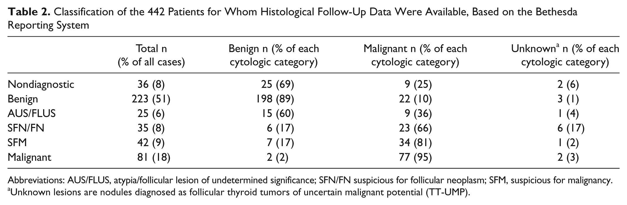
Abbreviations: AUS/FLUS, atypia/follicular lesion of undetermined significance; SFN/FN suspicious for follicular neoplasm; SFM, suspicious for malignancy.
Unknown lesions are nodules diagnosed as follicular thyroid tumors of uncertain malignant potential (TT-UMP).
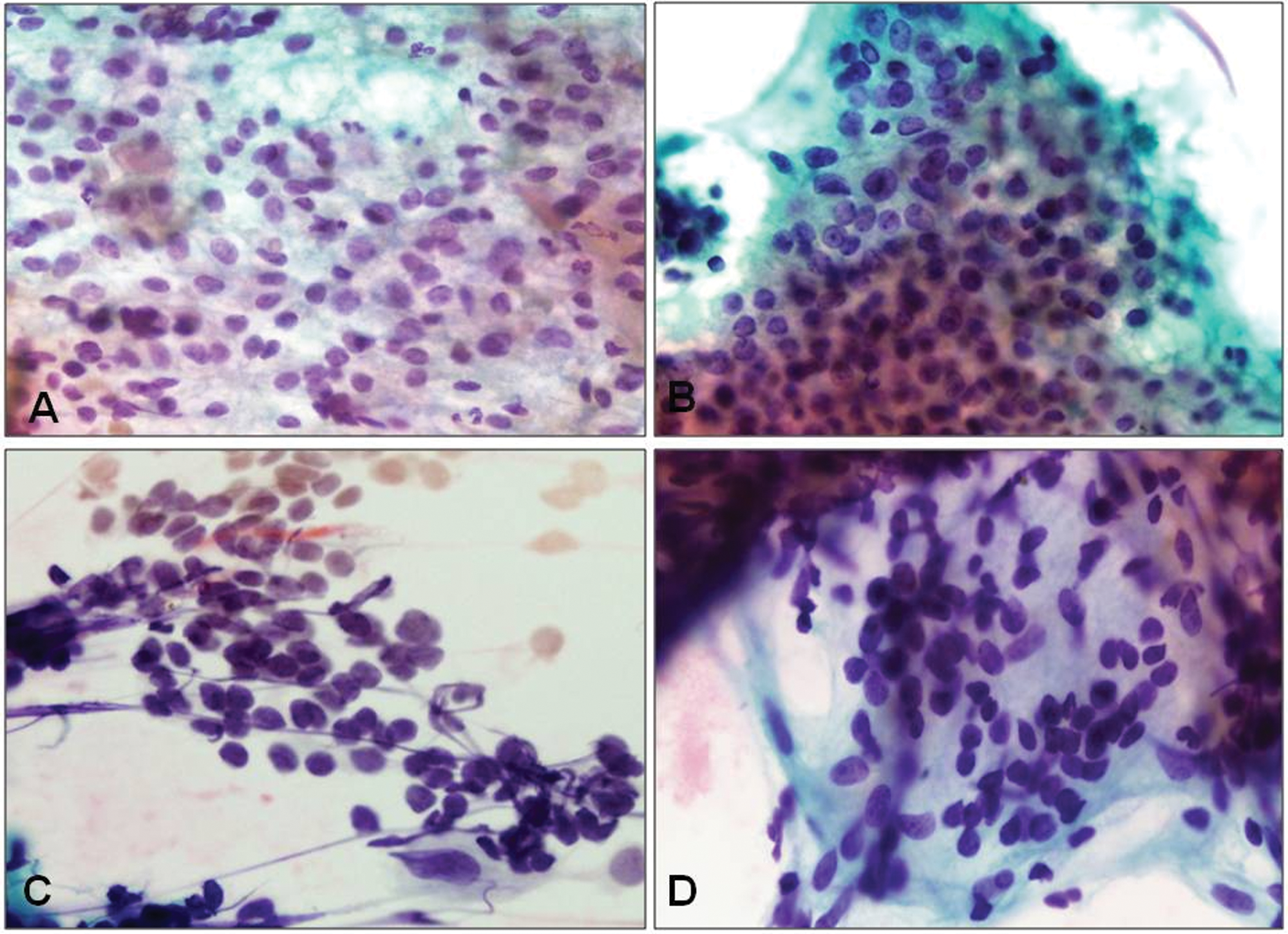
(A and B) This thyroid fine-needle aspiration (FNA) was interpreted as suspicious follicular cells due to hypercelluarity, the presence of cellular enlargement, and mild irregular nuclear contours by the in-house reporting system. However, reevaluation by the Bethesda reporting system resulted in a benign diagnosis. (C and D) The interpretation of this FNA specimen revealed a diagnosis of a follicular lesion/neoplasm by the in-house system. There were loosely cohesive microfollicles formed by enlarged follicular cells. Because of a lack of microfollicle predominance, cellular crowding, and overlapping, criteria that were overinterpreted in initial reading, this case was called as benign by the Bethesda system. Histologic follow-up of both cases displayed a hyperplastic nodule in thyroidectomy (Papanicolau stain 400×)
Histologic Follow-Up
Among the 442 thyroidectomy samples, 253 (57%) were benign and 177 (40%) malignant. However, in 12 patients (3%), histology indicated that the tumor could not be considered either benign or malignant, so the diagnosis was follicular thyroid tumor of uncertain malignant potential (TT-UMP). Of the 253 benign lesions, 72 (29%) were follicular adenoma/adenomatoid nodules, 105 (42%) hyperplastic nodules, 11 (4%) chronic lymphocytic thyroiditis (CLT), 64 (25%) goiter and CLT, and 1 paraganglioma. Of the 177 malignant tumors, 144 (83%) were papillary carcinoma (PTC), 6 (3%) follicular carcinoma (FC), 7 (4%) differentiated thyroid carcinoma not otherwise specified (DTCNOS), 7 (4%) medullary carcinoma (MC), 2 (1%) poorly differentiated carcinoma (PDC), 2 (1%) undifferentiated (anaplastic) thyroid carcinoma (ATC), 5 (3%) metastases, and 1 (1%) lymphoma.
A total of 10 patients underwent at least one repeat aspiration before thyroidectomy. Four of these patients had benign lesions, as shown by all aspiration data and also after thyroidectomy. Cytology classified 5 lesions as higher class and histological follow-up indicated the presence of neoplasms, 4 of which were malignant. One malignant lesion was diagnosed as SFM based on data from 2 aspirations before surgery.
Patients With Only Cytological Follow-Up
Among the 122 nodules that were followed-up by cytology only, 29 were originally classified as nondiagnostic, 80 B, 9 SFC, 2 FL/FN, and 2 SFM. Reclassification revealed 1 B and 9 AUS/AFLUS out of 13 initially suspicious lesions (including SFC, FL/FN, and SFM). Among the 29 nondiagnostic aspirations, 4 (14%) were diagnosed as nondiagnostic, 22 (76%) as benign, 1 (3%) as AUS/AFLUS, and 2 (7%) as SFN/FN in the follow-up aspiration. A total of 80 nodules that appeared benign on examination of initial aspirations were also benign on follow-up aspiration. Nine AUS/FLUS nodules were benign on repeat aspiration. Benign diagnoses on repeat aspiration were seen for 4 patients, 2 of whom had initially been diagnosed with SFN/FN and 2 with SFM.
Correlation Between Cytological and Histological Data
Table 2 shows the distribution of histological follow-up diagnoses according to the Bethesda system for the 442 thyroidectomy patients. The malignancy rates of nodules in various Bethesda categories were as follows: 25% for nodules nondiagnostic, 10% for benign, 36% for AUS/FLUS, 66% for SFN/FN, 81% for SFM, and 95% for malignant (Table 2). In the AUS/FLUS category, when data on patients with only cytologic follow-up were added, the final PPV was 27%.
Table 3 compares the 2 diagnostic schemes in terms of malignancy prediction for patients with follicular patterned lesions, and nodules in suspicious categories, based on histological follow-up data (n = 137). When cytologic diagnostic categories were employed, including the “follicular lesions,” “SFM,” and “malignant” categories of our in-house and Bethesda reporting systems, benign histologic diagnoses were evident in 34 (20%) and 15 patients (9%), respectively (Figure 3). This indicates that the use of the Bethesda system would lead to lesser number of patients who underwent surgery for nonmalignant nodules than would our in-house system.
Distribution of Follicular Patterned and Suspicious Lesions in Patients for Whom Histological Follow-Up Data Were Available (n = 137), Based on Cytologic Diagnoses Using the In-House and Bethesda Reporting Systems
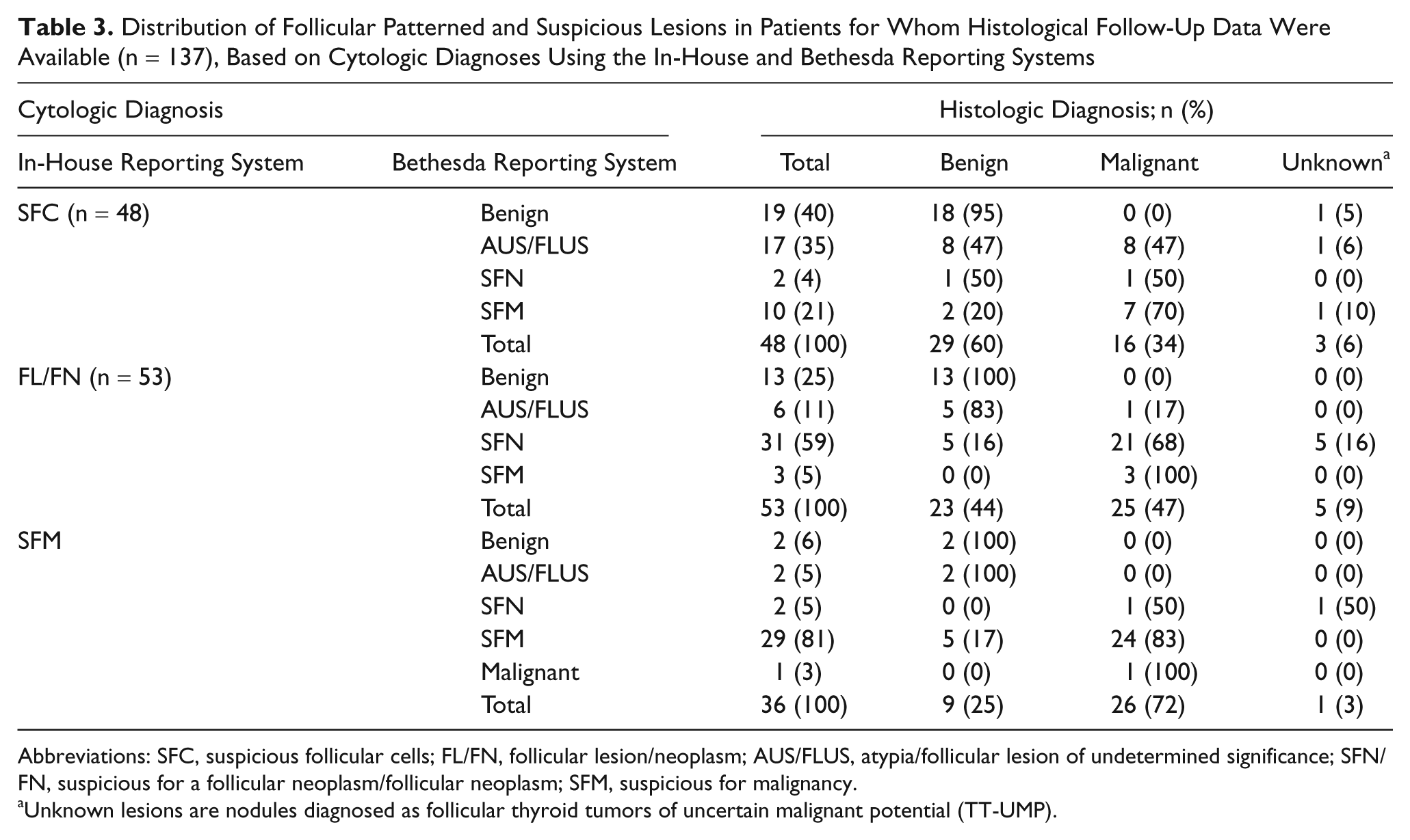
Abbreviations: SFC, suspicious follicular cells; FL/FN, follicular lesion/neoplasm; AUS/FLUS, atypia/follicular lesion of undetermined significance; SFN/FN, suspicious for a follicular neoplasm/follicular neoplasm; SFM, suspicious for malignancy.
Unknown lesions are nodules diagnosed as follicular thyroid tumors of uncertain malignant potential (TT-UMP).
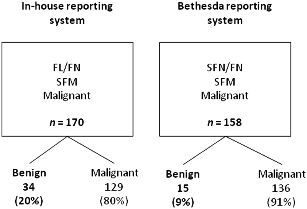
Malignancy rates in the suspicious and malignant cytologic categories diagnosed using both the in-house and Bethesda reporting systems. Note the decrease in the proportion of surgeries for nonmalignant nodules when the Bethesda reporting system is employed (20% vs 9%).
Sensitivity and specificity analyses were performed in 2 ways. First, we included only patients in the benign and malignant categories, as shown by both cytology and histology, and thus did not include those with TT-UMP. In this analysis, the sensitivity of both classification systems was 77% and the specificity 99%. Second, we included FL/FN and SFM patients as positive in both classification systems, and also considered TT-UMP to be histologically positive. In this analysis, the in-house and Bethesda sensitivities were 85% versus 85%; the specificities 87% versus 94%; the PPVs 78% versus 89%; the negative predictive values (NPVs) 91% versus 92%; and the diagnostic accuracies 86% versus 90% (Table 4).
Comparison of In-House and Bethesda Reporting System Diagnoses in the Identification of Thyroid FNAs as Neoplasms a
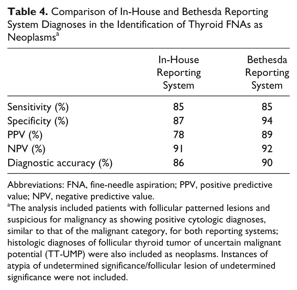
Abbreviations: FNA, fine-needle aspiration; PPV, positive predictive value; NPV, negative predictive value.
The analysis included patients with follicular patterned lesions and suspicious for malignancy as showing positive cytologic diagnoses, similar to that of the malignant category, for both reporting systems; histologic diagnoses of follicular thyroid tumor of uncertain malignant potential (TT-UMP) were also included as neoplasms. Instances of atypia of undetermined significance/follicular lesion of undetermined significance were not included.
Evaluation of Discrepancies
Of the 35 nodules for which cytologic findings and histological follow-up data showed discrepancies, 22 were cytologically benign but histologically malignant, 16 of which were identified as PTC, 2 as FC, 1 as DTCNOS, 2 as MC, and 1 as PDC in a papillary carcinoma background (Figure 4A). An additional three instances of benign cytology were diagnosed as TT-UMP on histological follow-up. A review of these data indicated that material was inadequate in 8 instances, very focal cellular atypia was evident in 7, technical artifacts were apparent in 2, and interpretation errors were evident in 5 instances.
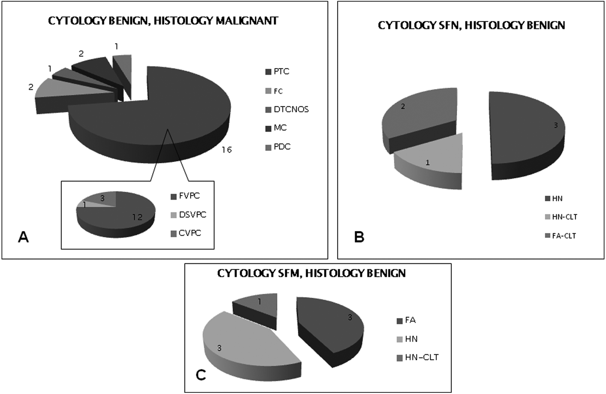
Distribution of the histologic diagnoses of patients showing discrepant features, according to the Bethesda terminology
Overall, 24 cases (15 with histologic, 9 with cytologic follow-up) out of 34 in AUS/FLUS category were detected to be benign and among 10 cases all with histologic follow-up 9 were malignant and 1 was TT-UMP. Hypocellularity was the case in 46% (11/24) of nonneoplastic and 100% (10/10) of neoplastic nodules. The remaining benign nodules displayed excessive blood and/or technical artifacts and/or mild cytologic atypia in a very few cells.
Six nodules in the SFN/FN group were classified as benign based on data obtained after thyroidectomy; 3 had CLT in the thyroid tissue and 3 showed the dominant nodules of multinodular goiter (Figure 4B). The cytological findings that resulted in SFN/FN diagnoses in these instances were high cellularity; predominance of a microfollicular pattern; and the presence of scant, dark, thick colloid. We also found that Hurthle cell differentiation sometimes accompanied CLT.
Seven nodules in the SFM category were benign, based on data obtained after thyroidectomy; 3 were hyperplastic; 1 was a CLT; and 3 were FA (Figure 4C). A review of the microscopy data from these post-thyroidectomy samples indicated that some nodules featured hyalinization, calcification, and intracystic papillary hyperplasia. Two nodules in the malignant category had benign lesions on follow-up. The reactive changes in the nodules were the same as seen in the instances of discrepancy within the SFN category.
Patient Age, Nodule Size, and Malignancy
Analysis of the relationship between patient age and malignancy indicated that malignancy rates were greater in patients younger than 40 years (P < .001; Table 5). The mean ages of patients with benign and malignant nodules were 52.4 ± 11 and 47.9 ± 15 years, respectively (P < .001). The mean diameter of benign and malignant nodules was 23.3 ± 13.2 and 18.9 ± 13 mm, respectively (P > .05). There was no significant difference in diameter between the diagnostic categories of the Bethesda system, and no significant difference in malignancy rate was evident between men and women (57% and 60%, respectively; P > .05).
Age and Nodule Size in Benign and Malignant Lesions
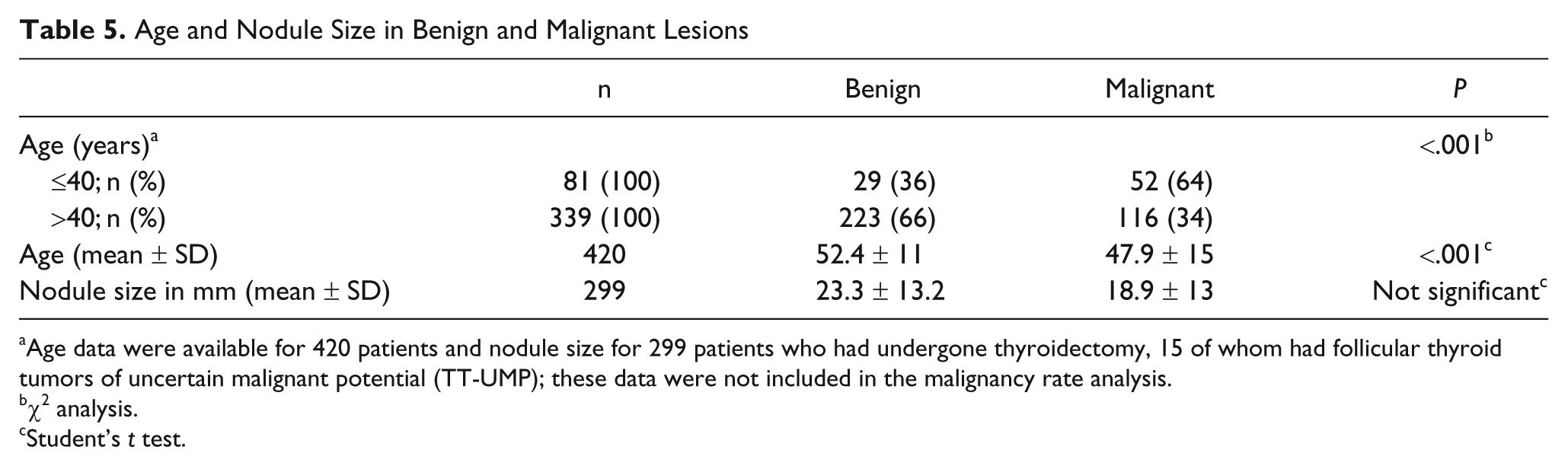
Age data were available for 420 patients and nodule size for 299 patients who had undergone thyroidectomy, 15 of whom had follicular thyroid tumors of uncertain malignant potential (TT-UMP); these data were not included in the malignancy rate analysis.
χ2 analysis.
Student’s t test.
Discussion
Our results indicate that the new Bethesda reporting system for suspected thyroid cancer, based on thyroid FNA cytology, is reliable and offers high sensitivity and specificity. A comparison between the Bethesda method and our in-house reporting system (used before the Bethesda system was promulgated) indicated that the diagnostic categories employed were similar but with few differences. The Bethesda system has 6 categories: nondiagnostic, benign, atypia/follicular lesion of undetermined significance (AUS/FLUS), suspicious for follicular neoplasm/follicular neoplasm (SFN/FN), suspicious for malignancy (SFM), and malignant.8,9 Our system had similar groupings, but some overlap of diagnoses within the AUS/FLUS, SFN/FN, and SFM categories was evident. Our data showed that the malignancy rates of nodules in each of the 6 Bethesda categories were 25%, 10%, 36%, 66%, 81%, and 95%, respectively. Cytological and histological analysis indicated that the Bethesda system had a sensitivity of 85% and a specificity of 94%, thus better than offered by our earlier system. The diagnostic accuracy afforded by the Bethesda criteria was higher (90% vs 86%), and surgeries for nonmalignant nodules were less (20% vs 9%), compared with use of our earlier system.
We found that patients younger than 40 years had higher rates of malignancy than did those older than 40 years. In contrast, several previous studies found a significant increase in the incidence of malignancy with age,15,16 and others reported that age did not affect the frequency of malignancy.10,17 However, some studies of follicular neoplasms found that patients younger than 40 years were more likely to have malignancies than were older patients.12,18 Although we found that malignancy was more common in younger patients, we could discern no effect of age on follicular neoplasm development. Nodule diameter is another possible indicator of malignancy. In our study, as in several previous works,12,17,18 no significant effect of nodule diameter on malignancy was evident.
Diagnostic terminology and morphologic criteria for thyroid FNA have not previously been described in detail, although various systems for classification of thyroid FNA have been reported.3-5 In our institution, we used 6 categories of thyroid FNA diagnosis: nondiagnostic, benign, suspicious follicular cells (SFC), follicular lesion (FL; with or without Hurthle cell differentiation), suspicious for malignancy (SFM), and malignant. The SFC criterion encompassed lesions with various morphologic features, including focal cellular atypia, reactive cells with prominent nucleoli, presence of 3-dimensional groups and/or microfollicles, and with evidence of moderate-to-high cellular material. Focal mild nuclear changes of PTC, without pseudo-inclusions, were included within this category. Classifications of “follicular lesion” or “neoplasm” were used for lesions with greater numbers of microfollicles, presence of remarkable discohesive cells, and scant and/or dense colloid. SFM was generally employed if PTC was suspected, as in the Bethesda classification.
Following promulgation of the Bethesda system, 9 reevaluation of our data indicated some changes in diagnostic category distribution. In the suspicious group of our in-house system (SFC), 40% of nodules were reclassified as benign, 4% as SFN, and 21% as SFM, using the Bethesda system. Similar changes were evident in diagnostic categories within the SFN/FN group. Although most FL/FN lesions remained in the same category, 25% were identified as benign. We showed that use of the Bethesda system led to a sharp distinction between lesions based on cytomorphology. The emphasis of the system on the predominance and detailed description of microfollicles within the SFN/FN category led us to classify 40% of our original SFC nodules and 25% of our FL/FN nodules as benign; histological follow-up confirmed that most were indeed benign. Pathologists disagree substantially on the significance of microfollicles, 19 and this may lead to differences in final diagnoses because the presence of microfollicles is the main criterion of the SFN/FN category. It has been suggested that the term suspicious be used only if malignancy is suspected. This is why the diagnoses of 10 of our nodules were changed from SFC to SFM.
The overall proportions of our patients in the 6 categories of the Bethesda system were 8% for nondiagnostic, 50% benign, 6% AUS/FLUS, 8% SFN/FN, 9% SFM, and 18% malignant. This distribution is similar to those previously reported in the literature.1,20-22 The higher proportions of patients in the SFM and malignant categories in the present study may be attributable to use of a highly selective population, which included only patients for whom cytological or histological follow-up data were available, and/or to the geographic uniqueness of our population. Using the Bethesda system, it is recommended that the frequency of patients in the AUS/FLUS category should be approximately 7%, almost the same as that of our study population. 9 False-negatives and false-positives are common when diagnostic tests, such as thyroid FNA, are employed.20,23 Our false-negative and false-positive rates using the Bethesda system were 5.0% and 0.4%, respectively.
Each category is assigned a risk of malignancy based on NCI terminology. These malignancy rates are less than 1% for benign, 5% to 10% for AUS/FLUS, 20% to 30% for SFN, 50% to 75% for SFM, and 100% for malignant.8,9 The malignancy rates in each category are higher than those previously reported in the literature, especially for lesions previously considered indeterminate. In the current study, the malignancy rates were 36% for AUS/FLUS, 66% for SFN, and 81% for SFM. When we included cytological follow-up data on AUS/FLUS lesions, the malignancy risk of this category of patients decreased to 27%. The most striking malignancy rate in our population was in patients in the AUS/FLUS category; the rate was much higher than expected.8,20,22,24 However, some previous reports have also found higher malignancy rates in patients in this category.1,25-27 We believe that the higher incidence of malignancy observed by us is attributable principally to our study design. We included only patients for whom cytological or histological follow-up data were available, and we made an additional effort to classify patients into definite diagnostic categories. The major indication for surgery in our study population was abnormal clinical findings combined with a high suspicion of malignancy, based on physical examination and imaging studies.
Thyroid tumors of uncertain malignant potential (TT-UMP) are nodules with nuclear features similar to those of PTC or capsular invasion, but which cannot ultimately be classified as FVPTC or FC.28,29 Although the TT-UMP category is generally not accepted, a review of pathology reports on thyroidectomies led us to classify 3.4% of our patients as suffering from TT-UMP. A previous study found a similar percentage (2.9%) of TT-UMP patients. 30 Because the work is outside the scope of the present study, we did not review slides of these patients and we consider it reasonable to not include such nodules in estimation of the thyroid FNA malignancy rate. However, we did include TT-UMP patient data when we tested the ability of thyroid FNA to identify neoplasms.
A previous study from our institution reported similar sensitivity and specificity rates to those described above (76% and 86%, respectively) for thyroid FNA detection, using our in-house reporting system, when a selected group of patients with multinodular goiter were examined. 31 In the current study, we compared our in-house system with the Bethesda criteria. We found a remarkable decrease in the number of patients undergoing surgery because of nonmalignant nodules (20% vs 9%) when the Bethesda system was employed. Furthermore, the Bethesda criteria offer a higher specificity and PPV than does our earlier system (83% vs 93% and 80% vs 90%, respectively). Thus, the Bethesda system successfully creates well-defined categories predicated on evidence-based management guidelines.
We performed a detailed analysis of instances where discrepancies between cytological and histological data were evident. On all, 22 cytologically benign cases, nearly 70% of which displayed sampling bias, had malignant tumors on thyroidectomy. The histological subtype distribution of these tumors indicated that most were FVPTC, but 3 were TT-UMP. This is comparable to information previously reported. 32 Furthermore, various studies have found that FVPTC was the most common malignancy in nodules cytologically identified with follicular neoplasms.16,33,34 Thyroid pathology studies have also found that inter- and intraobserver reproducibility in thyroid follicular lesion diagnosis varies even among experts.35,36 Thus, it was not surprising that discordance was evident between cytological and histological data on patients with follicular patterned lesions (FVPTC, FA, and FC). In the present study, we show that the discrepancy in the SFN category was mostly attributable to the presence of highly cellular smears, the predominance of microfollicles and dense colloid, and the presence of CLT in the background. Nodules diagnosed as benign based on examination after thyroidectomy, but as SFM or malignant on the basis of previous cytology, showed hyalinization, calcification, and intracystic papillary hyperplasia, which may together result in reactive atypical nuclear changes that could lead to cytological misinterpretations.
However, some problems remain with the Bethesda system. First, the AUS/FLUS category includes a heterogeneous group of abnormalities, ranging from reactive changes to malignancies. This category may become a “trash can” for difficult-to-classify thyroid FNAs. The NCI suggests that care must be taken in assigning patients to this category. The use of AUS/FLUS category varied widely between institutions and no specific cytomorphologic criteria have been described. 24 However, it was shown that elimination of AUS/FLUS category resulted in a decrease in sensitivity of thyroid FNA and also in an increase in rates of false-negative and false-positive cases. 25 Although there are no well-defined cytomorphologic criteria in AUS/FLUS category the cytologic factors contributing this category were described as sparse cellularity, presence of blood obscuring cytomorphologic features. 9 We also demonstrated that some of the cases in this category had malignancies in their follow-up and most of those showed hypocellularity rather than clotting. Similar to the other studies, we recommend the use of AUS/FLUS terminology.8,37,38 We conclude that it might be speculated the cases falling into this category because of hypocellularity should be considered more suspicious than the ones with excessive blood obscuring cytomorphologic features. It is important to use the same terminology and standard follow-up for AUS/FLUS category in triaging patients, since it has been shown that reaspirations from these patients resulted in benign diagnoses.1,39 Second, it is inevitable that benign nodules displaying hypercellularity and predominance of microfollicular pattern, will be placed in the SFN/FN category, because diagnosis of follicular carcinoma requires the identification of capsular or vascular invasion. Third, further studies are needed to test the reproducibility of the Bethesda system when used to interpret thyroid FNA cytology. Evaluation of inter- and intraobserver reproducibility, using the Bethesda system, was not an aim of the present study.
Although our current study was multidisciplinary, several limitations are apparent. First, the work was retrospective and included data collected before the Bethesda system was promulgated (in 2007); NCI management guidelines were thus not employed. Second, we did not include data on patients for whom cytological or histological follow-up data were not available. Thus, it is difficult to assess the overall incidence of the various thyroid FNA categories, and thyroid malignancy rates, in our local population.
In conclusion, we showed that FNA is most accurate when used to triage patients with thyroid nodules and that the Bethesda reporting system for thyroid FNA has high sensitivity, high specificity, and a high PPV. AUS/FLUS category, which is the most problematic one in Bethesda reporting system, can be separated into subgroups according to the factors causing difficulties in the interpretation of cytomorphologic features. There is a need of accumulation of cases in this category to do further evaluations and to make comments.
Footnotes
The authors declared no potential conflicts of interest with respect to the research, authorship, and/or publication of this article.
The authors received no financial support for the research, authorship, and/or publication of this article.