






Abstract
Multiple vascular patterns are presented in tumors of the central nervous system (CNS), including microvascular hyperplasia, branching capillaries, numerous capillaries without definite pattern, hyalinized vessels, and angiomatous area. These vascular patterns play important roles in pathological diagnosis of brain tumors. Because of insufficient recognition of the significance of the various vascular patterns, only a few of them have been applied in pathological diagnosis, leading to missed diagnosis and diagnostic errors. Microvascular hyperplasia can present in multiple brain tumors but display different diagnostic values. Otherwise, varied brain tumors characterized by branching capillaries or vascular pattern mimicking branching capillaries should be given careful consideration. Therefore, a familiarity of these tumors and their vascular pattern is essential for general pathologists. This study reviews the value of various kinds of vascular patterns for pathological diagnosis of brain tumors, constructs a framework for better understanding, and provides a novel perspective for general pathologists.
Multiple vascular patterns are presented in tumors of the central nervous system (CNS), including microvascular hyperplasia, branching capillaries, numerous capillaries without definite pattern, hyalinized vessels, and angiomatous area, which play important roles in diagnosis. Because of insufficient recognition of the features of the various vascular patterns, only a few of them have been applied in pathological diagnosis. General pathologists are familiar with the value of microvascular hyperplasia for pathological grading in astrocytoma. Nevertheless, microvascular hyperplasia is not all always an anaplastic feature in other brain tumors. As microvascular proliferation is the inherent characteristic of pilocytic astrocytoma and is insensitive for prognosis evaluation, ignoring the feature will lead to misdiagnosis and overtreatment. Branching capillaries are the characteristic of oligodendroglioma and central neurocytoma, but many other tumors, including clear cell ependymoma, clear cell meningioma, cellular hemangioblastma, and metastatic clear cell renal carcinoma, also present vascular architecture mimicking branching capillaries. Familiarity with the histological and immunohistochemical features of those tumors will help reduce diagnostic errors. Here we construct a framework for better understanding of the significance of vascular pattern in the diagnosis of CNS tumors for practical diagnostic application by the general pathologist.
Microvascular Proliferation
Microvascular proliferation does not imply an increased number of blood vessels but takes on a specific meaning referring to the morphologic finding of tufted aggregates of proliferating endothelial cells and pericytes/smooth muscle cells.1,2 The most exaggerated form of microvascular hyperplasia is called glomeruloid body. According to the value of microvascular hyperplasia for pathological grading, CNS neoplasms can be categorized into the following groups: (a) World Health Organization (WHO) grade III or IV tumors—glioblastoma, anaplastic oligodendroglioma, anaplastic oligoastrocytoma, and anaplastic ependymoma; (b) WHO grade I tumors—pilocytic astrocytoma and dysembryoplastic neuroepithelial tumor (DNT); and (c) central neurocytomas with “anaplastic” histological features.
WHO Grade III or IV Tumors
Infiltrative astrocytic neoplasms are by far the most common malignant brain tumors in adults. Along with more biological progression in astrocytic neoplasms, the significance of angiogenic dysregulation has also become increasingly apparent. Low-grade diffusely infiltrating astrocytomas (WHO grade II) have a vessel density that is comparable to or only slightly greater than adjacent normal brain (Figures 1A and B). 1 As neoplasms progress from diffusely infiltrating astrocytomas to anaplastic astrocytoma (WHO grade III), cell density, nuclear anaplasia, and cellular proliferation increase; however, changes in vascular density are only mild (Figure 1C), and areas of necrosis are not present.1-3 The most dramatic changes are seen when anaplastic astrocytomas progress to glioblastoma (WHO grade IV), which often have extensive and extreme levels of microvascular hyperplasia (Figure 1D).1-4

(A) Hematoxylin and eosin (H&E) staining (100×) of normal brain tissue. (B) H&E staining (100×) showing that no changes in vascular architecture and vessel density are found between normal brain and infiltrating astrocytoma (WHO grade II). (C) H&E staining (100×) showing that in anaplastic astrocytoma (WHO grade III) vessels are more numerous than in infiltrating astrocytoma. (D and E) H&E staining (100×) showing that in glioblastoma (WHO grade IV), microvascular hyperplasia, including tufted and glomeruloid vascular proliferation, are seen. (F) H&E staining (100×) of pilocytic astrocytoma. Microvascular hyperplasia is often noted in pilocytic astrocytoma. However, this feature is not associated with more aggressive biologic behavior
Oligodendroglioma (OD) is the third most common tumor of glial origin. Of these, roughly 70% are low-grade OD (WHO grade II), whereas 30% are anaplastic OD (WHO grade III). 5 Anaplastic OD is defined as an oligodendroglioma with focal or diffuse histological features of malignancy, including mitotic figures, high cellular density, irregular cells, and microvascular hyperplasia.5-8 In addition, microvascular proliferation may be present in anaplastic oligoastrocytoma. 1
Histopathologically, ependymal tumors comprise low-grade tumors, that is, ependymoma (WHO grade II), myxopapillary ependymoma (WHO grade I), and subependymoma (WHO grade I), and high-grade tumor, that is, anaplastic (malignant) ependymoma (WHO grade III). 1 Anaplastic ependymoma, as defined by WHO, is an ependymal tumor with histological evidence of anaplasia, for example, high cellularity, variable nuclear atypia, marked mitotic activity, and often prominent microvascular hyperplasia.9-11 Ho et al 12 proposed that anaplastic ependymoma could be diagnosed by the presence of any 2 of the following 4 parameters: mitoses ≥4/10 high-power field, hypercellularity, endothelial proliferation, and necrosis. However, optimal histologic criteria for the grading of ependymomas and their correlation with the clinical behavior are still an open issue. 13
From the above discussion, it can be concluded that microvascular hyperplasia is the malignant histological feature of astrocytoma, ependymoma, oligodendroglioma, and oligoastrocytoma. Hence it should be evaluated in the grading of these tumors. Besides, glomeruloid proliferations similar to those observed in primary high-grade neoplasms are frequently observed in the brain adjacent to metastases. 1
WHO Grade I Tumors
Pilocytic astrocytoma is the most common pediatric CNS glial neoplasm. Histopathologically, the tumors are highly vascular and frequently have markedly glomeruloid vessels (Figure 1E), which mimic some of the features of glioblastoma.1,14 However, pilocytic astrocytomas have a noteworthy benign biologic behavior corresponding to WHO grade I. In pathological diagnosis, a major difference between pilocytic astrocytoma and fibrillary astrocytoma is the microvascular hyperplasia, highly vascularized stroma, and great propensity for cyst formation in the former. A recent report found that pilocytic astrocytomas showed a different vessel architecture compared with glioblastoma: Pilocytic astrocytoma had fewer but wider microvascular hyperplasia compared with glioblastoma and turnover of endothelial and tumor cells were relatively lower in pilocytic astrocytoma. 15 Very rarely, a pilocytic astrocytoma may undergo malignant transformation with an aggressive histologic appearance, including hypercellularity, cellular atypia; multiple mitoses per high-power microscopic field, and necrosis with pseudopalisading, such a tumor is called an anaplastic (malignant) pilocytic astrocytoma.1,16,17
Dysembryoplastic neuroepithelial tumor is a rare low-grade mixed neuronal and glial tumor, usually seen in young adults and associated with pharmacologically intractable, complex partial or generalized seizures. The favored locations of this tumor are the temporal or frontal lobes. 18 Microscopically, DNT is characterized by multinodular proliferation of oligodendrocyte-like cells that appear to be arranged along bundled axons and capillaries (often perpendicular to cortical surface) and are separated by a myxoid matrix in which well-differentiated neurons seem to float.1,18,20 Sometimes DNTs show microvascular proliferations, but this feature is not associated with more aggressive biologic behavior.17,21
Compared with tumors in group 1, microvascular proliferation is the inherent characteristic of pilocytic astrocytoma and DNT, and it is insensitive for prognosis evaluation.
Central Neurocytomas With “Anaplastic” Histological Features
Although central neurocytomas are generally considered to be benign tumors (WHO grade II), some cases with “anaplastic” histological features such as cellular pleomorphism, mitotic activity, microvascular proliferation, and necrosis have been reported. However, it seems that histological atypia alone is not a useful predictor of clinical outcome.1,22-24 The correlation between histology and biologic behavior is far from clear.
The pattern of vascular proliferation is also present in nonneoplastic lesion, for instance, and this should not be ignored. For example, glomeruloid microvascular proliferation is a common event secondary to infarction, which may suggest a malignant glioma to the inexperienced pathologist.
Branching Capillaries
A delicate branching network of capillaries producing a “chicken-wire” pattern indicates a series of CNS tumors, including oligodendroglioma (OD), central neurocytoma (CNC), and extraventricular central neurocytoma (ENC). In addition, clear cell ependymoma (CCE), clear cell meningioma (CCM), cellular hemangioblastma (HB), and metastatic clear cell renal carcinoma (ccRCC). These tumors frequently present branching vessels that separate tumor cells into nests or clusters. Interestingly, morphologic observation of cells in tumors mentioned above usually reveals distinctive manifestations: small, round, with slightly enlarged round nuclei, a small amount of eosinophilic cytoplasm, and notable perinuclear halo. Thus, the morphology is described as “fried egg appearance” or oligodendrocyte-like cells (OLCs). Because of similar vascular architecture and monotonous population of OLCs, these tumors are often confused. The summation of these features generates a “honeycomb” pattern, which makes the diagnosis confusing. Here we recommend a framework to distinguish these tumors based on favored locations, histological features, and a panel of immunohistochemical markers (Table 1), which are discussed one by one in the following.
Differential Diagnosis of Highly Vascular Tumor of CNS
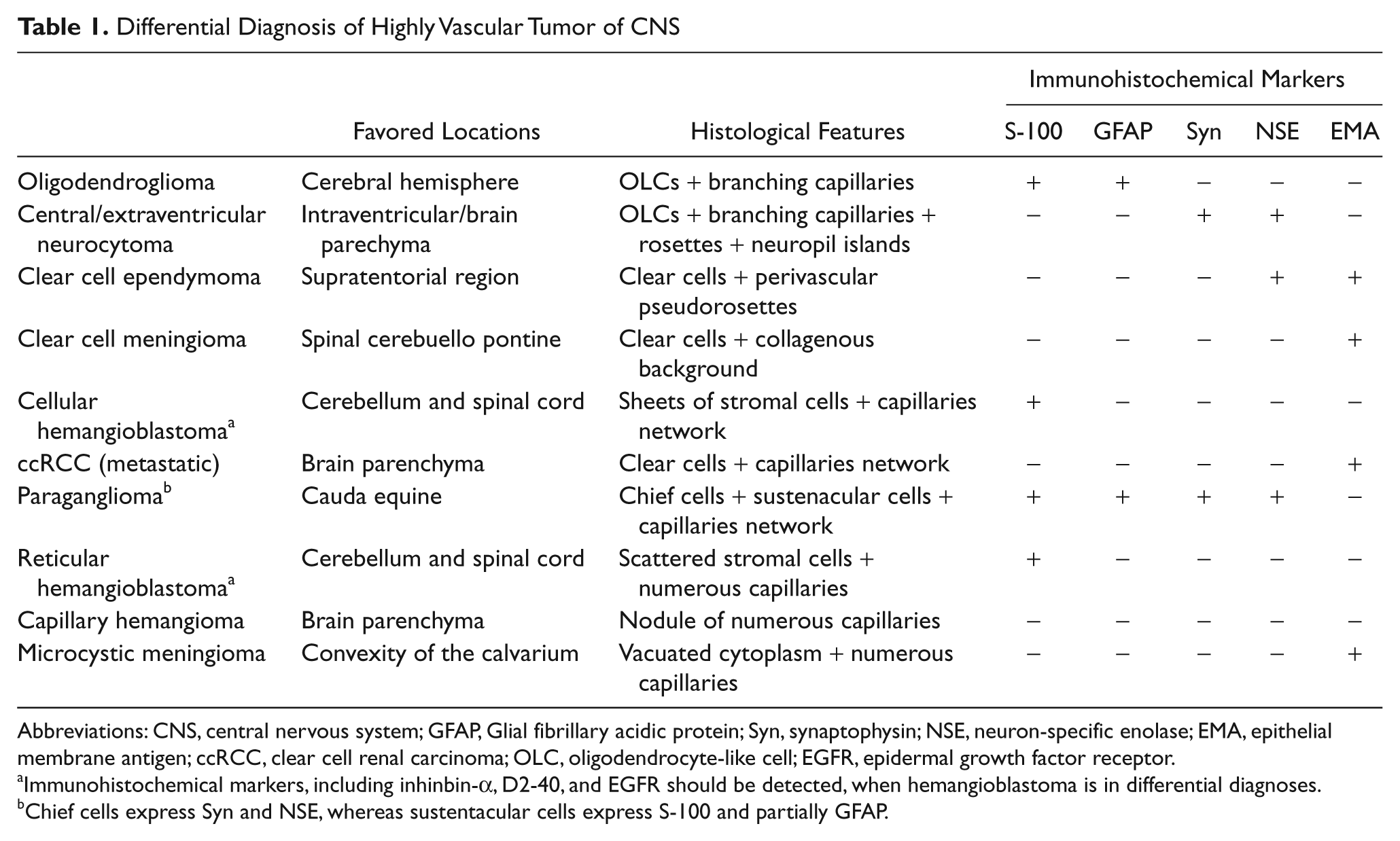
Abbreviations: CNS, central nervous system; GFAP, Glial fibrillary acidic protein; Syn, synaptophysin; NSE, neuron-specific enolase; EMA, epithelial membrane antigen; ccRCC, clear cell renal carcinoma; OLC, oligodendrocyte-like cell; EGFR, epidermal growth factor receptor.
Immunohistochemical markers, including inhinbin-α, D2-40, and EGFR should be detected, when hemangioblastoma is in differential diagnoses.
Chief cells express Syn and NSE, whereas sustentacular cells express S-100 and partially GFAP.
(a) OD is one of the most common glial tumors and often arises in the cerebral hemisphere, especially frontal lobe. It can be diagnosed at any age with peak incidence in 30- to 40-year-olds. 1 The tumor mass is typically located in the white matter, but extension into the cerebral cortex is common.25,26 Microscopically, ODs present pure honeycomb appearance because of dense network of branching capillaries and OLCs (Figure 2A). Microcalcifications in the tumor tissue and/or the surrounding brain are common. ODs are consistently positive for S-100 protein (S-100), oligodendrocyte transcription factor 2 (Olig-2), and microtubule-associated protein-2 (MAP2), whereas they are negative for epithelial membrane antigen (EMA), synaptophysin (Syn), and neuron-specific enolase (NSE).25,27 GFAP-positive minigemistocytes and so-called gliofibrillary oligodendrocytes are frequently found. 1 (b) CNC is a well-differentiated neuronal neoplasm and is characterized by its intraventricular location. The average age of patients at diagnosis is about 28 years.22,23 The histopathologic appearance of CNC can be similar to that of ODs, including small uniform cells with rounded nuclei and scant cytoplasm resembling perinuclear halos (“fried egg” appearance) and delicate branching capillaries. However, alternating fibrillary and cellular areas and a tendency to form ill-defined rosettes in the latter are features different from ODs. Immunoreactivity against NSE and Syn confirms the neuronal nature of the neoplasm. GFAP staining focally can be found in CNC. It may derive from bipotential precursor cells of the periventricular germinal matrix, which are capable of both neuronal and glial differentiation. 23 (c) ENC is a new entity in the 2007 WHO Classification of Tumors (WHO grade II), which are defined as neurocytic neoplasms occurring within brain parenchyma without any apparent association with the ventricular system. 1 ENCs share key histological and immunohistochemical features with the more common CNCs but exhibit a wider morphologic spectrum. The incidence of ganglion cell differentiation is more than 60% in ENC, whereas it is much lower in CNC.28,29 (d) CCE is an uncommon CNS tumor with a predilection for the supratentorial region in children, with only occasional reports describing alternate sites of origin, including cervical spinal cord, brain surface, and cerebellum.30-33 Histologically, CCE is characterized by sheets of cells with rounded nuclei and prominent, clear, and perinuclear halos (Figure 2B). This histological appearance may closely mimic ODs, CNCs, HBs, and ccRCC; however, all CCEs harbor at least focal perivascular pseudorosettes, which help establish the correct diagnosis.30-33 Nevertheless, true ependymal canals and rosettes were mostly absent. Immunohistochemically, the tumor cells showed positivity for GFAP and EMA. Clues for the pathological diagnosis include the following: Unlike ODs, which are diffusely infiltrative, CCEs generally are circumscribed sharply. Although HBs have discrete borders and may contain GFAP positive cells, they also contain abundant reticulin, and their lipidized stromal cells are multivacuolated, features not present in CCE. Furthermore, unlike CCE, HBs typically are negative for EMA. Immunohistochemistry is helpful in identifying CNCs/ENCs, which, unlike ependymal tumors, are positive for Syn. (e) CCM is one of the rarest histologic forms of meningiomas. CCMs are usually seen in younger patients and have a predilection for spinal or cerebuello pontine locations, while intracranial location has also been reported.34-36 CCM are potentially aggressive and placed in the category of WHO grade II. 1 The imaging features of CCM are not different from conventional meningiomas. Histologically, CCM appears as a solid cellular neoplasm mostly composed of clear glycogen rich cells with a collagenous background; sometimes rich branching vessels are presented (Figure 2C). Occasional whorl formation may be seen; however, psammoma bodies are not typical of this variant.34,35 The tumor cells were immunopositive for EMA and vimentin but negative for S-100, GFAP, and Syn. (f) Cellular HB: HB is a slowly growing, highly vascular tumor of adults, occurring in the cerebellum, brain stem of spinal cord. HBs are characterized histologically by 2 main components—stromal cells and abundant vascular cells. Cellular and reticular variants are distinguished on the basis of the abundance of the stromal cell component. 1 Cellular HBs are characterized by a sheet of stromal cells and abundant of delicate branching network of capillaries, and usually misdiagnosed as CCE.32,33 Interestingly, stromal cells are always large and vacuolated but in the cellular HB stromal cells are usually with ground-glass cytoplasm, resembling OLC to a certain extent (Figure 2D). The stromal cells are immunopositive for S-100, Inhibin-α, and D2-40 but negative for EMA and GFAP. (g) Metastatic ccRCC: Particular difficulty was presented in distinguishing between a primary “clear cell” tumor in CNS and a metastasis from ccRCC. No valid distinction could be made on histological appearance. Immunohistochemical studies may be helpful in this setting. In most of the cases, the ccRCC showed immunopositivity for EMA and CD10, and immunonegativity for S-100, Inhibin-α, and D2-40.37-39

Various kinds of brain tumors with oligodendrocyte-like cells (OLCs) and branching capillaries (or abundant vessels mimicking branching capillaries)
Paraganglioma, which is densely vascularized, should also be distinguished from tumors mentioned above. In the CNS, paragangliomas are almost exclusively located in the cauda equine and correspond to WHO grade I. It generally occurs in adults, with a slight male predominance.1,40 Praganglioma is a well-differentiated neuroendocrine neoplasm, composed of chief cells arranged in nests or lobules (zellballen) and surrounded by a single layer of sustenacular cells. Imunohistochemical stainings confirm the neuroendocrine nature of this tumor by demonstrating strong expression of Chromogranin (CgA) and Syn in the chief cells. The sustentacular cells interact with antibodies against S-100 and partially GFAP.1,40
In addition, DNT and pilocytic astrocytoma should not be overlooked in the differential diagnosis. OLCs could be observed in all tumors mentioned above, but branching network of capillaries is absent in DNT and pilocytic astrocytoma. 41 In pilocytic astrocytoma, OLCs are positive for GFAP. In the cortical DNT component, most OLCs are strongly reactive for S-100 protein and a few are positive for GFAP but show no immunoreactivity for neuronal markers.
Numerous Capillaries Without Definite Pattern
Another vascular architecture characterized by numerous small thin-walled vessels without definite pattern also appears in CNS tumors, and typical entities include reticular HB, capillary hemangioma (CH), and microcystic meninioma. (a) Reticular HB is more common than its cellular variant. It is characterized by abundant capillaries and scattered large and vacuolated stromal cells (Figure 3A). Evidence of foamy stromal cells, which are immunopositive for S-100, Inhibin-α, and D2-40, is the key point for differential diagnosis. (b) CHs rarely occur in the brain or spinal cord.1,42-44 Histological examination of the CNS CH revealed a lobular architecture with lobules that were separated by fibrous tissue septa and fed by thick-walled arteries. The lobules were composed of numerous, tightly packed, capillary-sized vessels that were lined by a single layer of cytologically benign endothelial cells. Blood vessels varied widely in size from small lumina lined with plump endothelial cells to dilated vessels lined with flattened endothelium. (c) Microcystic meningioma is a distinct morphological variant of meningioma and shows benign character corresponding to WHO grade I. Microscopically, it is characterized by loose texture and microcysts with formation of large extracellular spaces containing edematous fluid (Figure 3B), occasionally with typical meningiomatous whorls.45,46 The tumor cells have stellate and vacuolated cytoplasm with long cytoplasmic processes and present diffuse immunoreactivity for vimentin, EMA, and S-100, but negative for cytokeratin and GFAP.

(A) H&E staining (200×) of reticular hemangioblastoma showing numerous capillaries and scattered stromal cells. (B) H&E staining (200×) of microcystic meningioma showing multiple extracellular and intercellular cystic spaces, scattered pleomorphic nuclei, and numerous capillaries. (C) H&E staining (100×) of extraventricular neurocytoma showing marked hyalinized vessels. (D) H&E staining (100×) of papillary glioneuronal tumor showing thickened vessels lined by single layer of astrocytes
In the 10 different kinds of highly vascularized tumors in CNS mentioned above, different diagnosis seems difficult only according to the morphology. Therefore, many factors, such as favored locations, histological features, and a panel of immunohistochemical markers, should be taken into account comprehensively during diagnosis (Table 1).
Hyalinized Vessels
Abundant vessels exhibited marked thickening and hyalinization with obliteration of the lumina is another feature of vessels in CNS tumors. Some low-grade CNS tumors, such as papillary glioneuronal tumour (PGNT), ependenoma, CNC/ENC (Figure 3C), CCM, and pilocytic astrocytoma, frequently have marked hyalinized vessels.
PGNT is a new entity included in the WHO classification that typically corresponds to WHO grade I. 1 It manifests over a wide age range (mean 27 years) and preferentially locates the temporal lobe. Microscopically, it is a biphasic tumor with pseudopapillary configuration and focal solid pattern: (a) The cores of the pseudopapillae show thickened vessels lined by single as well as stratified layers of GFAP-positive astrocytes (Figure 3D). (b) The solid component of the tumor is formed by sheets of Syn-positive neurocytes, large neurons, and intermediate size “ganglioid” cells.1,47-49 Gliofibrillary background in the intervening areas shows GFAP-positive staining.
Astroblastoma is another rare tumor of uncertain histopathological origin and unpredictable clinical behavior, which occurs mainly among children or young adults. Astroblastomas may be found throughout the CNS, with the cerebral hemispheres being the most common site. It is grossly well demarcated and shows histologically characteristic perivascular pseudorosettes. Different from the perivascular pseudorosettes in ependymoma, those in astroblastoma have short and thick cytoplasmic processes with blunt-ended footplates attaching to basal lamina of blood vessels. A second characteristic histological feature of astroblastomas is the presence of preminent vascular hyalinization. Dystrophic calcifications may also be found. Immunohistochemically, the tumor cells show diffuse strong positivity for GFAP, S-100, vimentin, and NSE, and focal positivity for EMA and CAM 5.2, while showing negativity for Syn, neurofilament protein (NF), pan-cytokeratin, and high molecular weight keratin.1,50-53
Angiomatous Areas
The presence of angiomatous areas consisting of thin- or thick-walled vessels is also frequent in CNS tumors.
Angiomatous meningioma is a rare type of meningioma composed of vascular and meningothelial elements, corresponding to grade I. 1 Two histological subtypes are identified: the macrovascular and microvascular subtypes. 54 The former predominantly exhibits large vessels (diameter of >50% of all vessels larger than 30 µm) usually with marked fibrosis, occasionally leading to lumen obliteration and clusters of vessels resembling cavernous angioma. In contrast, the microvascular subtype is characterized by a dense mesh of delicate capillary vessels and venules (diameter of >50% of all vessels smaller than 30 µm). In the microvascular subtype, microcystic changes and foamy tumor cell cytoplasms are usually encountered, the latter resembling histological features of HB. 55 Meningothelial elements help establish the correct diagnosis.
Except for angiomatous meningioma, the presence of angiomatous area is also frequent in pleomorphic xanthoastrocytoma, CNC, HB, neurilemmoma, and piloastrocytoma.
Conclusions
As mentioned above, some CNS tumors have their distinctive vascular patterns, whereas other tumors show multiple vascular architectures, such as pilocytic astrocytoma, microcystic meningioma, CCE, CCM, HB, and CCN/ECN. A better understanding of their features can lead to considerable reduction of diagnostic errors. However, the current situation is not so encouraging. A dominant component of future research should be to understand the significant roles of various vascular patterns during diagnosis of CNS tumors.
Footnotes
The author(s) declared no potential conflicts of interest with respect to the research, authorship, and/or publication of this article.
The author(s) received no financial support for the research, authorship, and/or publication of this article.