
Abstract

The images in this article are taken from the biopsy of a 76-year-old man who presented with a solitary ulcerated lesion on the left nipple. Histopathologic examination revealed a typical nodular basal cell carcinoma (BCC; Figure 1A and B). Below the main nodular aggregations of the neoplastic cells, smaller islands of basaloid cells between densely cellular connective tissue were identified (Figure 1C). Focally, the neoplastic cells were noted to spread directly into the lactiferous duct (Figure 1D).
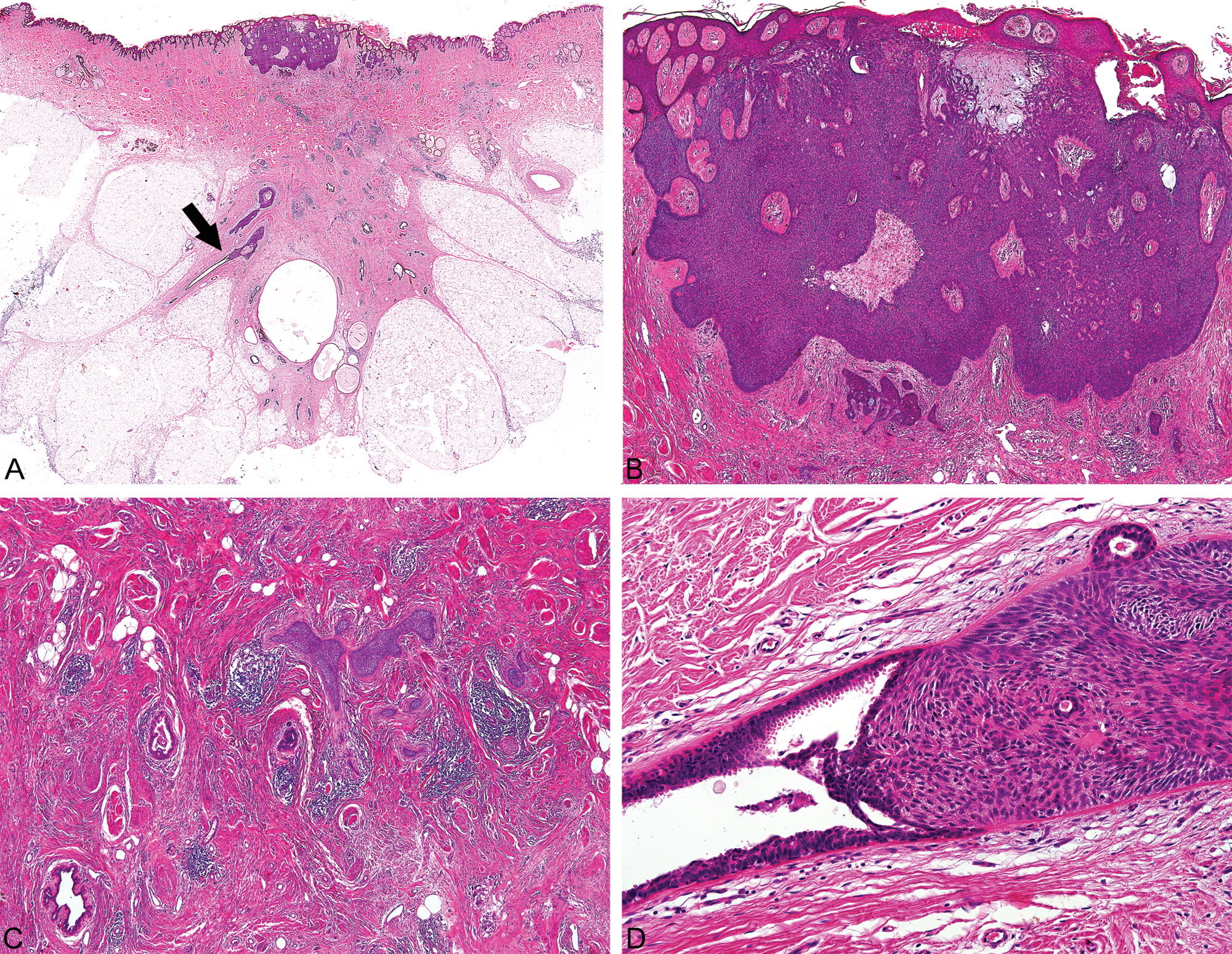
A, Low-power view of the tumor showing proliferation of basaloid cells arising from the epidermis, infiltrating the dermis and extending into a lactiferous duct (arrow). B, Solid basaloid tumor islands with the peripheral cell layer showing a palisade arrangement of the nuclei typical for basal cell carcinoma. C, Small basaloid cell islands separated by densely cellular connective tissue. D, Tumoral invasion into the lactiferous duct
BCC most commonly involves sun-exposed sites. Its location on the nipple–areola complex is extremely rare. To date, 36 cases (including the present case) of BCC involving the nipple–areola complex have been reported, with a male predominance. 1 This can perhaps be related to the fact that men are more likely to receive sun exposure to the nipple and areola than women. The main significance of the above illustrated changes is the tumoral intraductal extension into the lactiferous duct, which can lead to deep soft tissue tumor involvement not commonly seen in BCCs outside the nipple. Potentially the chance to metastasize is enhanced. In the previously 35 reported cases, the tumor involving the underlying lactiferous ducts was found only in 3 cases.2-4 Despite the intraductal extension there was no recurrence in 16 and 24 months after surgery in 2 cases2,3 and for the remaining case no follow-up data are available. In a limited biopsy specimen, BCC involving lactiferous ducts should be distinguished from rare mammary carcinomas with basaloid morphology as well as from unusual trichoblastomas located in the breast. 5
Footnotes
The author(s) declared no potential conflicts of interest with respect to the research, authorship, and/or publication of this article.
The author(s) received no financial support for the research, authorship, and/or publication of this article.