






Abstract
A 44-year-old woman presented with right flank mass of 6 months duration. A right side renal tumor was diagnosed, and a radical nephrectomy was performed. Histopathological examination showed chromophobe renal cell carcinoma (CRCC) with sarcomatoid transformation. The sarcomatous component contained large pleomorphic lipoblasts. The CRCC was positive for Hale’s colloidal iron stain, whereas the sarcomatous component was negative. Based on histopathological and immunohistochemical findings, a diagnosis of sarcomatoid CRCC with heterologous liposarcomatous differentiation was made. To the authors’ knowledge, this is the second reported case of a sarcomatoid CRCC where the sarcomatous component displayed features of liposarcoma. The case has been reported for its rarity.
Introduction
Chromophobe renal cell carcinoma (CRCC) was first described in humans by Thoenes et al in 1985. 1 CRCC has a better prognosis than conventional renal cell carcinoma. However, it is known that CRCC can undergo sarcomatous transformation, and when such a transformation occurs, the prognosis is grave. 2 Though there are several reports of sarcomatoid CRCC, there is only 1 case previously reported with liposarcomatous features. 3 Our case is hence the second reported case of sarcomatoid CRCC with heterologous liposarcomatous differentiation.
Case Report
A 44-year-old woman presented with right flank mass of 6 months duration. On examination, she had a large mass involving right hypochondria and lumbar region. Radiological examination showed right renal mass containing fat and calcific attenuation areas. With a radiological differential diagnosis of renal cell carcinoma/liposarcoma, the patient was taken up for surgery. A right radical nephrectomy was performed.
The radical nephrectomy specimen measured 19 cm × 17 cm × 8.5 cm. Cut section showed a large grey-white firm growth with yellowish areas measuring 11 cm × 11 cm × 6 cm, almost involving the entire cut surface and infiltrating perinephric fat (Figure 1). Normal renal tissue was seen at the lower pole. Three lymph nodes were also identified, which appeared to be involved by the neoplasm. Microscopy showed a neoplasm with dual components, CRCC and a high-grade sarcomatous component with spindle cells and large lipoblasts with multivacuolated cytoplasm and pleomorphic indented nuclei (Figure 2A and B). The different components were intermingled in areas. The CRCC component showed cells arranged in alveolar pattern and adenomatous pattern. Individual cells were polygonal with well-defined cell membrane, having faintly granular pale eosinophilic cytoplasm with raisinoid nucleus. In areas, perinuclear halos could be appreciated (Figure 3A). The CRCC was positive with Hale’s colloidal iron stain, whereas the sarcomatous component was negative (Figure 3B). The CRCC component was positive for cytokeratin and epithelial membrane antigen. Vimentin and CD10 were negative in the CRCC component and positive in sarcomatous areas (Figure 4A-D). All the lymph nodes showed metastasis from sarcomatoid component.
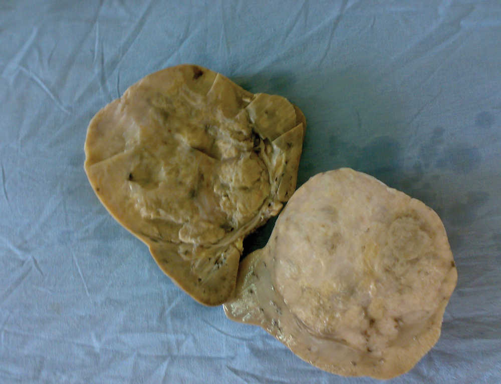
Cut surface of kidney showing large grey-white growth with yellowish areas

(A) A neoplasm with carcinomatous areas, intermingled with sarcomatous spindle cell areas and liposarcomatous areas (hematoxylin–eosin [H&E], 200×). (B) Higher power view of liposarcomatous area (H&E, 400×)
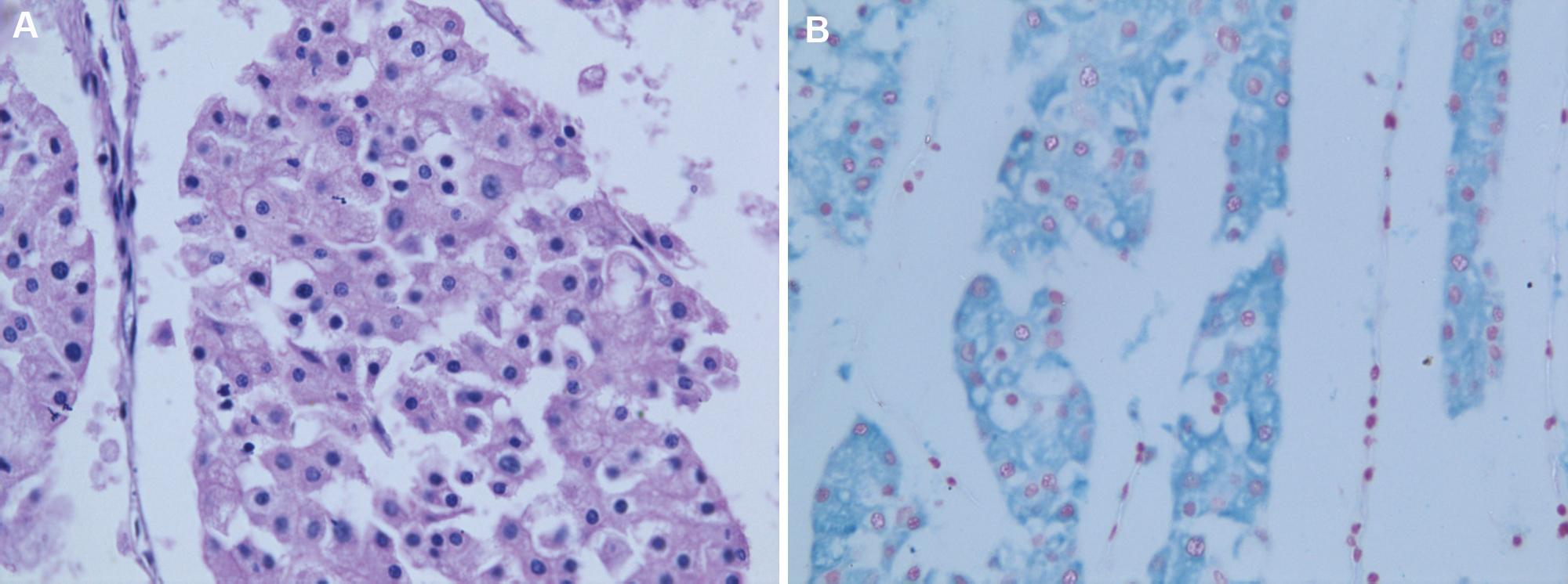
(A) Higher power view of carcinomatous area showing histopathologic features of chromophobe renal cell carcinoma (hematoxylin–eosin [H&E], 400×). (B) Positive Hale’s colloidal iron stain
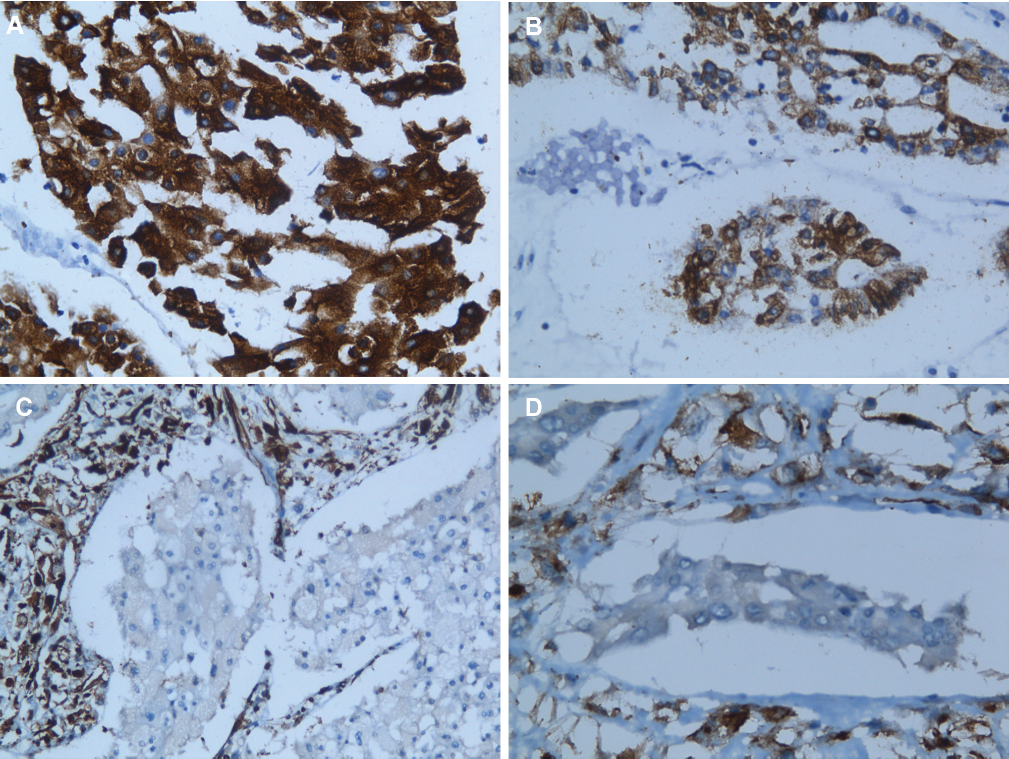
Immunohistochemical (IHC) findings
Discussion
CRCC is a distinct type of renal cell carcinoma. The histopathological features useful in the diagnosis of CRCC are accentuated cell borders, hyperchromatic wrinkled nucleus, and perinuclear halos. Histopathological features along with immunohistochemical findings help reach a diagnosis of CRCC. Studies have shown that the most sensitive and specific marker for CRCC is parvalbumin, followed by immunohistochemical profile EMA+/vimentin−. 4 This pattern of EMA+/vimentin− was observed in our case also. Conventional renal cell carcinoma is known to co-express cytokeratin and vimentin and is positive for CD10. CRCC has a characteristic alveolar pattern. In our case, apart from the alveolar pattern, an adenomatous growth pattern also could be appreciated. The microcystic and adenomatous pattern of CRCC was first described by Michal et al in 1998. 5
The tendency for sarcomatous transformation is well recognized in all types of renal cell carcinoma. However, whether CRCC has an increased frequency of sarcomatoid transformation than other subtypes is controversial. In a recently published study by Przybycin et al, 6 wherein they studied 203 cases of CRCC, an opinion has been raised that CRCC has better prognosis than papillary and clear cell renal cell carcinoma and that CRCC has a lesser chance of undergoing sarcomatous transformation than other subtypes of renal cell carcinoma. 6 The sarcomatous component can be of various morphological types. Cases of sarcomatoid CRCCs displaying chondrosarcomatous and osteosarcomatous differentiation were reported by Itoh et al 7 and Quiroga-Garza et al. 8 Hes et al 9 reported a case with rhabdomyosarcomatous differentiation. Petersson et al 3 was the first to report CRCC with heterologous liposarcomatous areas. Our case is the second reported case with liposarcomatous differentiation.
Most CRCCs are hypodiploid, in contrast to conventional renal cell carcinomas. 10 CRCC is characterized by loss of multiple chromosomes 1, 2, 6, 10, and 17. It has been recently suggested that the hypodiploid neoplastic cells in CRCC may undergo polyploidization, resulting in gain of chromosomal material, and this together with genetic instability causes sarcomatous differentiation in these tumors.2,11
The differential diagnosis to be considered when liposarcomatous component is obtained in core biopsies is retroperitoneal liposarcoma involving kidney and rarely lipoid urothelial carcinoma. A liposarcoma arising in angiomyolipoma is also a remote possibility. 12 In our case, the presence of CRCC along with the high-grade sarcomatous component with lipoblasts helps exclude primary liposarcoma. The rare lipoid urothelial carcinoma can be easily excluded by its different microscopy and immunohistochemical profile. 13
To conclude, sarcomatoid CRCC with heterologous liposarcomatous component is extremely rare and is known to carry a poor prognosis. Our patient expired 1 month after the surgery.
Footnotes
Declaration of Conflicting Interests
The author(s) declared no potential conflicts of interests with respect to the research, authorship, and/or publication of this article.
Funding
The author(s) received no financial support for the research, authorship, and/or publication of this article.