






Abstract
Cotyledonoid dissecting leiomyoma is a rare benign uterine tumor whose gross and radiological appearance may raise the possibility of a malignancy. The authors summarize herein the clinical, radiological, and pathological features of the 41 previously reported cases. The patients typically presented with menorrhagia or symptoms that were ultimately relatable to the presence of a pelvic mass. The median patient age was 46 years (range 23-73). The average tumor size was 15.4 cm (range 4-41); most were exophytic, multinodular, occasionally cystic masses with a congested, spleen or placenta-like color, protruding over the uterine serosa and, variably, the broad ligaments and adjacent organs. In most cases, the exophytic component was contiguous with the intramural dissecting leiomyomatous components. Histological features suggestive of malignancy, such as cytological atypia, necrosis, or increased mitotic activity were absent. Intravascular growth, as assessed histologically, was present in 20% of reported cases, and this finding is apparently devoid of clinical significance based on limited data. Reported information on radiological features is limited. However, these lesions are typically isointense to myometrium on T1-weighted MRI and are in general less heterogeneous on T2 and postcontrast-imaging than sarcomatous lesions. The tumor may display extension to but not frank invasion of surrounding organs. Ultrasound features are nonspecific. Follow-up information was available in 25 (61%) of 41 reported patients: none experienced a tumor recurrence or metastases during the follow-up period, which ranged from 1 month to 41 years. This affirms the benign nature of this tumor, its alarming gross appearance and growth patterns notwithstanding.
Introduction
Leiomyomas of the uterus may display a variety of growth patterns other than the typical appearance of a well-circumscribed, uniformly expansile tumor. 1 Leiomyomas with such unusual growth patterns include diffuse leiomyomatosis of the uterus, leiomyoma with vascular invasion, intravenous leiomyomatosis (IVL), benign metastasizing leiomyoma, parasitic leiomyomas, disseminated peritoneal leiomyomatosis, dissecting leiomyomas, and leiomyomas with a nodular growth pattern and perinodular hydropic degeneration. 2 Leiomyomas with a dissecting growth pattern were defined in 1 study as smooth-muscle tumors that microscopically display tongues of tumoral tissue extending significantly (at least 5 mm) between fascicles of the myometrium adjacent to the dominant mass. 3 These tumors are probably synonymous with the infiltrating leiomyomas as described by Hendrickson and Kempson almost 20 years previously. 1 Dissecting leiomyomas of the uterus may display extrauterine extension or be confined to the uterus.1-4
Cotyledonoid dissecting leiomyoma (CDL) is a distinctive variant of dissecting leiomyoma that is characterized by a reddish, exophytic, placenta-like gross appearance and by its frequent extrauterine extension into the adjacent tissues and the pelvic cavity.5-33 CDL was originally described as “grapelike leiomyoma” by David et al 5 in 1975 and as “proliferating pelvic angioleiomyomatosis (red seaweed lesion)” by Sternberg 6 in 1979. Sternberg tumor and CDL (the latter based on its placental-like appearance) are both appellations that were initially applied by Roth et al 7 in their 1996 report of 4 cases that formally defined the lesion as a distinct clinicopathological entity. In total, approximately 43 cases of CDL have been reported, including 41 in the English language literature (Table 1).
Summary of Reported Cases of Cotyledonoid-Dissecting Leiomyoma in the English-Language Literature a

Abbreviations: TH, total hysterectomy; TAH, total abdominal hysterectomy; BSO, bilateral salpingo-oophorectomy; TVH, total vaginal hysterectomy; ANED, alive with no evidence of disease; DNED, dead with no evidence of disease; SO, salpingo-oophorectomy; LSO, left salpingo-oophorectomy; NA, information not available.
Excludes the case reported by Gowri et al 34 because of insufficient clinicopathological information on the tumor.
Clinical Presentation
Of the CDL patients outlined in Table 1, 40 ranged in age from 23 to 73 years (mean 45, median 46 years). The patients typically presented with abnormal uterine bleeding (most frequently menorrhagia) and/or symptoms that were ultimately relatable to the presence of a pelvic mass (including constipation, abdominal pain, weight gain, and palpable abdominopelvic masses5-28,30-33). Rare presentations included uterine prolapse, infertility, stress urinary incontinence, urinary retention, and rectal bleeding. In 3 patients, the tumors were incidental discoveries during the diagnostic workup for, or management of, other medical issues.10,12,17
Radiological Features
The ultrasonographic attributes of CDL are not notably distinct from those of conventional leiomyomas, and it is characterized by one or more bulky or lobulated masses with irregular borders.12,19,23 Preda et al 24 detailed the radiographic features of a case of CDL as evaluated by MRI. On T1-weighted sequences, the lesion was homogeneously isointense (relative to the surrounding myometrium) and showed marked contrast enhancement in the images obtained postcontrast. T2-weighted sequences were more heterogeneous, which the authors attributed to hyperintense stromal components and confluent tumor nodules that were isointense to the surrounding myometrium. 24 Our general impression of the reported cases is that preoperative imaging was not suggestive of the eventual diagnosis and very often raised the index of suspicion for a malignant process. In 1 recently encountered case at Vanderbilt University Medical Center, a large mass was demonstrated to be arising from the uterus, extending predominantly anteriorly and laterally with some extension to the superior margin of the bladder and adjacent bowel above the uterus. This lesion showed features differing from conventional leiomyomas in the adjacent uterus. Morphological differences included the lack of well-defined margins and the presence of a bizarre lobulated appearance. Signal characteristics also provided a contrast, though less striking, to adjacent well-circumscribed leiomyomas with isointensity on T1 relative to the myometrium. T2 signal characteristics were more variable, with modest heterogeneity, increased signal as compared with adjacent leiomyomas and decreased signal with respect to the myometrium. Following contrast administration, mass enhancement was robust, paralleling the adjacent focal leiomyomas with minimal heterogeneity and near isointense signal relative to the myometrium. The tumor showed extension to but not frank invasion of surrounding organs; a fat plane, albeit small, was seen between the mass and adjacent structures such as the urinary bladder and bowel. There was no evidence of intravascular extension despite the aggressive appearance of this lesion. These features may allow the radiologist to suggest CDL or another less aggressive process in the differential, although biopsy or resection would certainly be required for a definitive diagnosis. (Figures 1A-1C)
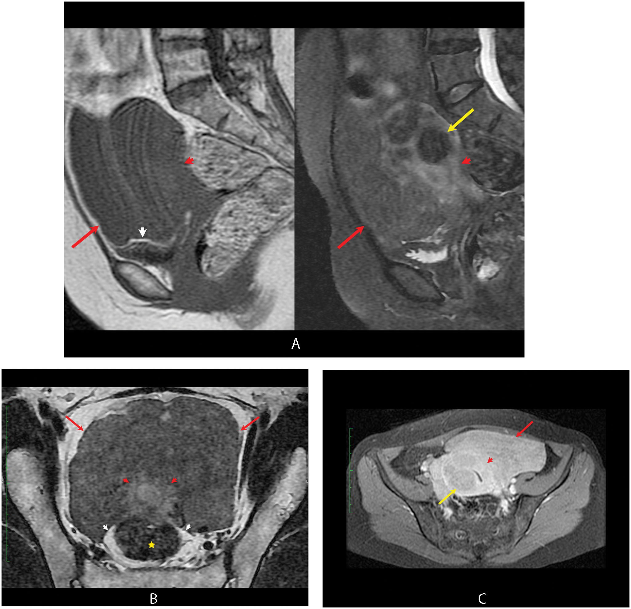
A. Sagittal T1 without fat saturation (left) and T2 with fat saturation (right) weighted MR images of the pelvis display the signal characteristics of the cotyledonoid-dissecting leiomyoma (CDL; red arrow). The mass is isointense on T1-weighted imaging to both the myometrium (red arrowhead) and adjacent leiomyomas (not indicated because they cannot be distinguished from the myometrium). On T2 MR imaging (right), the mass is slightly hypointense to the myometrium (red arrowhead) and isointense to hypointense to adjacent conventional leiomyomas (yellow arrow). B. Oblique axial T2 image without fat saturation shows the lobulated nature of the CDL (red arrows) and the relationship to the rectum (yellow star) and the uterus (red arrowhead). Note the fat plane between the CDL and the rectum (white arrowhead). Note also that the fat plane becomes less distinct between the rectum and uterus as a result of the scan plane. C. Axial T1-weighted image with fat suppression illustrates near isointense enhancement of the CDL relative to the adjacent myometrium (red arrowhead) and slightly hyperintense signal relative to conventional leiomyomas (yellow arrow). This sequence also showed no definite “invasion” of surrounding structures
Gross Pathology
A typical case of CDL is pictured in Figure 2. In this particular case, the uterus displayed multiple bulky, exophytic, bulbous, red-brown masses protruding from the serosal surface in the left uterine cornu and involving the broad ligament. The largest mass measured 11 × 6 × 4.5 cm3. The myometrium harbored numerous white-tan, whirled, nodular masses. Subserosal conventional leiomyomata were also present. The exophytic masses protruding from the serosa of the uterus were in continuity with intramural masses.
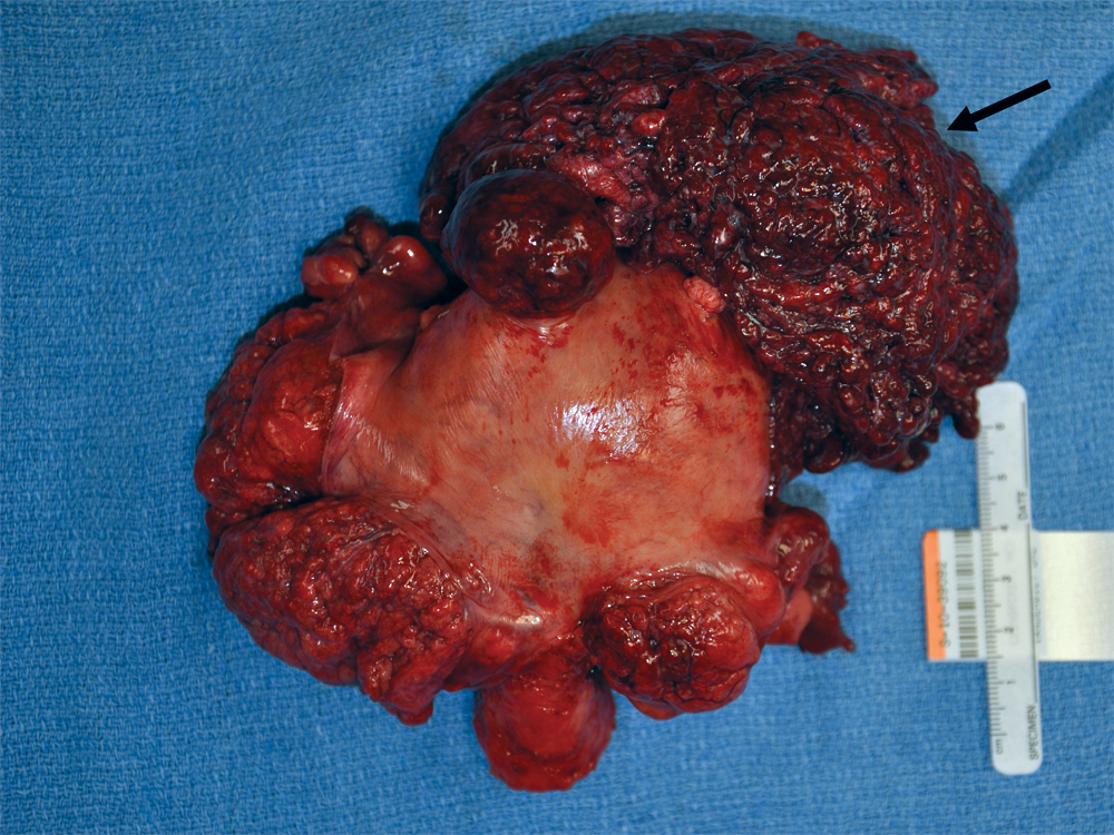
Gross appearance of cotyledonoid-dissecting leiomyoma, showing the exophytic (cotyledonoid) red bulbous mass protruding from the uterine serosa
Across a variety of reports, the described macroscopic appearance of CDL has been notably similar to this basic profile. CDL is characterized by exophytic, multinodular, occasionally cystic masses with a congested, spleen or placenta-like color, involving the uterine surfaces and, variably, the broad ligaments, the adnexal organs, the larger abdominopelvic cavity, the retroperitoneum, and the surfaces of the uterine cervix and bladder. The bulbous protrusions that constitute the mass were described as being “loosely attached to their neighbors by thin adhesions and delicate vessels” by Roth et al, 7 and Fukunaga et al 27 noted that small veins are characteristically present in the interlobular regions. The nidus of the mass is frequently in the uterine cornual region, and bilaterality is common. This exophytic mass is often contiguous with one or more uterine intramural leiomyomatous masses,7,27 but the focus of continuity may not be immediately apparent, resulting in the potential appearance of a separate adnexal mass. 14 Additionally, rare examples of CDL that presumably originated from subserosal smooth muscle may lack a parent leiomyomatous mass or an intramural dissecting component.11,17,18,33 The reported tumors have ranged in size from 4 to 41 cm (average 15.4, median 12 cm; Table 1). Conventional leiomyomas, which may be numerous and widely distributed, are frequently present. The gross appearance of the intramural component is variable and is largely a function of the type of leiomyoma that is so classified. A conventional intramural dissecting leiomyoma may have a solid appearance with a whorled, tan, cut surface that is distinguishable from a conventional leiomyoma only by its infiltrative edges. However, they may also be notably edematous, variably cystic, and/or only focally infiltrative.
Histopathology
The typical microscopic appearance of the cotyledonoid component of CDL is of poorly cohesive round nodules of benign smooth muscle that are surrounded by loose connective tissue with congested vessels (Figure 3A). When tissue sections are able to capture a nonfragmented portion of the cotyledonoid component, nodules of variably cellular smooth muscle can be seen separated by hydropic fibrous tissue, an appearance that is reminiscent of perinodular hydropic degeneration. (Figure 3B). Numerous vessels of varying caliber may be seen in the central portions of the nodules, 7 and florid edema may significantly separate the vessels. 14 Hydropic change of varying extents were stated to be present in 29 cases, and no information on hydropic change was given in the remaining 12. Constituent cells are most frequently spindled smooth-muscle cells lacking cytological atypia (Figure 3C), but a single case of an epithelioid CDL has been described. 25 The spindle cells show a higher level of disorganization than is typical for a conventional leiomyoma, and there is often stromal hyalinization (Figure 3D).7,14,27 Neurilemmoma-like cellular margination or “swirling” is not uncommon, and adipocytic differentiation was present in 1 case. 27 None of the reported cases displayed an increased mitotic index (>5 mitotic figures/10 high-power microscopic fields) or tumor cell necrosis. Approximately 90% of the reported cases of CDL had an intramural dissecting component. This component comprises infiltrating, sinus-like, haphazard tongues of smooth muscle in the background myometrium (Figure 3E). As noted by Roth et al, 7 in a given tissue section, the tongues appeared as “rounded, ovoid, or elongated processes.” These changes may be focal, at the edge of a conventional leiomyoma, or may comprise the entire intramural mass. Some authors have required that the dissecting component extends for at least 5 mm. 7 Three cases were associated with endosalpingiosis22,27,33 and 2 with endometriosis,27,33 but it is unclear if these associations were entirely fortuitous. One uterine tumor, interpreted as a CDL with an “adenoleiomyomatous” element, extended into the adnexa resulting in a cystic mass lined by a tuboendometrial epithelium. 14 Intravascular growth, typically focal (Figure 3F), was present in 8 (20%) of the reported 41 cases.14,21,27,30

A. Typical microscopic appearance of the exophytic component, comprising discohesive nodules surrounded by loose connective tissue with congested vessels. B. Nodules of smooth muscle surrounded by hydropic fibrous tissue. C. Characteristic appearance of the benign smooth muscle cells of cotyledonoid-dissecting leiomyoma (CDL)—there is a general disorganization of the cells, but they lack cytological atypia. D. Cotyledonoid nodule with a large focus of hyalinization (asterisk). E. Dissecting component of CDL (asterisk) between lateral aggregates of normal myometrial fibers; cotyledonoid component in lower field (arrow). F. Focus of lymphovascular space involvement in CDL
Pathological Differential Diagnosis
The most significant differential diagnostic considerations for CDL, especially if encountered in a biopsy, include hydropic leiomyomas and leiomyoma with perinodular hydropic degeneration (LPHD), myxoid lesions, IVL, low-grade endometrial stromal sarcoma (LGESS), perivascular epithelioid cell tumors (PEComas), and leiomyosarcomas.
Hydropic Leiomyomas, LPHD, and IVL
Hydropic leiomyomas are characterized by a prominent accumulation of intercellular fluid that may be so extensive as to cause cystic degeneration. 35 As previously noted, CDLs commonly display hydropic change. Pure hydropic leiomyomas, however, do not display intramural dissecting or extramural cotyledonoid components. The typical gross appearance of a hydropic leiomyoma is a circumscribed mass with a tan, solid, occasionally cystic cut surface. 35 LPHDs are characterized by nodules of variably hydropic smooth muscle surrounded by markedly hydropic stroma.35-37 When LPHDs are extrauterine, they may have a gross and microscopic appearance that renders them essentially indistinguishable from a CDL. As discussed below, we believe that CDL and LPHD are related lesions. IVL is defined by an intravascular growth pattern of benign smooth muscle outside of the confines of a main leiomyomatous mass.38-40 The extensive perinodular hydropic change in LPHD can result in some overt degeneration around individual nodules, and as such, they may appear to be in vascular spaces, simulating IVL. As previously noted, approximately 20% of CDLs display intravascular growth; those lesions where the intravascular growth is outside the main tumoral mass should be said to display an IVL component. Those lesions in which the intravascular growth is within the body of the tumor should be stated as displaying vascular invasion. It is probable that CDL, LPHD, and IVL are pathogenetically related. 7 All 3 tumors may display a mural dissecting growth pattern, extrauterine extension, a cotyledonoid appearance, lymphovascular invasion, and tumoral hydropic change (Table 2). Indeed, in the largest study of uterine dissecting, noncotyledonoid leiomyomas reported to date, 3 of the 8 cases so classified, 4 were intramural dissecting leiomyomas, 3 were examples of IVL, and 1 was a multinodular leiomyoma with hydropic degeneration. This overlap has also resulted in the description of lesions that are “intermediate” or that combine characteristics of any 2 or all of these lesions. Nevertheless, apart from the aforementioned exceptions, both LPHD and CDL should be distinguished from IVL because the latter has a well-defined clinicopathological profile, including rare, isolated metastases and extension through the vena cava and into the heart, with cardiac manifestations that may be seen many years after extirpation of the original uterine tumor.38,41 Additionally, IVL has been associated with recurrence, and although the recurrence rate is low, 40 it has nevertheless not been documented in the other 2 tumors.
A Comparison of the Clinicopathological Features That Have Been Described for Intravenous Leiomyomatosis, Leiomyoma With Perinodular Hydropic Degeneration, and Cotyledonoid-Dissecting Leiomyoma
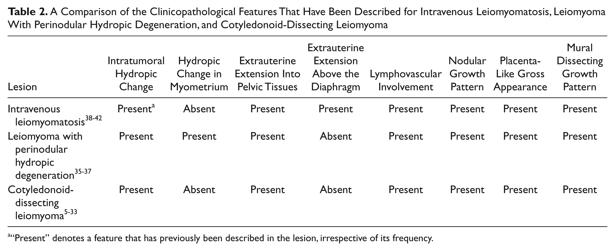
“Present” denotes a feature that has previously been described in the lesion, irrespective of its frequency.
Low-Grade Endometrial Stromal Sarcoma
The dissecting pattern of growth in CDLs within the myometrium as well as their frequently high vascularity are areas of morphological overlap with LGESSs. CDLs may also display significant hydropic change, 4 and accordingly, mimic myxoid LGESS. 43 The constituent cells of conventional LGESS are oval to fusiform and have a diffuse growth pattern, whereas the cells of CDLs are predominantly spindled and are configured in variably disorganized fascicles. Numerous thin-walled arterioles are typical of stromal neoplasms. The vessels of CDLs may be as numerous, but they are typically of a thicker caliber and are less uniformly distributed. In the absence of overt smooth-muscle differentiation, most LGESS have a CD10-positive/desmin-negative/h-caldesmon-negative immunophenotype, whereas most smooth-muscle neoplasms display the converse.
Perivascular Epithelioid Cell Tumors
Some PEComas may display an infiltrative growth pattern within the uterus that is reminiscent of dissecting leiomyoma.44,45 PEComas comprise admixures of epithelioid and spindle cells, whereas CDLs are mostly constituted of spindle cells, although 1 case of an epithelioid CDL was reported by Soleymani Majd et al. 25 PEComas are defined in part by a mixed myogenic/melanocytic immunophenotype and are positive for desmin in 49% of cases and for melanocytic markers (such as HMB-45 and Melan-A) in 100% and 24% of tested cases, respectively .45 Conventional leiomyomas are of course typically desmin positive and are known to display some expression of melanocytic markers, typically in a weak and/or focal pattern for the latter.46-48 Features favoring PEComa include a plexiform vascular pattern, presence of the tuberous sclerosis complex in the patient, degenerative multinucleated or “spider” cells, and diffuse expression of melan-A. 45 Additionally, tumor necrosis, cytological atypia, and a high mitotic index—features that characterize a potentially aggressive subset of PEComas—have not been described in CDLs.
Leiomyosarcoma and Other Lesions
Although the gross appearance of CDLs may raise the definite possibility of malignancy during their intraoperative evaluation, their distinctive microscopic appearance allows them to be clearly defined as benign, in that CDLs do not display morphological features diagnostic of conventional spindle-cell leiomyosarcoma (diffuse cytological atypia, a high mitotic index, and tumor cell necrosis). 1 Myxoid leiomyosarcomas, however, may not display any of these features in a biopsy and may only be distinguishable from hydropic leiomyomas by the recognition of the myxoid, nonedematous nature of the intercellular material and/or lymphovascular invasion. 49 Other myxoid lesions that are also worthy of consideration in a biopsy include the rare uterine myxomas,50,51 myxoid leiomyomas, 52 and inflammatory myofibroblastic tumor of the uterus. 53 Myxomas are exceptionally rare at this location and are characterized by a hypocellular proliferation of spindle to stellate cells in a myxoid background. Immunohistochemically, they display no smooth-muscle differentiation. Myxoid leiomyomas are also uncommon, may be associated with pregnancy, are grossly circumscribed, and are microscopically characterized by spindle cells separated by myxoid (nonedematous) material. However, the myxoid change may be focal. Leiomyomas in patients treated with progestins may display hydropic and/or myxoid changes adjacent to foci of infarct-type necrosis. 54 Unlike CDLs, neither myxoid leiomyomas nor myxomas should display an intramural dissecting or extramural cotyledonoid component. Some features of inflammatory myofibroblastic tumors that may cause them to mimic CDLs include an infiltrative growth pattern into the myometrium, a myxoid background, and microscopic patterns (hypocellular, fascicular, or hyalinized). Unlike CDLs, inflammatory myofibroblastic tumors uniformly express the anaplastic lymphoma kinase protein, typically have a lymphoplasmacytic infiltrate 53 and do not display extramural cotyledonoid components.
The etiopathogenesis of CDLs remains largely unknown. In their seminal report, Roth et al 7 speculated that CDLs originate from the corpus, then extend along the muscular vessels of the middle portion of the myometrium into the broad ligament. At extrauterine sites, venous drainage may become hampered, and congestion may develop. In the cotyledonoid portion, the widely dispersed muscular fibers, the fine fibrous matrix, the high vascularity of discrete areas, and the overlying vascular congestion all combine to result in a placenta-like appearance.
Treatment, Prognosis, and Outcome
Follow-up information was available for 25 (61%) of the 41 reported patients. Of these patients, 6 had been initially treated with a tumor resection only or a subtotal hysterectomy. The 19 others were treated with a total hysterectomy and (at minimum) a unilateral salpingo-oophorectomy; 2 others received only a total hysterectomy. None of the patients for whom follow-up information was available experienced a tumor recurrence or metastases during the follow-up period, which ranged from 1 month to 41 years. This favorable outcome is apparently independent of the presence or absence of conventionally adverse pathological prognostic factors, such as broad extrauterine extension. CDL with lymphovascular invasion did not appear to be prognostically distinct from the other cases in the group, but there were only 8 cases with this finding.14,21,27,30 Follow-up information was available in 5 of the 6 patients who were treated with tumor resection only, and none had experienced a recurrence or metastases at 10 months (30), 15 months (7), 16 years (9), 2 years (12), and 5 months (20) after their procedures. These findings affirm that CDL is a benign tumor that has minimal, if any, recurrence or metastatic potential after its complete resection.
Summary
CDLs are rare, benign uterine tumors whose gross or radiological appearance may simulate a malignant process. They are most frequently associated with a mural dissecting leiomyomatous component in the uterus and, at the microscopic level, are readily separable from lesions in the differential diagnosis. Familiarity with this distinctive tumor is important: the macroscopic attributes of this tumor are sufficiently distinctive as to allow the gross pathologist to at least suggest the diagnosis before microscopic examination (eg, during intraoperative evaluation). Similarly, some radiological features may allow the radiologist to suggest CDL or another nonaggressive process in the differential prior to a biopsy or definitive resection. A review of the outcomes for previously reported cases confirmed that CDL is a benign tumor.
Footnotes
Declaration of Conflicting Interests
The author(s) declared no potential conflicts of interest with respect to the research, authorship, and/or publication of this article.
Funding
The author(s) received no financial support for the research, authorship, and/or publication of this article.