
Abstract

Liesegang rings, first described in 1896, are among the most fascinating structures in physical chemistry. 1 These rings, formed by an ill-understood mechanism whereby various minerals and substances aggregate and segregate, can be observed in the in vitro and in vivo settings. 2 In the latter, the rings have been described in cysts of various tissues—including eyelids, kidney, and breast.3-5 In the breast, Liesegang rings have been reported in cytological and histological material derived from cysts, 5 abscesses, 6 and in association with secretory (ie, lactational-like) changes. 7
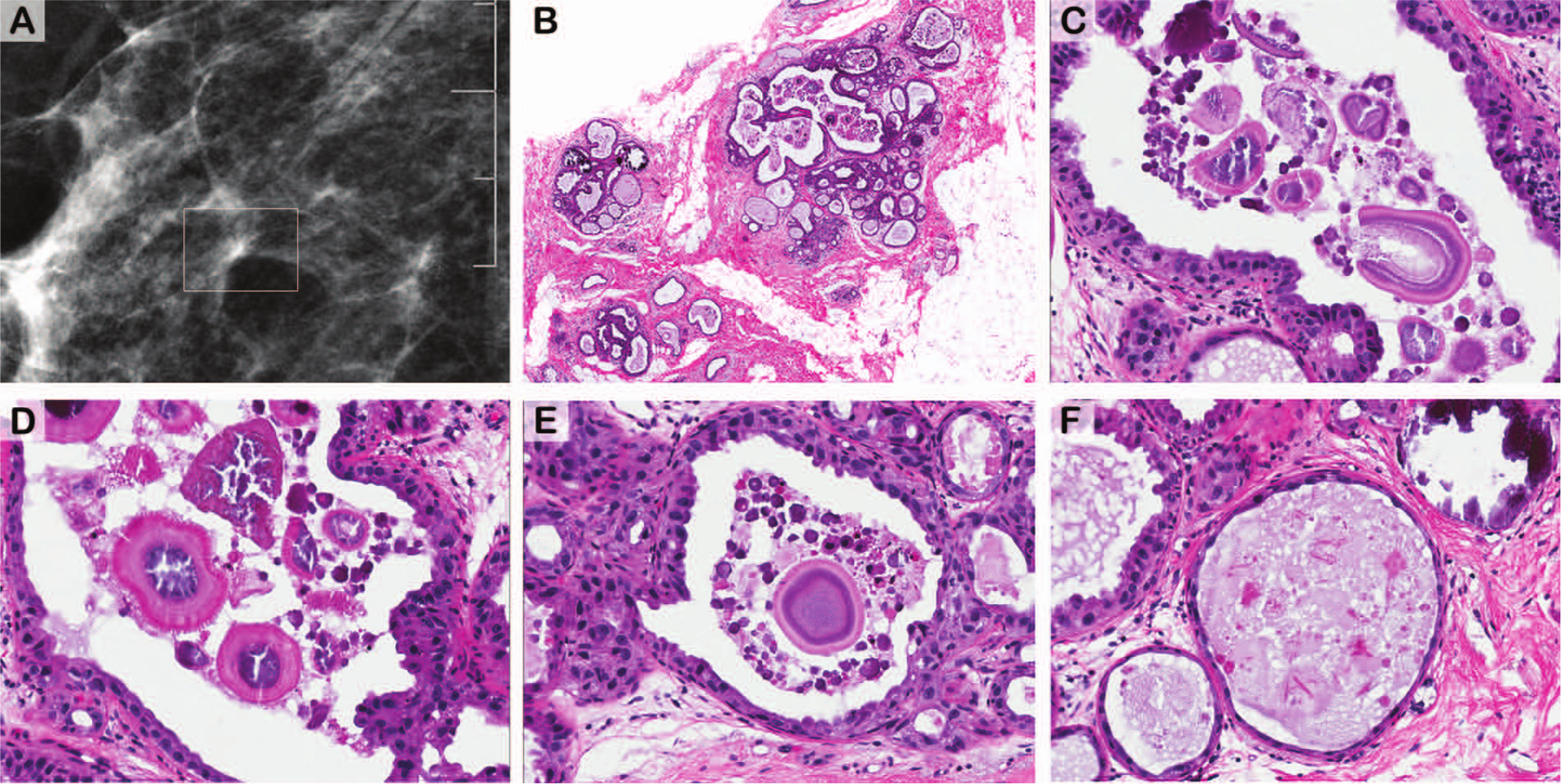
Here, we illustrate the case of an 82-year-old woman who was found to have a 3-mm cluster of microcalcifications amid focal density on screening mammography (Figure 1A). The ensuing stereotactic needle core biopsy showed cystically dilated glands lined by largely inactive metaplastic apocrine epithelium (Figure 1B). Intracystic Liesegang rings with characteristic acellular cores, radial striations, and concentric laminations—in various states of organization—were evident (Figure 1C-E). Incipient forms of these rings, and calcifications (the ostensible target), were present (Figure 1F).
Liesegang rings in apocrine cysts of the breast.
Liesegang rings have been mistaken for Dioctophyma renale, the giant kidney worm, elsewhere.4,8,9 This mistake would be improbable in the breast. More likely, such visually appealing objet de vertu would remain unrecognized.