
Abstract
Introduction. Induction of tumor lymphangiogenesis by vascular endothelial growth factor (VEGF)-C and VEGF-D promotes metastasis in many human cancers. Aim. The aim of this study was to examine the role of VEGF-C and VEGF-D in lymphangiogenesis and lymph node metastasis in patients with cutaneous melanoma. Materials and Methods. Fifty-four melanoma specimens (18 with lymph node metastasis, 36 nonmetastatic) were investigated by immunostaining for VEGF-C, VEGF-D, and for lymphatic endothelial marker D2-40. VEGF-C and VEGF-D expression was assessed as a percentage and intensity of stained tumor cells, tumor-associated macrophages and fibroblasts. The quantification of lymphangiogenesis was conducted by computer-assisted morphometric analysis. Results. The expressions of both VEGF-C and VEGF-D in tumor cells were significantly higher in lymph node metastatic melanomas compared with nonmetastatic melanomas (P = .015 VEGF-C; P = .005 VEGF-D). There was no statistically significant difference between metastatic and nonmetastatic melanomas regarding the expression of VEGF-C and VEGF-D in macrophages and fibroblasts. Metastatic melanomas showed a significantly higher intratumoral and peritumoral lymphatic vessel density (LVD) compared with nonmetastatic melanomas (P = .000 intratumoral, P = .000 peritumoral). Melanomas with VEGF-C positive tumor cells showed a significantly higher intratumoral and peritumoral LVD compared with VEGF-C negative tumor cells group of melanomas (P = .006 intratumoral, P = .010 peritumoral). VEGF-C expression in macrophages, fibroblasts, as well as VEGF-D expression in tumor cells, macrophages, and fibroblasts, showed no correlation with the intratumoral and peritumoral LVD. Conclusions. Our findings show the significance of VEGF-C in tumor cells in the induction of intratumoral and peritumoral lymphangiogenesis. This study suggests that both VEGF-C and VEGF-D in tumor cells promote lymph node metastasis, and that the immunohistochemical analysis of expression can be a useful tool for predicting clinical behavior of cutaneous melanoma.
Introduction
Cutaneous melanoma has the propensity for early metastatic spread via lymphatic vessels to regional lymph nodes. The molecular mechanisms, which lead to metastasis have not yet been completely defined and it is difficult to suppose which tumor will metastasize. The recent identification of lymphatic growth factors and receptors, together with the discovery of lymphatic specific markers have provided important new insight into the formation of tumor-associated lymphatic vessels and their active contribution to lymphatic tumor spread.1,2
Scientific research of the lymphatic vessels invasion process and its metastasis led to the conclusion that tumors producing a higher level of lymphangiogenic factors behave more aggressively than tumors negative for those factors. The major lymphangiogenic factors in melanoma are vascular endothelial growth factor (VEGF)-C and VEGF-D. VEGF-C and VEGF-D released by melanoma cells and tumor-associated stromal cells, bind to VEGF-receptor-3 (VEGFR-3) on lymphatic endothelial cells, and induce proliferation and growth of tumor-associated lymphatic vessels (tumor lymphangiogenesis). They also induce enlargement of tumor-associated lymphatic vessels within and surrounding these tumors. This process advances the invasion of lymphatic vessels by tumor cells and metastatic spreading to the draining lymph nodes.2-4
Numerous clinicopathological studies have found a positive association between VEGF-C and VEGF-D expression, tumor lymphangiogenesis, and lymph node metastasis in many human cancers.5-11 Some data also suggest that VEGF-C and VEGF-D expression may be a useful prognostic marker for the risk of lymph node metastasis in cutaneous melanoma.12-15
In this study, we examined the role of lymphangiogenic factors in the progression of melanoma. The aim was to determine whether VEGF-C and VEGF-D expression in melanoma cells, tumor-associated macrophages and fibroblasts is correlated with tumor lymphangiogenesis and lymph node metastasis in patients with cutaneous melanoma.
Materials and Methods
Patients were identified retrospectively after reviewing the complete medical documentation in the Oncology Clinic, Clinical Center of Banja Luka. Eighteen patients with primary cutaneous melanoma with lymph node metastasis were matched with 36 patients without metastasis. Each metastatic melanoma was matched with 2 nonmetastatic controls for tumor thickness and presence of ulceration, and in most cases closely matched for Clark’s level, histological type, and anatomical site of primary melanoma.
There were no significant differences between clinicopathological characteristics of patients, except in gender (Table 1). Patients who did not develop metastasis for a long period, over 5 years, after the melanoma excision (mean follow-up time was 90.28 months, standard deviation months 20.81) were involved within the group of patients with nonmetastatic melanoma. Ten patients died from metastatic melanoma.
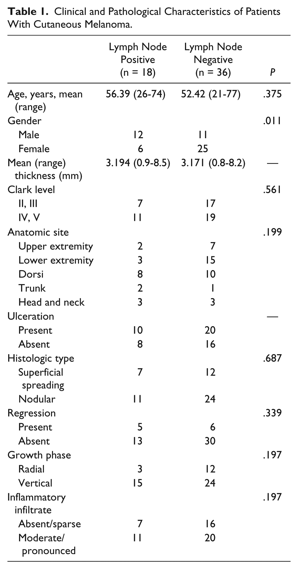
Clinical and Pathological Characteristics of Patients With Cutaneous Melanoma.
Conventionally processed formalin-fixed paraffin-embedded melanoma tissue specimens were taken from the Department of Pathology archives, Clinical Center of Banja Luka.
Immunohistochemistry
Immunohistochemical staining was performed using the streptavidin-biotin peroxidase conjugate method. Three micrometers thick paraffin sections were deparaffinized in xylol, rehydrated through a graded ethanol series and water and submitted to microwave antigen retrieval with the pressure cooker method in 0.01 M citrate buffer, pH 6.0.
After inactivation of endogenous peroxidase activity with 3% hydrogen peroxidase in water for 5 minutes, sections were washed in phosphate buffered saline (PBS 0.1 M). Sections were then incubated with primary anti-VEGF-C and anti-VEGF-D goat polyclonal antibodies (anti-VEGF-C (N-19), Santa Cruz Biotechnology Inc, Santa Cruz, CA, sc-7133; anti-VEGF-D (N-19), Santa Cruz Biotechnology Inc, Santa Cruz, CA, sc-7603) at a 1/100 of dilution overnight at 4°C in a humid chamber. Subsequently, the sections were incubated with biotinylated secondary antibodies (LINK, DAKO LSAB+/HRP, Cat. No. K0690) for 20 minutes at room temperature in a humid chamber, and then sections were incubated with streptavidin-peroxidase for 20 minutes at room temperature in a humid chamber (streptavidin HRP, DAKO LSAB+/HRP, Cat. No. K0690). Sections were visualized with diaminobenzidine (DAB+) and washed in water, counterstained with Mayer’s hematoxylin, dehydrated, and mounted.
The D2-40 antibody was used to decorate lymphatic endothelium, and melanoma cells were identified using S-100 antibody. The Envision double labeling system (DakoCytomation, HRP-AP, Code K5361) was used for double staining. Sections were incubated with the first primary D2-40 monoclonal mouse anti-human antibody (DAKO, Cat. No. M3619, clone D2-40) at 1/100 of dilution for 45 minutes at room temperature. Subsequently, the sections were incubated with HRP polymer, and then with the brown chromogen diaminobenzidine (DAB+). On completion of the first reaction, a double stain block solution was applied for 3 minutes at room temperature, and the sections were incubated with a second primary S-100 polyclonal rabbit antibody (Dako, Cat. No. Z0311) at 1/100 of dilution for 30 minutes at room temperature in a humid chamber, and then incubated with a secondary linking polymer antibody (Rabbit/Mouse-LINK). On rinsing with PBS, sections were incubated with alkaline phosphatase–labeled polymer, and treated with a substrate–chromogen solution (Permanent Red). Sections were counterstained with Mayer’s hematoxylin and mounted in aqueous mounting medium. Negative controls were performed by substituting the primary antibody with nonimmune serum.
Evaluation of Growth Factor Expression
Two observers without knowledge of the clinical data assessed immunostained sections. Staining intensity was scored semiquantitatively as negative (absent or weak), moderate or strong staining. Specimens were considered positive for statistical evaluation when cells were clearly moderately or strongly stained. The quantity of VEGF-C and VEGF-D was assessed as a percentage of positively stained cancer cells, macrophages, and fibroblastic stromal cells per total number of cancer cells, macrophages, and fibroblasts, and assigned to 1 of 3 subgroups (<20%, 20% to 50%, >50%).
Computer-assisted Morphometric Vessel Analysis
Lymphatic vessel density (LVD) was defined as the number of vessels per square millimeter. Sections were examined using an Olympus CX21 microscope at 100× magnification seeking the fields with the highest LVD (hot spots). Digital images were taken at 400× magnification on 6 hot spots per section—3 intratumoral and 3 peritumoral within an area of 200 µm2 from the tumor border. LVD was determined with Image Tool software and “count and tag” program addition. The mean values of the measurements in 3 fields were used for statistical analysis. Two observers, blinded to the lymph node status, performed every quantification.
Statistical Analysis
All statistical tests were performed using SPSS software (version 22). The continuous variables were presented as mean, range, and standard deviation. The qualitative nominal variables were presented as frequencies and percentages. To examine differences between numeric variables the Student’s t test and Mann-Whitney U test were used. Frequency difference testing was estimated with the χ2 test. Correlations between variables were analyzed by the Spearman’s ρ test. Kaplan-Meier survival analysis was used to obtain overall and disease-free survival curves that were compared using the log-rank test. P < .05 was considered significant.
Results
We observed VEGF-C and VEGF-D expression in tumor cells, tumor-associated macrophages and fibroblastic cells. Immunohistological localizations of VEGF-C and VEGF-D were cytoplasmic. Staining intensity of VEGF-C and VEGF-D among different specimens varied from complete absence of staining, to weak, moderate, and strong staining. In cases with positive staining, the number of cells ranged from a few to almost all of the cells. In most cases, staining in tumor cells was diffuse, but in some specimens the staining intensity was heterogeneous among tumor areas. Both in metastatic and nonmetastatic melanomas, staining in macrophages was stronger, and with a higher percentage than in tumor cells. Negative staining of both factors in macrophages was present in a small number of specimens, unlike that of fibroblasts that showed mainly weak staining or the absence of staining. Staining in fibroblasts was at the base of tumor and around tumor nests. (Figure 1).
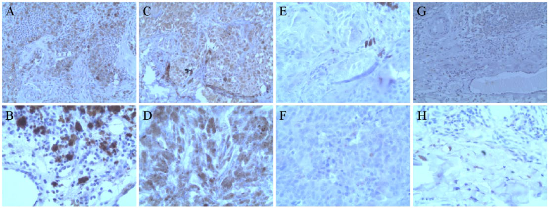
Immunohistochemical expression of vascular endothelial growth factor (VEGF)-C and VEGF-D in cutaneous melanoma specimens: moderate VEGF-D staining in macrophages (200× magnification) (A) and strong VEGF-C staining in macrophages (400× magnification) (B); moderate VEGF-D staining in tumor cells (200× magnification) (C) and strong VEGF-D staining in tumor cells (200× magnification) (D); VEGF-C negative staining in tumor cells (400× magnification) (E) and VEGF-D negative staining in tumor cells (200× magnification) (F); VEGF-D negative staining in fibroblasts (200× magnification) (G) and VEGF-C positive staining in fibroblasts (400× magnification) (H).
In all, 88.88% (16/18) of lymph node metastatic melanomas and 47.22% (17/36) of nonmetastatic melanomas showed a positive expression of VEGF-C in tumor cells. A total of 77.77% (14/18) of metastatic melanomas and 36.11% (13/36) of nonmetastatic melanomas showed a positive expression of VEGF-D in tumor cells. The expressions of both VEGF-C and VEGF-D in tumor cells were significantly higher in metastatic melanomas compared with nonmetastatic melanomas (Spearman’s ρ test: r = −0.329, P = .015, VEGF-C; r = −0.373, P = .005, VEGF-D) (Tables 2 and 3).
Association Between VEGF-C Expression and Lymph Node Status and Other Clinicopathological Characteristics.
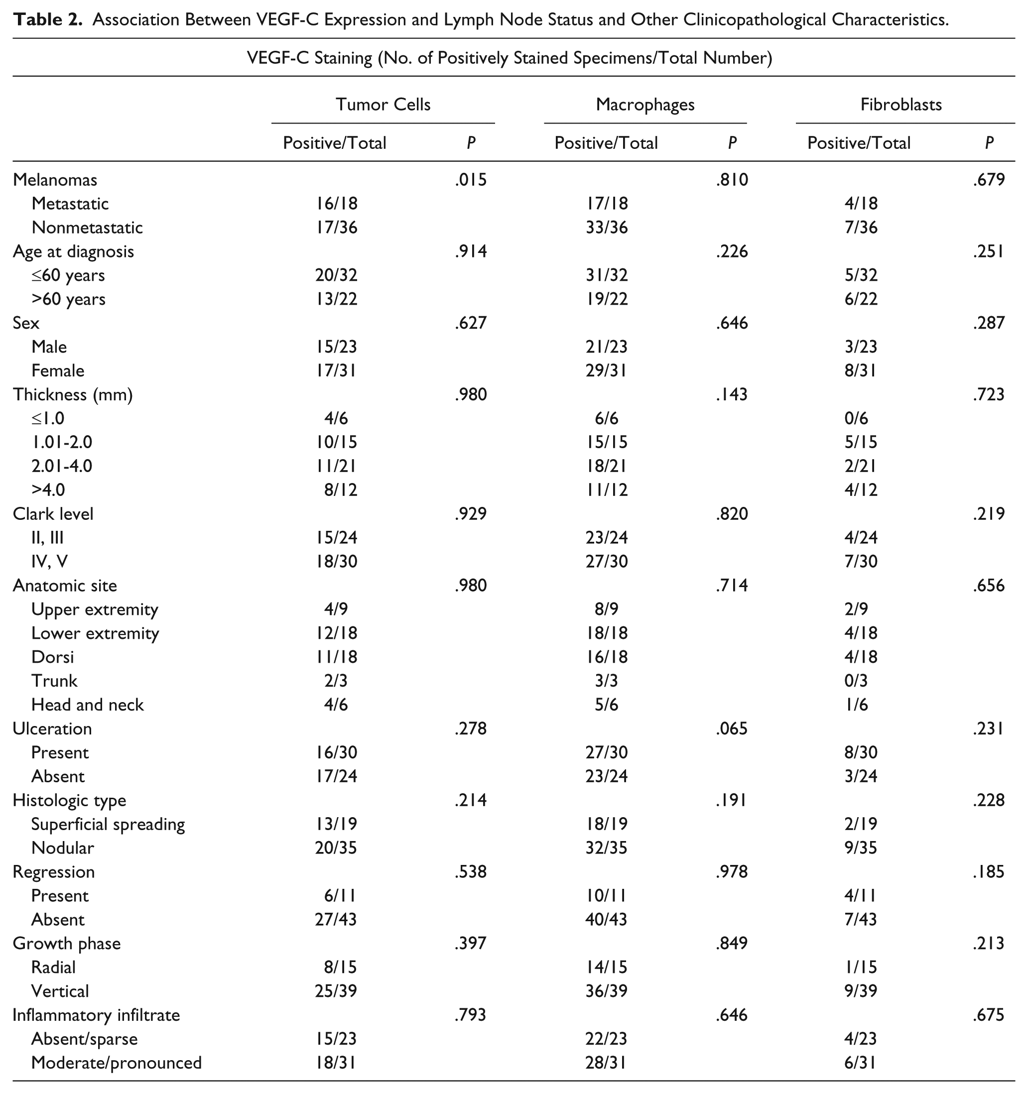
Association Between VEGF-D Expression and Lymph Node Status and Other Clinicopathological Characteristics.
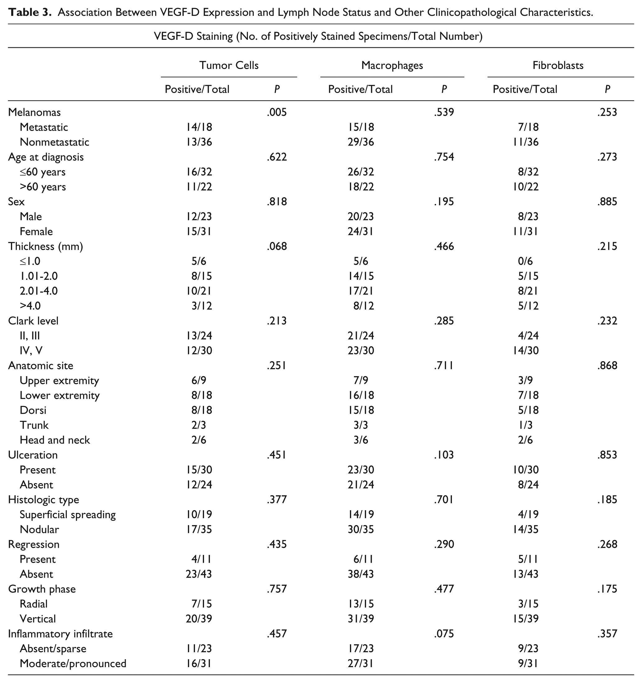
There was no statistically significant difference between metastatic and nonmetastatic melanomas regarding the expression of VEGF-C and VEGF-D in macrophages and fibroblasts (Tables 2 and 3). VEGF-C and VEGF-D expression in tumor cells, macrophages and fibroblasts were not associated with other clinicopathological parameters (Tables 2 and 3).
Patients with VEGF-C positive tumor cells had significantly shorter survival times compared to those with VEGF-C negative tumor cells (log rank, χ2 = 4.082, P = .043). VEGF-C positive tumor cells were not predictive of a disease-free survival (log rank, χ2 = 0.313, P = .909). VEGF-D positive tumor cells showed no significant correlation with patients’ overall survival (log rank, χ2 = 1.882, P = .170) or disease-free survival (log rank, χ2 = 0.529, P = .467) (Figure 2).
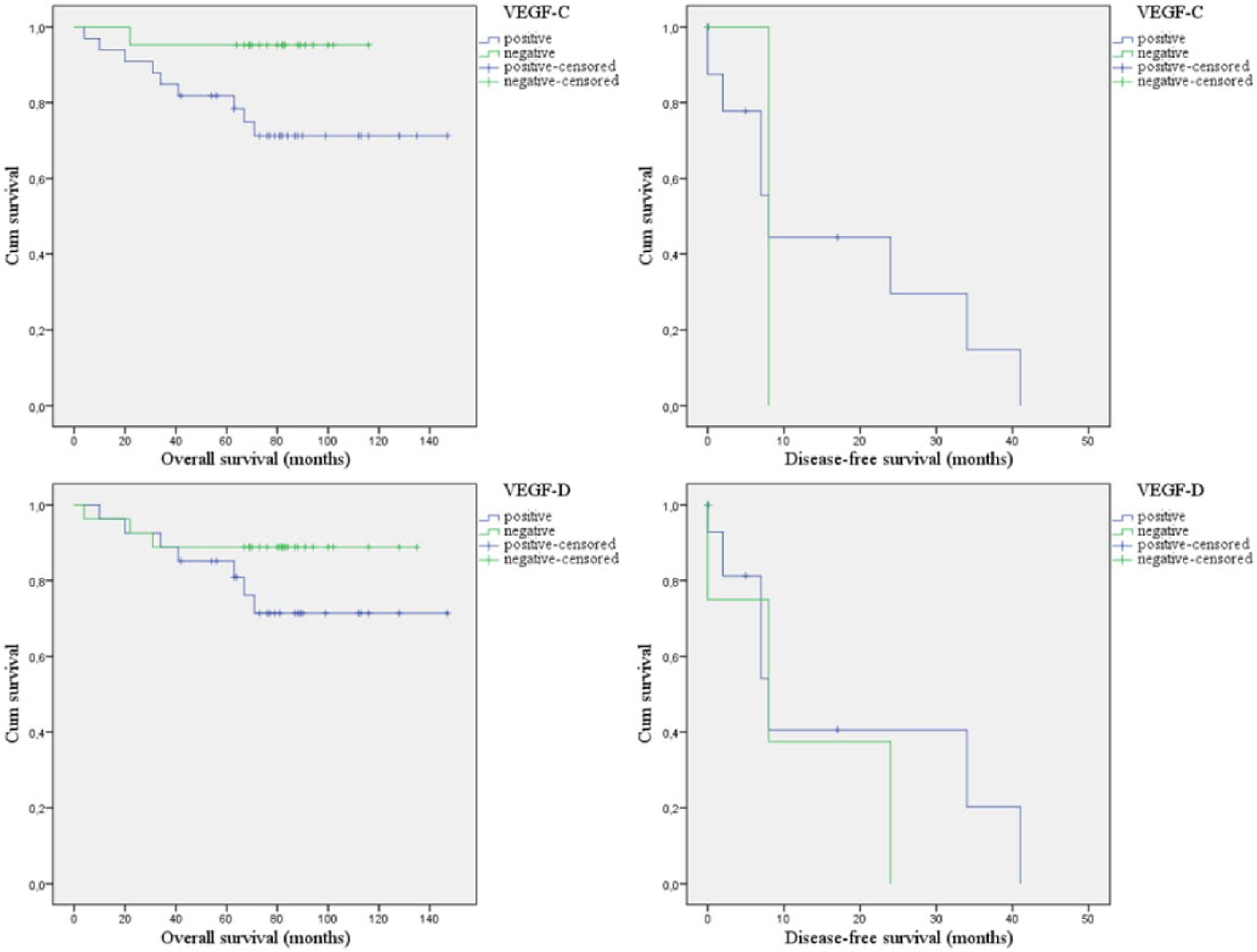
Kaplan-Meier survival analysis depending on vascular endothelial growth factor (VEGF)-C and VEGF-D expression in tumor cells. Positive expression of VEGF-C was significantly associated with shorter overall survival (log rank, P = .043), there was no significant association with disease-free survival (log rank, P = .909). Positive VEGF-D expression was not significantly associated with overall (log rank, P = .170) and disease-free survival (log rank, P = .467).
Immunohistochemistry of D2-40/S-100 showed stained lymphatic vessels in the intratumoral and peritumoral area (Figure 3). Metastatic melanomas showed a significantly higher intratumoral and peritumoral LVD compared with nonmetastatic melanomas (Mann-Whitney U test, P = .000 intratumoral, P = .000 peritumoral) (Table 4). Melanomas with VEGF-C positive tumor cells showed a significantly higher intratumoral and peritumoral LVD compared with VEGF-C negative tumor cells group of melanomas (Mann-Whitney U test, P = .006 intratumoral, P = .010 peritumoral) (Table 4). VEGF-C expression in macrophages, fibroblasts, as well as VEGF-D expression in tumor cells, macrophages and fibroblasts, showed no correlation with the intratumoral and peritumoral LVD (Table 4).
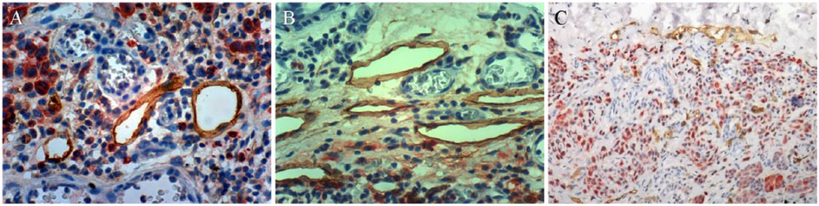
Double immunostaining D2-40/S-100 in cutaneous melanoma (D2-40 brown lymphatic vessels): intratumoral lymphatic vessels (400× magnification) (A); peritumoral lymphatic vessels (400× magnification) (B); peritumoral lymphatics are larger, with an open lumen compared with a narrow lumen of intratumoral lymphatics (100× magnification) (C).
The Quantitative Results of Peritumoral and Intratumoral Lymphatic Vessel Density in Metastatic and Nonmetastatic Melanomas, and Correlation Between VEGF-C, VEGF-D Expression, and LVD.
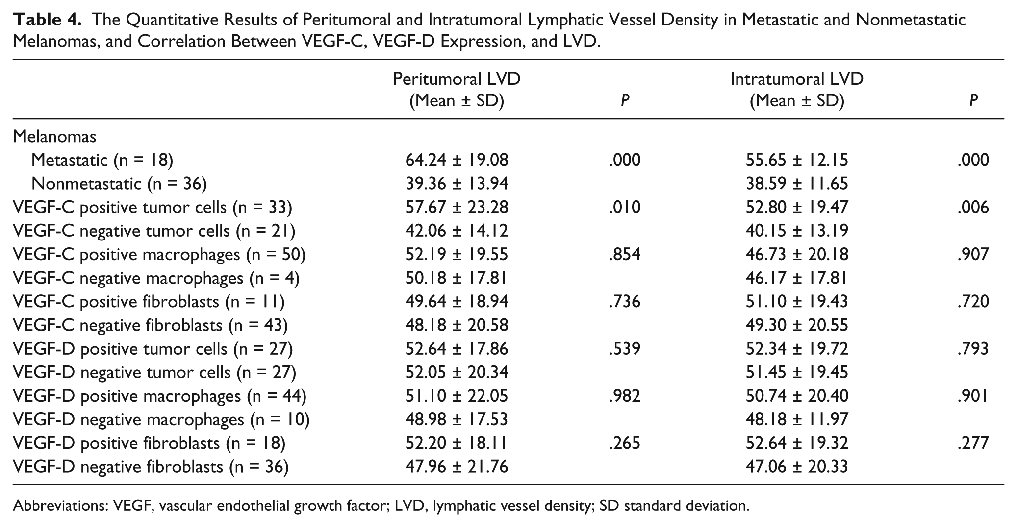
Abbreviations: VEGF, vascular endothelial growth factor; LVD, lymphatic vessel density; SD standard deviation.
Discussion
The significance of VEGF-C and VEGF-D expression in relation to lymph node metastasis and patient outcome has been reported in many different tumor types.5,16-18 However, the relationships between VEGF-C and VEGF-D expression, lymphangiogenesis, and lymph node metastasis in cutaneous melanoma are still controversial. Few studies have reported prognostic significance of VEGF-C in melanoma.13-15,19 We found only one study, which suggests that both VEGF-C and VEGF-D are involved in peritumoral lymphangiogenesis and lymphatic metastasis in cutaneous melanoma. 12
The first study by Dadras et al, 20 as well as the study by Massi et al, 21 showed that the expression of VEGF-C protein is more frequent in tumor cells of metastatic melanomas than in nonmetastatic melanomas. However, this difference was not statistically significant in either of these 2 studies. Dadras et al 20 additionally researched the expression of VEGF-D in tumor cells; however, they did not detect any. Furthermore, Shields et al 22 found no qualitative differences in the intensity of staining for VEGF-C and VEGF-D in metastatic versus nonmetastatic melanomas.
The second study of Dadras et al 19 determined moderate to high levels of cytoplasmic VEGF-C in the majority of sentinel lymph node (SLN) positive melanomas compared with nonmetastatic melanomas. This difference was significant, and the authors suggested that tumor cell expression of VEGF-C is useful to predict metastasis to the SLN. There was no correlation between VEGF-D levels and SLN metastasis. 19
Schietroma et al 23 also suggested that VEGF-C expression in primary melanoma may be predictive of lymph node metastatic dissemination. However, they did not find a correlation between VEGF-D expression and lymph node metastasis. Similarly, Cianfarani et al 15 found a correlation between VEGF-C expression in melanoma cells and SLN metastasis, but they did not determine a correlation between VEGF-C expression and peritumoral lymphatic vessel count.
Unlike the previous research, Liu et al 12 suggested that both VEGF-C and VEGF-D expression may be clinically useful indicators for prognostic evaluation in patients with cutaneous melanoma. They determined that VEGF-C and VEGF-D expression in melanoma cells are significantly correlated with both peritumoral lymphangiogenesis and lymph node metastasis, and that both factors are independent indicators for overall survival and disease-free survival. 12
The results of our study showed that both VEGF-C and VEGF-D expression in tumor cells are significantly correlated with lymph node metastasis. Our findings show that VEGF-C and VEGF-D expression in tumor cells can be used as a reliable prognostic factor, as with the increase of expression the risk of metastasis increases significantly.
In addition, we found that only VEGF-C expression in tumor cells is significantly correlated with both intratumoral and peritumoral lymphangiogenesis. We also evaluated the prognostic significance of VEGF-C expression in tumor cells for overall survival.
Considering the studies that proved that tumor stromal cells are a significant source of lymphangiogenic factors,8,24-26 we examined VEGF-C and VEGF-D expression in tumor-associated macrophages and fibroblasts. We found macrophages to be a stronger source of both lymphangiogenic factors than tumor cells. However, we did not find a correlation between the expression in stromal cells, lymphangiogenesis, and lymph node metastasis.
Contrary to our research, two studies pointed to a significant contribution of stromal cells in the neoplastic dissemination of cutaneous melanoma.13,14 Boone et al 14 found that both VEGF-C expression in tumor-associated macrophages and tumor cells were significantly associated with the SLN status. In the study by Gallego et al, 13 only the expression of VEGF-C in the fibroblasts was significantly associated with the SLN status, with no correlation of VEGF-C expression in tumor cells, macrophages, fibroblasts with intratumoral or peritumoral LVD. Because of such results, the authors suggested that VEGF-C has additional qualitative action on lymph vessels, causes their dilatation and thus leads to the dissemination of tumor cells. 13
Our findings support previous studies that found a significant correlation between VEGF-C expression in tumor cells, tumor lymphangiogenesis and lymph node metastasis.7,9,11,12
Besides our study, we found only the study of Liu et al 12 that points out a significant correlation between VEGF-D expression in tumor cells and lymph node metastasis in cutaneous melanoma. Liu et al 12 also found a significant correlation of both VEGF-C and VEGF-D expression and peritumoral lymphangiogenesis. Our study did not find any correlation between VEGF-D expression in tumor cells and peritumoral or intratumoral lymphangiogenesis.
VEGF-C may significantly increase metastasis by increasing the number of intratumoral and peritumoral lymphatic vessels, as it increases the density of entry sites for tumor cells.25,27 VEGF-D can have a different role, maybe a qualitative role, in the development of lymphatic metastasis. This role can include dilatation of vessels and stimulation of tumor cells to enter the lymphatics.13,25
This study shows the significance of VEGF-C in tumor cells in the induction of intratumoral and peritumoral lymphangiogenesis. Also, our study shows that both factors play an important role in the lymph node progression of cutaneous melanoma, and that the immunohistochemical analysis of VEGF-C and VEGF-D expression in tumor cells can be used as a diagnostic method for predicting clinical behavior of cutaneous melanoma.
Induction of tumor lymphangiogenesis by lymphangiogenic factors is the process that proceeds to metastasis and necessitates therapy, thus slowing down or blocking metastatic dissemination of a tumor.2-4
Footnotes
Declaration of Conflicting Interests
The author(s) declared no potential conflicts of interest with respect to the research, authorship, and/or publication of this article.
Funding
The author(s) received no financial support for the research, authorship, and/or publication of this article.