






Abstract
The patient was baby B of a diamniotic-dichorionic twin pregnancy. He was delivered at 28 weeks’ gestation via cesarean section due to oligohydramnios, intrauterine growth restriction, and absent diastolic flow with intermittent reversal of diastolic flow. The birth weight was 660 g (<10th percentile). The Apgar scores were 6 at 1 minute and 7 at 5 minutes. The newborn was admitted to the neonatal intensive care unit. The placental examination revealed evidence of maternal vascular malperfusion.
The neonatal course was notable for respiratory distress syndrome. Initially, he received total parenteral nutrition. His first feeding was on day of life 8, and full enteral feeds were achieved on day of life 26. He received phototherapy due to conjugated hyperbilirubinemia. On day of life 66, he was noted to have slightly increased work of breathing, decreased energy, and occasional desaturations. He was transfused for symptomatic anemia. Later, he was found vomiting formula. His abdomen was markedly distended and rigid. X-ray revealed portal venous gas and pneumatosis intestinalis. Code blue was initiated; however, he did not respond to maximal resuscitative efforts.
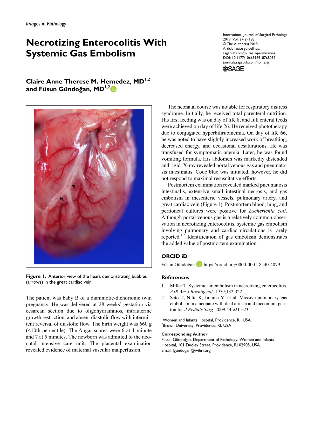
Postmortem examination revealed marked pneumatosis intestinalis, extensive small intestinal necrosis, and gas embolism in mesenteric vessels, pulmonary artery, and great cardiac vein (Figure 1). Postmortem blood, lung, and peritoneal cultures were positive for Escherichia coli. Although portal venous gas is a relatively common observation in necrotizing enterocolitis, systemic gas embolism involving pulmonary and cardiac circulations is rarely reported.1,2 Identification of gas embolism demonstrates the added value of postmortem examination.
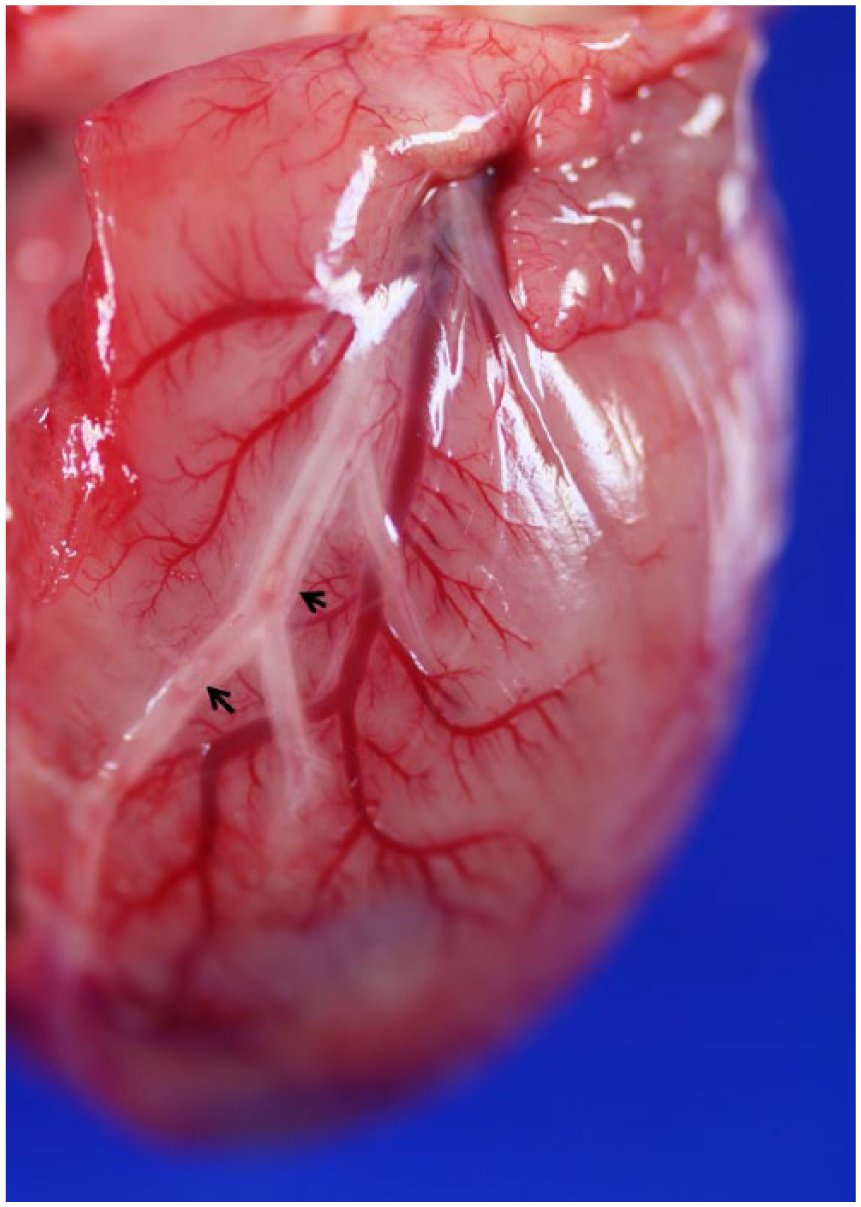
Anterior view of the heart demonstrating bubbles (arrows) in the great cardiac vein.
