






Abstract
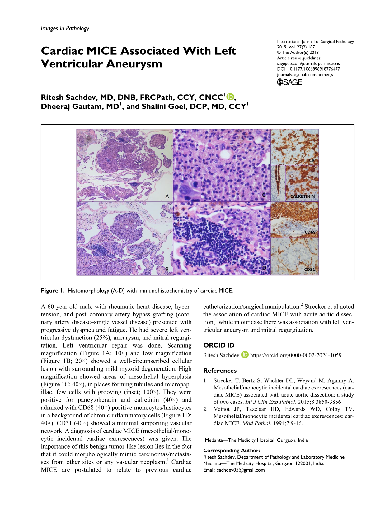
A 60-year-old male with rheumatic heart disease, hypertension, and post–coronary artery bypass grafting (coronary artery disease–single vessel disease) presented with progressive dyspnea and fatigue. He had severe left ventricular dysfunction (25%), aneurysm, and mitral regurgitation. Left ventricular repair was done. Scanning magnification (Figure 1A; 10×) and low magnification (Figure 1B; 20×) showed a well-circumscribed cellular lesion with surrounding mild myxoid degeneration. High magnification showed areas of mesothelial hyperplasia (Figure 1C; 40×), in places forming tubules and micropapillae, few cells with grooving (inset; 100×). They were positive for pancytokeratin and calretinin (40×) and admixed with CD68 (40×) positive monocytes/histiocytes in a background of chronic inflammatory cells (Figure 1D; 40×). CD31 (40×) showed a minimal supporting vascular network. A diagnosis of cardiac MICE (mesothelial/monocytic incidental cardiac excrescences) was given. The importance of this benign tumor-like lesion lies in the fact that it could morphologically mimic carcinomas/metastases from other sites or any vascular neoplasm. 1 Cardiac MICE are postulated to relate to previous cardiac catheterization/surgical manipulation. 2 Strecker et al noted the association of cardiac MICE with acute aortic dissection, 1 while in our case there was association with left ventricular aneurysm and mitral regurgitation.
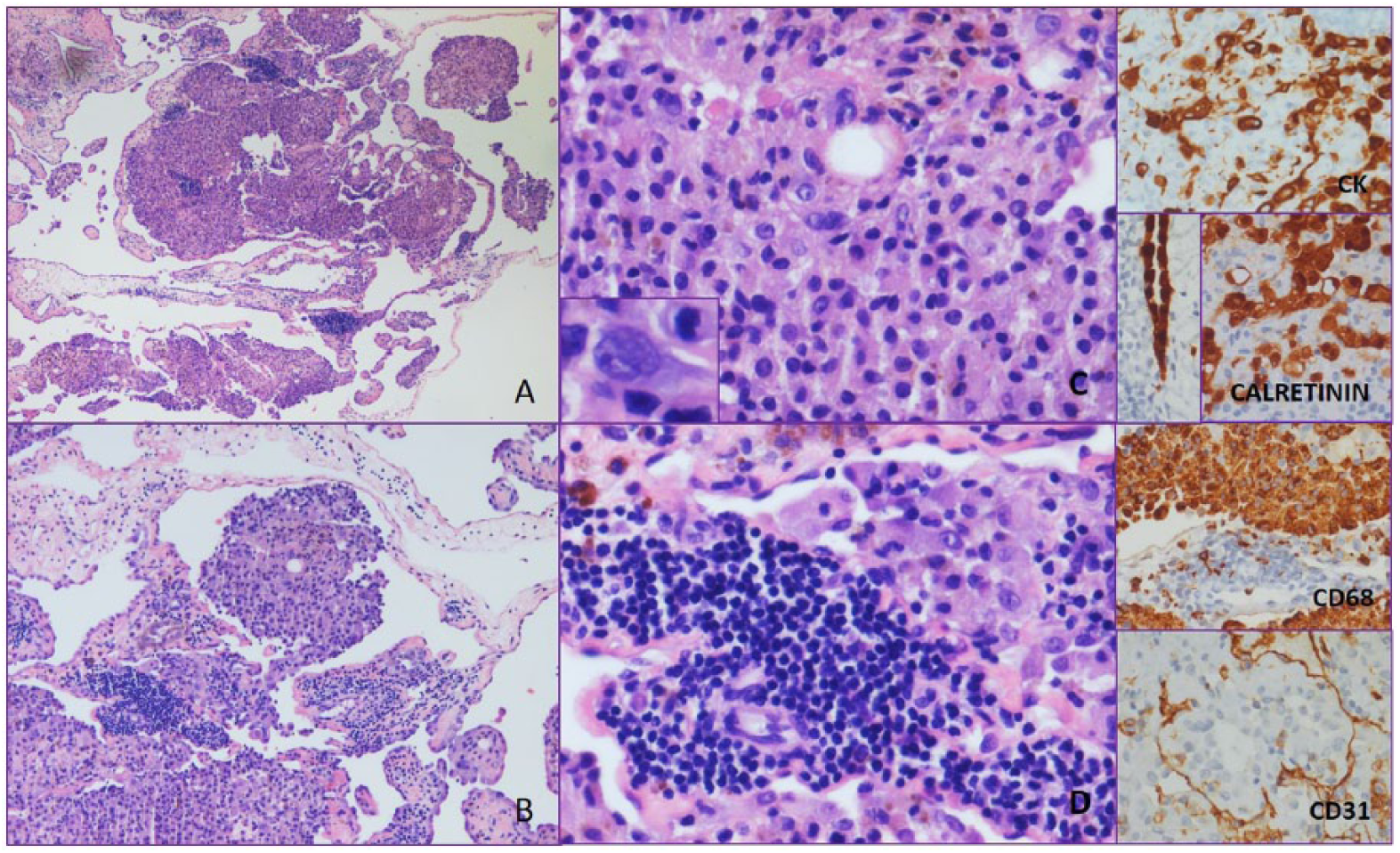
Histomorphology (A-D) with immunohistochemistry of cardiac MICE.
