






Abstract
The colonic epithelium is a perfect functional barrier, separating the interior of the intestinal lumen from the sterile inner layers forming the intestinal wall. This functional barrier consists of absorptive enterocytes, goblet cells, Paneth cells, and enteroendocrine cells that undergo continuous cycles of renewal. 1
The mucus covering the intestinal epithelium promotes the progression of the fecal contents, provides the first line of innate defense of the colonic mucosa, and is secreted by goblet cells lining the intestinal glands called crypts of Lieberkühn.1,2
The goblet cell morphology is shaped by the distended theca containing the mucin granules located below the apical membrane. Goblet cells synthesize bioactive secretory mucin glycoproteins, trefoil factor peptides, resistin-like molecule β, and Fc-γ binding protein. 2 Crypts open into surface epithelium or into innominate grooves, are surrounded by pericryptal fibroblast sheath, and also contain precursor cells, endocrine cells, and Paneth cells in right sided colon. 2
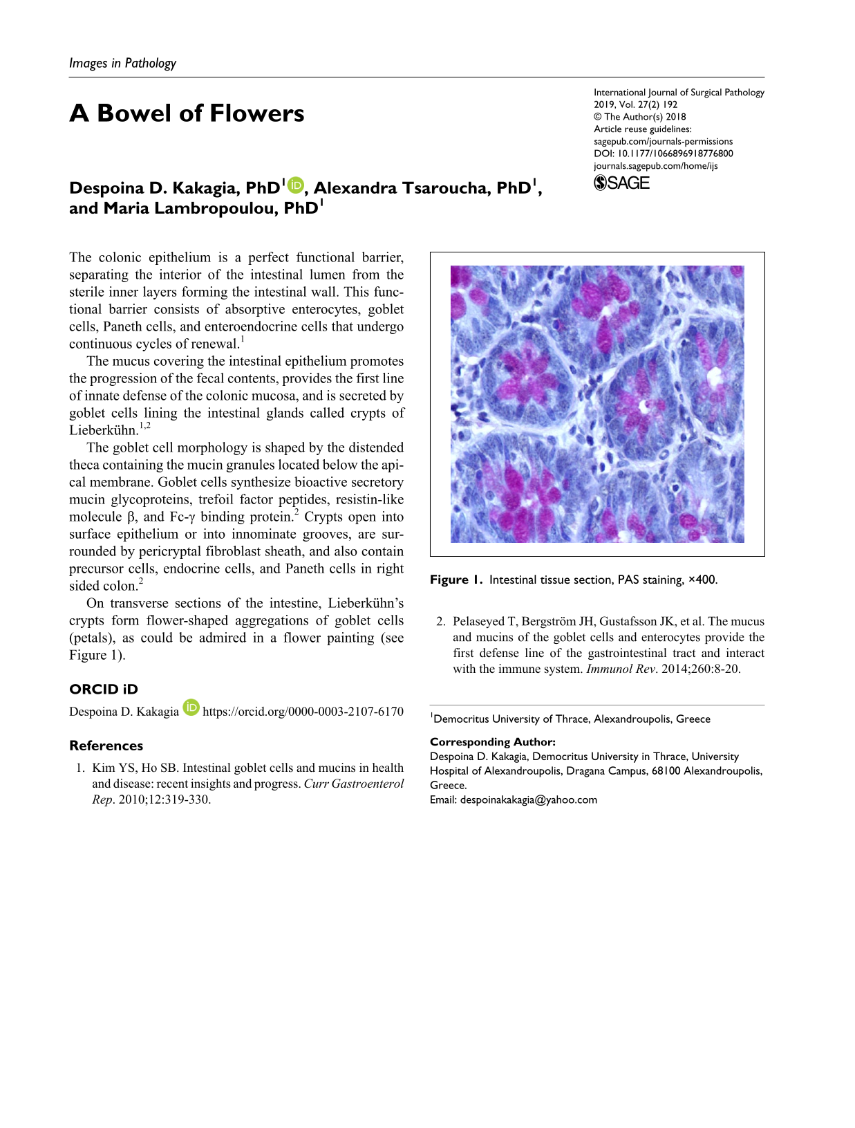
On transverse sections of the intestine, Lieberkühn’s crypts form flower-shaped aggregations of goblet cells (petals), as could be admired in a flower painting (see Figure 1).
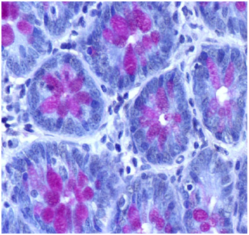
Intestinal tissue section, PAS staining, ×400.
