






Abstract
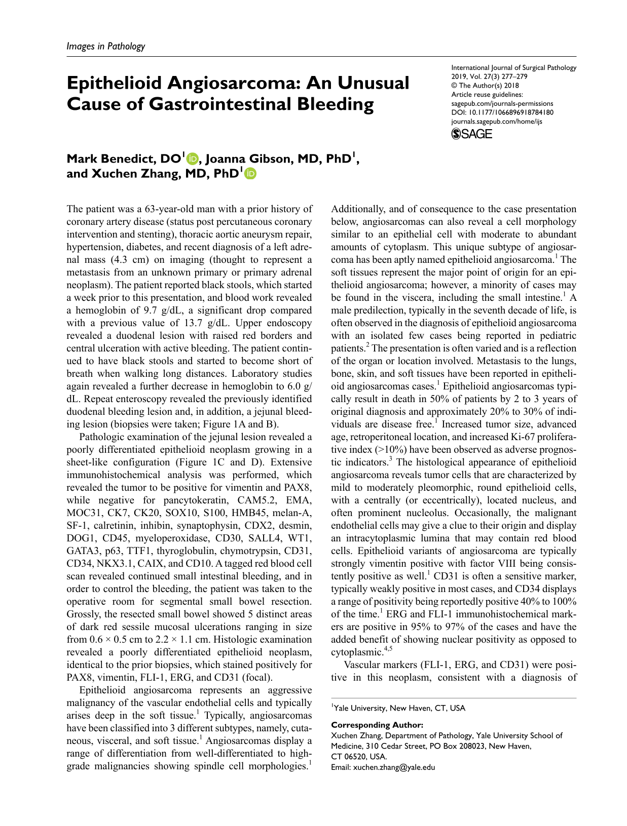
The patient was a 63-year-old man with a prior history of coronary artery disease (status post percutaneous coronary intervention and stenting), thoracic aortic aneurysm repair, hypertension, diabetes, and recent diagnosis of a left adrenal mass (4.3 cm) on imaging (thought to represent a metastasis from an unknown primary or primary adrenal neoplasm). The patient reported black stools, which started a week prior to this presentation, and blood work revealed a hemoglobin of 9.7 g/dL, a significant drop compared with a previous value of 13.7 g/dL. Upper endoscopy revealed a duodenal lesion with raised red borders and central ulceration with active bleeding. The patient continued to have black stools and started to become short of breath when walking long distances. Laboratory studies again revealed a further decrease in hemoglobin to 6.0 g/dL. Repeat enteroscopy revealed the previously identified duodenal bleeding lesion and, in addition, a jejunal bleeding lesion (biopsies were taken; Figure 1A and B).

(A) Endoscopic image revealing a raised centrally ulcerated lesion within the duodenum. (B) Endoscopic image revealing a mass lesion within the jejunum. (C) Microscopic image of small bowel revealing a primarily submucosally based lesion (hematoxylin-eosin [H&E] stain; 40×). (D) Higher power image of “C” revealing large pleomorphic cells with moderate amounts of cytoplasm (epithelioid), prominent nucleoli, and intermixed red blood cells (H&E stain; 200×).
Pathologic examination of the jejunal lesion revealed a poorly differentiated epithelioid neoplasm growing in a sheet-like configuration (Figure 1C and D). Extensive immunohistochemical analysis was performed, which revealed the tumor to be positive for vimentin and PAX8, while negative for pancytokeratin, CAM5.2, EMA, MOC31, CK7, CK20, SOX10, S100, HMB45, melan-A, SF-1, calretinin, inhibin, synaptophysin, CDX2, desmin, DOG1, CD45, myeloperoxidase, CD30, SALL4, WT1, GATA3, p63, TTF1, thyroglobulin, chymotrypsin, CD31, CD34, NKX3.1, CAIX, and CD10. A tagged red blood cell scan revealed continued small intestinal bleeding, and in order to control the bleeding, the patient was taken to the operative room for segmental small bowel resection. Grossly, the resected small bowel showed 5 distinct areas of dark red sessile mucosal ulcerations ranging in size from 0.6 × 0.5 cm to 2.2 × 1.1 cm. Histologic examination revealed a poorly differentiated epithelioid neoplasm, identical to the prior biopsies, which stained positively for PAX8, vimentin, FLI-1, ERG, and CD31 (focal).
Epithelioid angiosarcoma represents an aggressive malignancy of the vascular endothelial cells and typically arises deep in the soft tissue. 1 Typically, angiosarcomas have been classified into 3 different subtypes, namely, cutaneous, visceral, and soft tissue. 1 Angiosarcomas display a range of differentiation from well-differentiated to high-grade malignancies showing spindle cell morphologies. 1 Additionally, and of consequence to the case presentation below, angiosarcomas can also reveal a cell morphology similar to an epithelial cell with moderate to abundant amounts of cytoplasm. This unique subtype of angiosarcoma has been aptly named epithelioid angiosarcoma. 1 The soft tissues represent the major point of origin for an epithelioid angiosarcoma; however, a minority of cases may be found in the viscera, including the small intestine. 1 A male predilection, typically in the seventh decade of life, is often observed in the diagnosis of epithelioid angiosarcoma with an isolated few cases being reported in pediatric patients. 2 The presentation is often varied and is a reflection of the organ or location involved. Metastasis to the lungs, bone, skin, and soft tissues have been reported in epithelioid angiosarcomas cases. 1 Epithelioid angiosarcomas typically result in death in 50% of patients by 2 to 3 years of original diagnosis and approximately 20% to 30% of individuals are disease free. 1 Increased tumor size, advanced age, retroperitoneal location, and increased Ki-67 proliferative index (>10%) have been observed as adverse prognostic indicators. 3 The histological appearance of epithelioid angiosarcoma reveals tumor cells that are characterized by mild to moderately pleomorphic, round epithelioid cells, with a centrally (or eccentrically), located nucleus, and often prominent nucleolus. Occasionally, the malignant endothelial cells may give a clue to their origin and display an intracytoplasmic lumina that may contain red blood cells. Epithelioid variants of angiosarcoma are typically strongly vimentin positive with factor VIII being consistently positive as well. 1 CD31 is often a sensitive marker, typically weakly positive in most cases, and CD34 displays a range of positivity being reportedly positive 40% to 100% of the time. 1 ERG and FLI-1 immunohistochemical markers are positive in 95% to 97% of the cases and have the added benefit of showing nuclear positivity as opposed to cytoplasmic.4,5
Vascular markers (FLI-1, ERG, and CD31) were positive in this neoplasm, consistent with a diagnosis of epithelioid angiosarcoma (Figure 2). Angiosarcomas are very rare tumors of the gastrointestinal (GI) tract. 6 The clinical presentation is variable and may include GI tract bleeding, anemia, intestinal obstruction, abdominal pain, nausea, weight loss, shortness of breath, weakness, and diarrhea. 6 According to one of the larger case series (27 cases of angiosarcoma involving the small intestine), the most common presentation is abdominal pain and GI bleeding, reported in approximately 37% of patients. 7 Overall, 85.2% angiosarcoma in the GI tract are primary to the small intestine and 14.8% are indeterminate as to primary origin. 7 Angiosarcomas are classified as well differentiated, poorly differentiated, and epithelioid. 6 Epithelioid angiosarcomas pose additional challenges as they can be misdiagnosed, given significant morphologic overlap compared with the poorly differentiated carcinomas, melanoma, epithelioid GI stromal tumor, epithelioid leiomyosarcomas, among other tumors with epithelioid morphology. 6 Although immunohistochemical analysis can be useful for the confirmation of vascular differentiation in angiosarcoma, antigen heterogeneity within tumors and small tissue samples may limit the ability to accurately diagnose the tumor. For example, in this patient’s tumor, the tumor in the original biopsy was negative for CD31 and CD34, leading to erroneous interpretation that vascular differentiation was not present. These markers are typically positive in 88% and 73% of angiosarcomas, respectively. However, in the resection specimen, focal staining for CD31 was seen in the tumor. In addition, arriving at the correct diagnosis in this patient was also challenging due to the original biopsy showing staining for PAX8 (subsequent resection specimen also revealed PAX8 positivity). PAX8, a nephric-lineage transcription factor, is an integral part of the development of the thyroid gland, kidney, and Mullerian system. 8 Immunohistochemical positivity with PAX8 has been seen in various malignancies and include renal cell carcinoma, ovarian carcinoma, thyroid carcinoma, endometrial carcinoma, cervical carcinoma, and squamous cell carcinoma. 8 Limited expression has also been observed in bladder carcinoma and lung carcinomas. 8 To date, there has been no literature reported case of angiosarcoma with positivity for PAX8. Originally vimentin and PAX8 expression on the biopsy with negativity for CD31 and CD34 lead to other differential diagnostic considerations in which PAX8 has been known to be positive, namely, renal in this case (coupled with an imaging report that revealed an adrenal mass, which may have actually been arising from the kidney, a fact that was unclear initially). Ultimately, the expression of nuclear markers FLI-1 and ERG on the small bowel resection confirmed the diagnosis of epithelioid angiosarcoma.

(A) Microscopic image of a small bowel epithelioid angiosarcoma with large pleomorphic cells, abundant cytoplasm, prominent nucleoli, and red blood cells with the malignant cells (hematoxylin-eosin [H&E] stain; 400×). (B) ERG immunostain revealing nuclear positivity supporting a vascular origin (200×). (C) Low-power view of PAX8 immunostain revealing nuclear positivity in the tumor cells (40×). (D) High-power view of PAX8 immunostain revealing nuclear positivity within this small bowel angiosarcoma (200×).
