






Abstract
Although pancreatic and gastric heterotopias are common findings in the gastrointestinal tract, heterotopic respiratory mucosa (HRM) in the rectum is extremely rare and has only been reported twice previously. We are presenting, to our knowledge, the third case of HRM in the rectum. A 56-year-old man with a history of chronic diarrhea presented for diagnostic colonoscopy, where he was found to have a rectal subepithelial nodule. He was subsequently referred to a tertiary medical center for further evaluation with rectal endoscopic ultrasound. Endoscopically, the nodule was hypoechoic, 2 to 3 mm in size, located in the submucosa, and did not appear to invade the muscularis propria. An uncomplicated endoscopic submucosal resection was subsequently performed. Microscopically, the nodule showed a multicystic complex lesion located in the submucosa, lined by ciliated pseudostratified columnar epithelium and surrounded by thin to moderately thick smooth muscle bundles and multiple lobules of seromucinous glands. There was associated acute and chronic inflammation. The rectum overlying the subepithelial lesion was lined by congested and edematous colonic mucosa and demonstrated no connection with the underlying cystic lesion. Immunohistochemical stains showed positive p63 basal cell staining in the respiratory epithelium of the lesion, while CDX2, TTF-1, and estrogen receptors were all negative. HRM is a benign nonneoplastic lesion with unclear etiology. Pathologists and gastroenterologists should be aware of this entity and consider it in their differential diagnosis for a subepithelial nodule in the rectum, keeping in mind that neoplastic processes can also develop in this location.
Keywords
Background
Heterotopia describes the presence of histologically normal tissue in an ectopic anatomic location, which coexists with the normal tissue at that anatomic location. Although pancreatic and gastric heterotopias are common findings in the gastrointestinal (GI) tract, heterotopic respiratory mucosa (HRM) in the rectum is extremely rare; this has only been reported twice previously. We are presenting, to our knowledge, the third case of HRM in the rectum.
Case Report
A 56-year-old Caucasian male with no significant past medical history presented with chronic diarrhea and defecatory syncope. Colonoscopy was performed, and the patient was found to have multiple histologically proven hyperplastic polyps and tubular adenomas in the ascending, descending, and sigmoid colon. A 1-cm subepithelial rectal nodule was also found and biopsied to show a few hyperplastic glands and a lymphoid aggregate with no dysplasia. He was subsequently referred to a tertiary medical center for rectal endoscopic ultrasound (EUS). The patient underwent flexible sigmoidoscopy with EUS to exclude other causes of subepithelial nodules such as GI stromal tumor or neuroendocrine tumor. EUS demonstrated a nodule that was hypoechoic, 2 to 3 mm in size, located in the submucosa, and did not appear to invade the muscularis propria (Figure 1). A multiband mucosectomy kit was used to create a pseudopolyp, and an uncomplicated endoscopic mucosal resection was subsequently performed. Microscopically, the nodule showed a multicystic complex lesion located in the submucosa composed of glands lined by ciliated pseudostratified columnar epithelium and surrounded by thin to moderately thick smooth muscle bundles and multiple lobules of seromucinous glands (Figures 2 and 3). There was associated acute and chronic inflammation. The rectum overlying the submucosal lesion was lined by congested and edematous colonic mucosa and demonstrated no connection with the underlying cystic lesion. No cytological atypia or mitotic figures were identified within the lesion. Immunohistochemical stains were performed: p63 (clone BC4A4, Biocare Medical, Concord, CA), CDX2 (clone DAKO, Agilent Technologies, DAKO, Santa Clara, CA), TTF-1 (clone 8G7G3/1, Agilent Technologies, Dako), and estrogen receptor (pharmDx kit, clone 1D5 and ER-2-123, Agilent Technologies, Dako). The lesion showed positive p63 basal cell staining in the respiratory epithelium, while CDX2, TTF-1, and estrogen receptors were all negative (Figures 4 and 5).
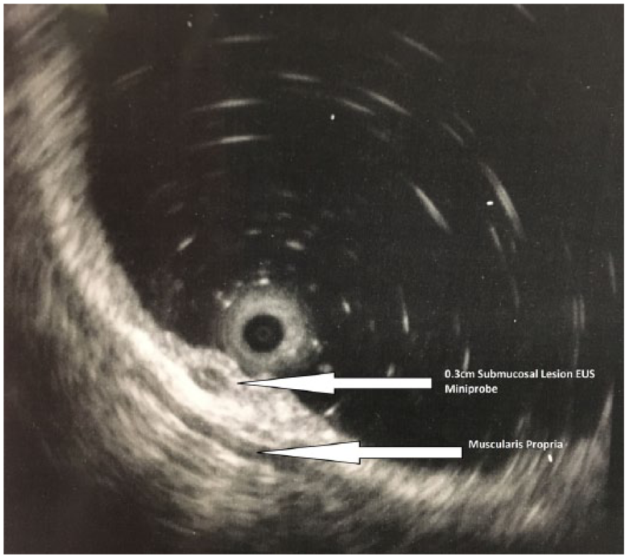
Hypoechoic nodule on endoscopic ultrasound (EUS).
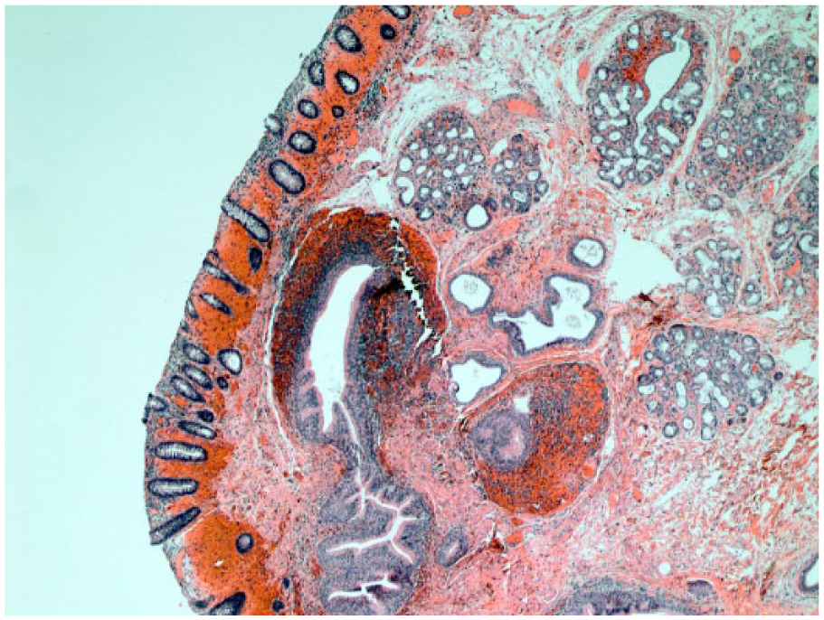
Subepithelial multicystic lesion, composed of glands lined by respiratory epithelium and seromucinous glands with congested overlying colonic mucosa (hematoxylin-eosin, original magnification ×40).
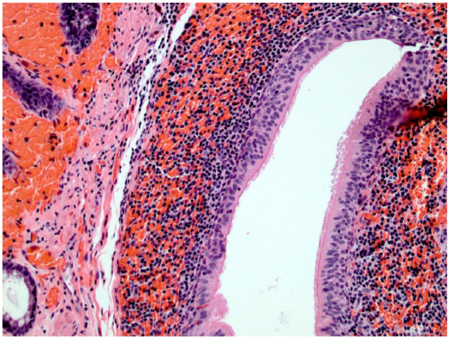
Ciliated pseudostratified columnar epithelium lining the glands and surrounded by chronic inflammation (hematoxylin-eosin, original magnification ×200).
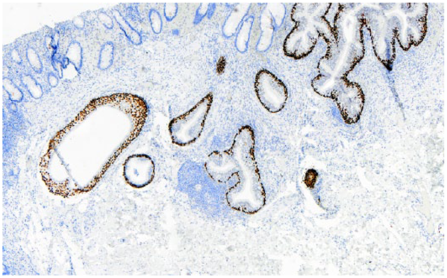
p63 stains the basal cells of the respiratory epithelium (p63, original magnification ×40).
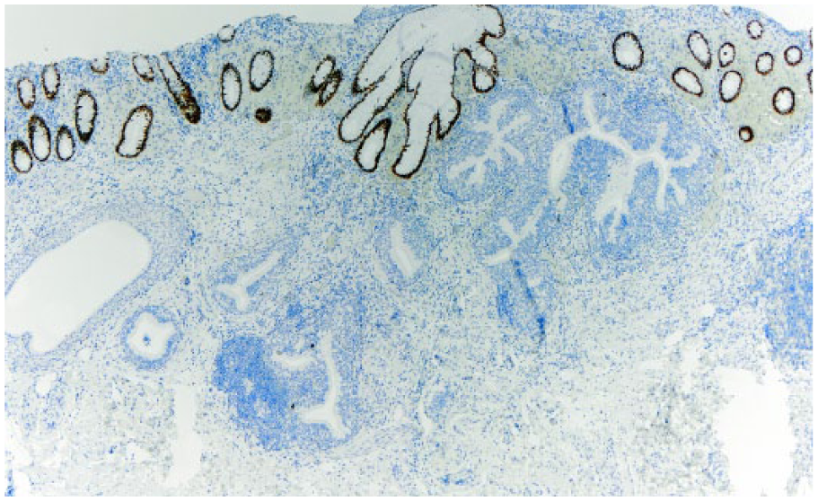
Negative CDX2 in the lesion with positive internal control within the colonic mucosa (CDX2, original magnification ×40).
Discussion
Heterotopia in the GI tract is a common histologic finding, for example, pancreatic heterotopia may be seen in the upper GI tract, especially the stomach. 1 However, heterotopia in the rectum is relatively rare. Our case is in a middle-aged Caucasian male and showed ciliated respiratory epithelium with seromucinous glands. No associated carcinoma was identified. To our knowledge, only 2 cases of HRM in the rectum have been reported in the literature.2,3 Both of these cases were described in middle-aged Japanese females, 50 and 38 years old, respectively.2,3
The first case, reported by Kawahara et al 2 in 2007, presented with hematochezia and was accompanied by rectal carcinoma. On endoscopy, there was a sessile subepithelial polypoid lesion (1.2 × 1.3 cm) with central depression, 2 cm away from the carcinoma component. Microscopically, their case showed an abrupt transition from a benign-appearing colonic mucosa to a branching tubular structure lined by a ciliated pseudostratified columnar and low columnar respiratory epithelium with associated seromucinous glands compatible with respiratory mucosa. The second case, reported by Ishida et al 3 in 2014, described an incidentally found subepithelial nodule (0.7 × 0.3 cm) on colonoscopy. Microscopically, their case showed a unilocular cystic lesion lined by pseudostratified ciliated epithelium and similar to our case showed acute inflammation and had no connection with the surface epithelium. However, the second described case lacked the presence of seromucinous glands and was composed entirely of respiratory epithelium. The second reported case showed positive p63 basal cell staining in the respiratory epithelium with a very low proliferation index based on the Ki-67 (Ki-67 < 1%). Estrogen receptors and progesterone receptors, synaptophysin, and chromogranin A were all negative in the lesion. 3 These immunophenotypic findings are identical to those in our case.
HRM has been reported in other organs including the eye, thyroid, mandible, tongue, manubrium sterni, skin, and uterus with or without a salivary gland component.4-9 Most of the reported HRM lesions were described as cystic lesions in the literature.
Gastric heterotopia is the most common reported epithelial heterotopia in the rectum and elsewhere. A systematic review for gastric heterotopia in the rectum by Iacopini et al 10 showed that oxyntic mucosa was most commonly seen in these lesions. Clinical presentation for these patients ranged from asymptomatic and incidentally diagnosed lesions to patients with nonspecific symptoms (such as bloating and abdominal discomfort) or specific anorectal symptoms (such as hematochezia). Congenital malformations were observed in 24% of the cases. Ectopic prostatic and pancreatic tissue as well as salivary gland tissue in the rectum typically occurs as an isolated finding and cases with associated gastric heterotopia have also been described in the literature.11-16
Heterotopic tissue anywhere in the body can have a potential risk of neoplastic transformation. There are reports of colonic adenocarcinoma arising from gastric heterotopia in the transverse colon and a case of invasive ductal adenocarcinoma arising from ectopic pancreatic tissue in the rectum.14,15 It is hypothesized that the number of malignant transformed cases might be underestimated due to the fact that the malignant tumor may obscure the presence of the heterotopic tissue. 15
For the pathologist, an important consideration discussed by Dinarvand et al, 16 given the rarity of such lesions in the rectum, is to exclude a possible floater or tissue contaminant by evaluating deeper levels, if necessary. A definitive diagnosis can usually be reached by seeing a mixture of 2 mucosal types (ie, colonic and respiratory). 16
The histogenesis of HRM in the rectum is still unclear. Several etiologies have been suggested including a metaplastic process as a response to inflammation or injury, fetal differentiation or developmental error, and pluripotent primitive cells lining the intestinal tract differentiating into a variety of epithelial types.3,9 The Müllerian hypothesis has been excluded based on the estrogen receptor/progesterone receptor negativity in such a lesion by Ishida et al and further confirmed in our case. 3
Conclusion
HRM is a benign nonneoplastic lesion with unclear etiology. Pathologists and gastroenterologists should be aware of this entity and consider it in their differential diagnosis of a subepithelial lesion in the rectum, keeping in mind that neoplastic processes can also develop in this location.
Footnotes
Authors’ Note
A portion of this study was presented at the College of American Pathologists annual meeting in 2017 in National Harbor, Maryland.
Declaration of Conflicting Interests
The author(s) declared no potential conflicts of interest with respect to the research, authorship, and/or publication of this article.
Funding
The author(s) received no financial support for the research, authorship, and/or publication of this article.
Ethical Approval
Not applicable, because this article does not contain any studies with human or animal subjects.
Informed Consent
Not applicable, because this article does not contain any studies with human or animal subjects.
Trial Registration
Not applicable, because this article does not contain any clinical trials.
