






Abstract
NUT (nuclear protein in testis) carcinoma (NC) is an aggressive carcinoma characterized by rearrangements of the NUT gene on chromosome 15q14. Histologically, it is a poorly differentiated carcinoma composed of monotonous, medium-sized, round cells with scant amphophilic or eosinophilic cytoplasm. Foci of abrupt keratinization are often seen. In this report, we compare the morphology of 2 cases of NC. The first case shows characteristic features of uniform, round epithelioid cells admixed with foci of abrupt keratinization. The second case demonstrates nests of epithelioid-polygonal cells that appear to be loosely cribriform within a mucoid stroma. Although considered rare, the actual incidence of NC may be underestimated, as it is likely that many go undiagnosed because the morphology deviates from what is typical. Our report demonstrates that NC should always be considered in any case of an undifferentiated carcinoma and should not be excluded if typical histologic and immunohistochemical features of squamous differentiation are lacking.
Keywords
Introduction
NUT (nuclear protein in testis) carcinoma (NC) is an aggressive carcinoma characterized by rearrangements of the NUT gene on chromosome 15q14.1-3 It was first identified in 1991 when 2 separate groups published case reports of intrathoracic high-grade carcinomas harboring a t(15;19) translocation.4,5 In two thirds of cases, a reciprocal chromosomal translocation occurs between NUT and BRD4 on chromosome 19. 2 The remaining one third of cases pair with BRD3 on chromosome 9 or with undefined fusion partners, known as NUT-variant carcinomas.2,6,7
Initially NC was thought to be a childhood cancer, but later studies have shown that they can develop in patients of any age.3,8 There is no gender or sex predilection. 1 Many studies report that NCs arise in the midline and affect the mediastinum/thymus, upper aerodigestive tract, nasal cavity, and sinus.1,8,9 However, cases have been reported in infradiaphragmatic and non-midline structures as well. 9 Patients usually present with mass-related symptoms and nonspecific symptoms, such as fever and weight loss.1,10 NC is locally aggressive and displays early hematogenous metastasis. 2 Prognosis is extremely poor, with a median overall survival of 6.7 months.2,9
Histologically, NC has been described as a poorly differentiated carcinoma composed of monotonous, medium-sized, round cells with scant amphophilic or eosinophilic cytoplasm.1,10 The nuclei have irregular contours with fine to vesicular chromatin and prominent nucleoli. 1 Foci of abrupt keratinization are often seen.1,2,6,9,10 Diagnosis can be confirmed by demonstration of the NUT translocation with conventional karyotyping, reverse transcriptase-polymerase chain reaction, or fluorescence in situ hybridization (FISH).1-3,6 Reverse transcriptase-polymerase chain reaction is less sensitive than FISH, however, as it cannot be used to diagnose NUT-variant carcinomas.1,10 Diagnosis can also be confirmed with NUT immunohistochemistry (IHC). In 2009, Haack et al 11 demonstrated that IHC staining with the monoclonal antibody C52 correlates with rearrangement of NUT. 11 The antibody demonstrates 87% sensitivity and 100% specificity and will detect all forms of NC, including BRD4-NUT, BRD3-NUT, and NUT variants.1-3,10 A positive result is described as diffuse, speckled/punctate nuclear staining in more than 50% of tumor cells. 1 In this report, we compare the morphology of 2 cases with a confirmed diagnosis of NC, one with a classic presentation and the other with unusual features.
Case Reports
Case 1
A 19-year-old Caucasian male with a history of irritable bowel syndrome presented to his pediatrician with a 1.5-month history of dry cough, chest tightness, hemoptysis, and shortness of breath. He denied fever, chills, and night sweats but experienced a decrease in appetite that led to a weight loss of 7 pounds. He was a former smoker with a 0.3 pack per day history and consumed alcohol. After treatment with an inhaler followed by antibiotics and steroids proved ineffective, a chest X-ray was performed and showed atelectasis of the right lower and middle lobe with associated mass. Computed tomography scan revealed mediastinal adenopathy, a 3.0 × 3.0 × 2.8 cm right hilar mass, and complete right lower lobe and partial right middle lobe collapse (Figure 1). Bronchoscopy was performed and revealed a large, obstructive, exophytic, fungating, raised mass in the middle portion in the bronchus intermedius. The mass was biopsied and a stent was placed in the right lower lobe of the lung.
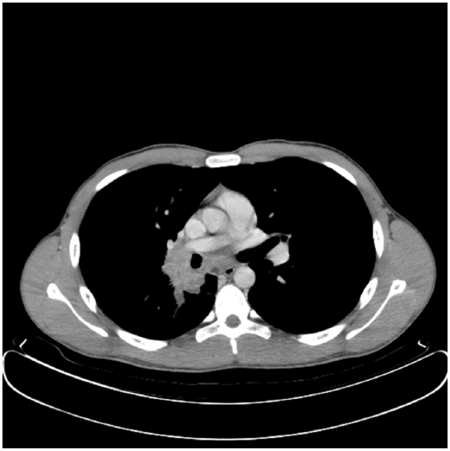
Initial chest computed tomography mediastinal window demonstrating the right hilar mass, endobronchial tumor, and subcarinal lymphadenopathy.
Histopathologic examination of the soft tissue revealed an invasive moderately to poorly differentiated keratinizing squamous cell carcinoma with areas of invasive basaloid tumor islands (Figure 2A and B). Tumor cells showed nuclear variability, moderate/high mitotic activity, and prominent nucleoli. IHC stains for p16INK4a demonstrated patchy positivity. High-risk human papilloma virus in situ hybridization, Epstein-Barr virus–encoded RNA in situ hybridization, and PAX8 were negative. Further IHC with NUT-C52 showed speckled nuclear staining in over 50% of tumor cells (Figure 2C and D).
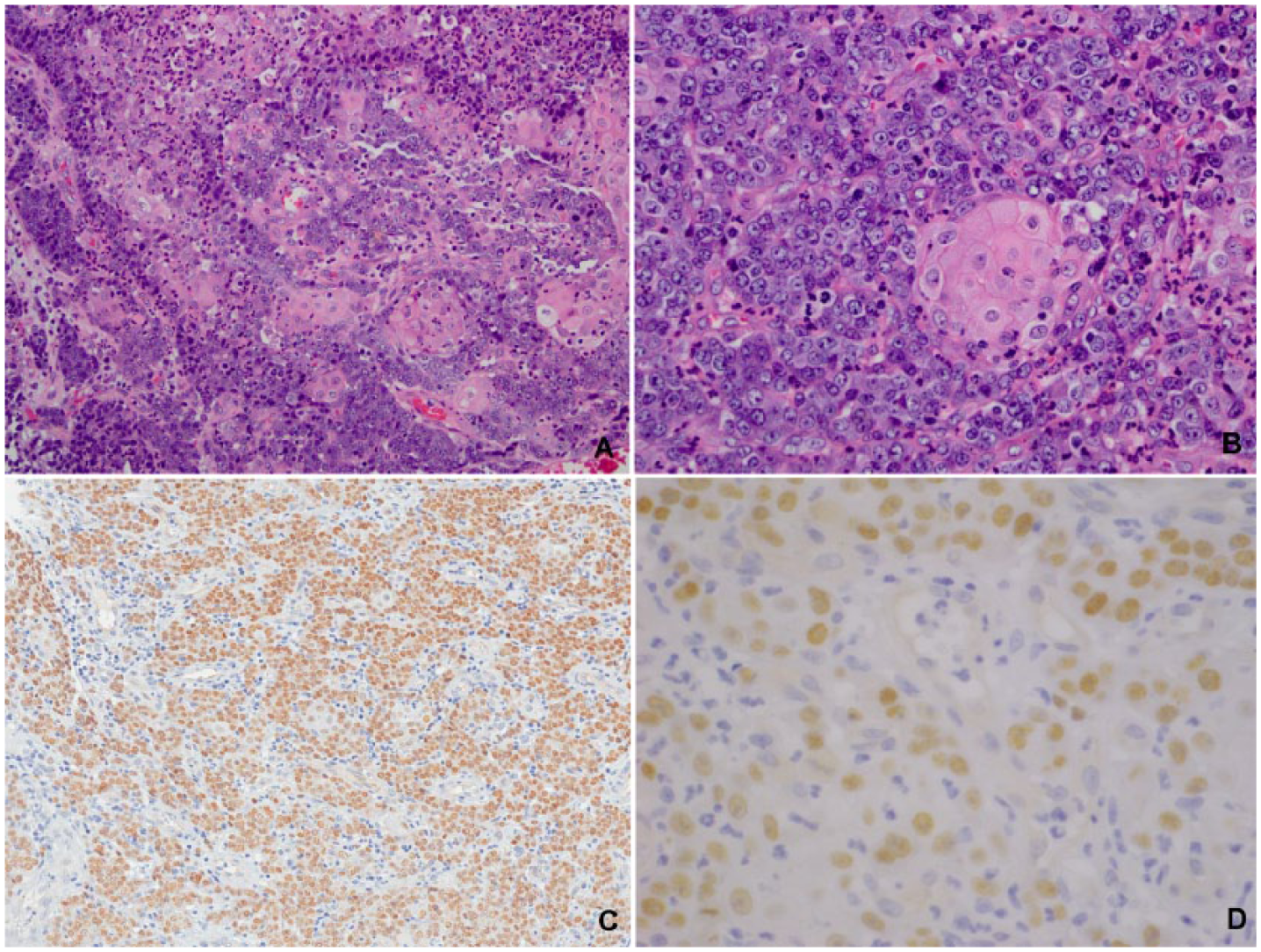
Histology and immunohistochemistry for Case 1. (A) Sheets of uniform, medium-sized, epithelioid cells with prominent nucleoli (10×). (B) Higher magnification showing focus of abrupt keratinization (20×). (C) NUT-C52 showing diffuse positive staining in >50% of tumor cells (20×). (D) Higher magnification of NUT-C52 showing speckled nuclear positivity (60×).
Magnetic resonance imaging of the brain revealed no evidence of metastasis. A positron emission tomography scan showed abnormal fluorodeoxyglucose uptake within the right hilar mass as well as metastases to subcarinal and level 4R mediastinal lymph nodes and the anterior fourth rib. A final diagnosis of Stage 4 T2N2M1 NUT carcinoma was made. The patient was treated with cisplatin/etoposide with concurrent radiation therapy. He succumbed to the disease 1½ years later.
Case 2
A 66-year-old male with a history of Parkinson’s disease was admitted to the intensive care unit for airway monitoring due to a subglottic mass. Associated symptoms included tachycardia, light-headedness, shortness of breath, stridor, sore throat, odynophagia, decreased appetite, weight loss of 10 to 20 pounds, hemoptysis, and leukocytosis. He denied chest pain, palpitations, fever, and chills. A former smoker with a 5 pack-year history, he denied recent alcohol use. The patient was started on intravenous steroids and nebulized racemic epinephrine. He was also started on intravenous aztreonam and vancomycin due to leukocytosis. Magnetic resonance imaging of the neck thyroid/trachea revealed a circumferential, subtly enhancing mass in the subglottic region, spanning at least 2.4 cm that resulted in near complete obliteration of the subglottic airway. The thyroid gland was heterogeneous and enlarged, representing tumoral infiltration. There was effacement of the fat plane between the esophagus and left posterior heterogeneous thyroid mass extension, which was concerning for invasion. Multiple enlarged cervical lymph nodes were identified bilaterally, suggestive of extensive metastatic involvement.
Approximately 1 week later, the patient was admitted for surgery. A large exophytic mass blocking 80% of the lumen was identified in the left subglottis and was subsequently debulked and partially resected. Histopathologic examination of the soft tissue demonstrated nests of tumor cells within a mucoid stroma. The nests appeared to be loosely cribriform (Figure 3A). The cells had basaloid features with scant cytoplasm and increased mitoses and apoptosis. Very focal comedonecrosis was identified. Focally, the tumor appeared to connect to the surface squamous epithelium, which appeared dysplastic. There were rare foci of squamous differentiation within the tumor (Figure 3B). Perineural invasion was not identified.
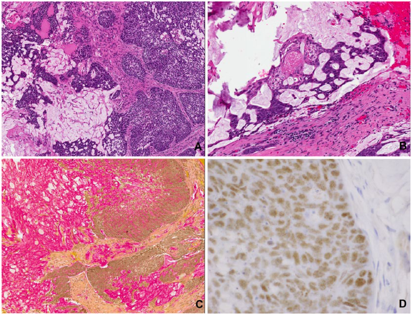
Histology and immunohistochemistry for Case 2. (A) Nests of basaloid and epithelioid-polygonal cells in a loosely cribriform pattern (10×). (B) Focus of abrupt keratinization (20×). (C) Mucicarmine staining (10×). (D) NUT-C52 showing speckled nuclear positivity (60×).
IHC stains for cytokeratin (CK) 5/6, carcinoembryonic antigen, epithelial membrane antigen, p63, p40, and p16INK4a demonstrated patchy positivity. Myoepithelial markers (calponin, S100, smooth muscle myosin heavy chain, GFAP, and smooth muscle actin), neuroendocrine markers (synaptophysin and chromogranin), CD34, and CDX2 were negative. There was no loss of INI-1. There was very focal positivity for CD117. Small nests, cords, and single cells were positive for CK7 and CK20, but definite lumina were not identified within the nests. The mucoid material was positive for mucicarmine (Figure 3C) and alcian blue at pH 2.5. The tumor cells displayed speckled nuclear positivity (Figure 3D) for NUT-C52 IHC in over 50% of tumor cells. Rearrangement of the NUT gene at 15q14 was confirmed with FISH analysis.
After the initial biopsy and debulking, the patient presented to the emergency room with stridor and respiratory distress within 2 weeks and underwent repeat debulking of the tumor and tracheostomy. He declined chemotherapy and radiation and opted for palliative care. He succumbed to the disease within a month of being transferred to our institution.
Discussion
In this report, we present 2 cases of NC. Both patients had large, obstructive, exophytic masses located in midline structures and metastasis, which are very characteristic of NC. The patients also presented with overlapping symptoms, including hemoptysis, shortness of breath, and decreased appetite that led to significant weight loss. Neither patient experienced fever or chills. While the patient in case 1 is 19 years old, the patient in case 2 is 66 years old, supporting the claim that NC can develop in patients of any age.3,8
The diagnosis of case 1 was confirmed with NUT IHC. The diagnosis of case 2 was confirmed with both NUT IHC and FISH analysis. Although FISH analysis is the gold standard for diagnosing NC, 12 the NUT monoclonal antibody is also reliable, as it is both highly sensitive and highly specific.1-3,10 Additionally, NUT protein expression is normally exclusive to the testes, thus expression outside the testes highly correlates with the diagnosis of NC. 3
According to Hellquist et al, 3 NC has characteristic morphological features that include an undifferentiated carcinoma with cells that may mimic a small round blue cell tumor admixed with squamous differentiation. While case 1 fits this description, case 2 has a very different morphology. Many of the cells in case 2 appeared to be forming pseudoglandular spaces with mucin production. Bellizzi et al 6 state that glandular differentiation has been reported in one NC. Rarely, mesenchymal differentiation has also been reported. 13 This has important implications, as some authors have suggested that NUT monoclonal antibody should be routinely included in IHC panels for analysis of all poorly differentiated carcinomas without glandular differentiation. 2 However, case 2 adds to the evidence that NUT monoclonal antibody may need to be included in the IHC panel for any poorly differentiated carcinomas in the appropriate clinical setting.
Most NCs react with antibodies to keratins, consistent with epithelial differentiation, and p63, suggesting squamous differentiation.1,10 In addition to CKs, p63, and p40, NC may express CD34 (>50% of cases), p16, TTF-1, and neuroendocrine markers.14-16 However, case 2 was focally positive for CK7 and CK20 and patchy positive for p63. Furthermore, Dickson et al 7 have demonstrated that visceral and soft tissue tumors may harbor the NUT gene fusion. These tumors exhibited morphologies that ranged from round, rhabdoid, and epithelioid cells to spindle-shaped cells, with focal to absent CK and p63. 7 None of the cases demonstrated overt squamous differentiation in the form of keratinization and occurred in locations that deviated from the midline. 7 It is unclear how these NUT mutation-associated visceral and soft tissue neoplasms are related to NC.
Although case 2 occurred in an anatomic structure that is characteristic of NC, the morphology and immunophenotype is very different from most NCs. Similar to some of Dickson et al’s 7 cases, case 2 had cells that were more epithelioid to polygonal with very sparse squamous differentiation. Given the differences in morphology and immunophenotype of case 2 from most NCs reported in the literature, we support Dickson et al’s 7 theory that NC is part of a spectrum of NUT-associated tumors. We also support the idea that NC should not be ruled out for cases that demonstrate patchy or focal positivity for CKs and p63, or cases that are completely negative for CKs and p63.
BRD4 is a member of the dual bromodomain family of proteins. BRD4-NUT oncoprotein blocks differentiation of cells and maintains proliferation. Targeted therapies, such as bromodomain inhibitors (BETi) and histone deacetylase inhibitors, which induce differentiation and growth arrest of NC cells, are now available as clinical trials. The former is available for adults and the latter has been associated with a good clinical response in the pediatric age-group. 17
Conclusion
NC is an aggressive malignancy with a tendency for early metastases that is inevitably fatal. 7 Although considered rare, the actual incidence of NC is unknown and may be underestimated. 7 Our report shows that NC may have classic features (case 1) or variant histology including mucinous differentiation (case 2). It is very probable the incidence of NC is higher, as many may go undiagnosed due to morphology deviating from what is typical. As a result, NC should always be considered in patients with a clinical presentation of clinically aggressive tumors, undifferentiated carcinoma cases, and should not be excluded if typical histologic and IHC features of squamous differentiation are lacking. 9
Footnotes
Declaration of Conflicting Interests
The author(s) declared no potential conflicts of interest with respect to the research, authorship, and/or publication of this article.
Funding
The author(s) received no financial support for the research, authorship, and/or publication of this article.
Ethical Approval
All reviews performed in this study with human participants were done in accordance with the ethical standards of the institutional and/or national research committee and with the 1964 Helsinki Declaration and its later amendments or comparable ethical standards.
Informed Consent
Not applicable, because this article does not contain any studies with human or animal subjects.
Trial Registration
Not applicable, because this article does not contain any clinical trials.
