






Abstract
Lichen sclerosus (LSc) with penile cancer is found in about two thirds of specimens. It has been hypothesized that LSc represents a precancerous condition. To qualify as such, in addition to cytological atypia and similarity with the invasive tumor, a spatial correlation between LSc and neoplastic lesions needs to be demonstrated. The purpose of this study was to evaluate such a spatial relationship. Circumcision (28 cases) and penectomy (81 cases) specimens were evaluated. All cases had LSc, penile intraepithelial neoplasia (PeIN), and/or invasive squamous cell carcinomas. We examined LSc in relation to invasive carcinoma, PeIN, and normal epithelia. Invasive squamous cell carcinomas, classified according to the World Health Organization criteria as non–human papillomavirus (HPV)-related and HPV-related PeIN, were present in 100 cases. Non-HPV-related (differentiated) PeIN was the most common subtype associated with LSc (89%). There were 5 spatial patterns identified: (1) LSc adjacent to PeIN (23%), (2) LSc adjacent and comprising PeIN (42%), (3) LSc next to and within invasive carcinomas (8%), (4) LSc throughout the sequence PeIN-invasive carcinoma (24%), and (5) LSc was separate (with normal tissue between the lesions) from PeIN and/or invasive carcinomas in a minority of cases (3%). LSc within the cancer was not previously described. In this series, we found 35 cases with LSc within invasive carcinomas. The striking continuous spatial relationship among LSc, PeIN, and/or invasive carcinoma as shown in this study may be a necessary (but not sufficient) condition for the hypothesis postulating LSc as a penile precancerous lesion.
Keywords
Introduction
The relationship of lichen sclerosus (LSc), precancerous lesions and invasive squamous cell carcinomas (SCCs) in anogenital sites is poorly understood. After having carefully reviewed the literature, we have encountered that the risk of LSc developing into invasive carcinoma is from 0% 1 to 12.5%. 2 The incidence of coexistence of cancer and LSc found ranged from 1% 3 to 50%. 4 There is a frequent association of LSc and carcinomas in the penis to penile intraepithelial neoplasia (PeIN) in resected specimens.5,6 Examining an adequate number of sections, LSc is found in about two thirds of resection specimens in relation to penile cancer. 7 LSc is found preferentially in association with non–human papillomavirus (HPV)-related subtypes of penile SCCs.8,9 The latter and recent molecular evidence10-12 support the hypothesis of LSc as a possible precancerous condition of non-HPV-related tumors. The examination of hundreds of cases of penile cancer, many of which had associated LSc, allowed us to observe what appeared to be a gradual continuity of LSc with adjacent neoplasia, in situ or invasive. Since the early recognition of a spatial proximity of in situ and invasive cervical carcinomas by Cullen et al in 1903, such a relationship is considered an important prerequisite to justify a precancerous lesion. 13 In order to observe such morphological relationship and to contribute to the hypothesis of LSc as a precancerous condition in detail, we designed a research to document the spatial topographic relation of LSc with PeIN and invasive carcinomas.
Material and Methods
Pathological materials from 109 cases of LSc associated with PeIN and/or invasive carcinomas in the same specimen were evaluated. They were circumcision (28 cases) and partial or total penectomy resection specimens (81 cases) from the archives of the Instituto de Patología e Investigación, Asunción, Paraguay. Morphological criteria for the diagnosis of LSc were previously described. 14 In invasive carcinoma, LSc was documented after the finding of a sclerotic hyalinized stroma at the infiltrating tumor front or within the carcinoma. PeIN and invasive carcinomas were separated as non-HPV and HPV-related, according to the recent World Health Organization classification. 8 We prepared a diagrammatic model of each case where LSc, PeIN, and invasive carcinomas were present in various relationships to help in the interpretation and spatial demonstration of generated data. Each of these lesions was represented by rectangles (LSc), squares (PeIN), and triangles (invasive cancer), respectively. The site of LSc was documented in relation to the other lesions. Lesions were continuous or separate. Separate lesions had normal tissues between them.
Results
General Pathological Features
LSc, PeIN, and invasive SCCs were found in the same specimen in 100 cases. In the remaining 9 cases, LSc and invasive carcinomas were documented but no PeIN was detected. Subtypes of PeIN associated with LSc, present in 100 cases, were differentiated in 89 cases, mixed differentiated + warty/basaloid in 8 cases, and warty/basaloid in 3 cases (Table 1). Morphological variants of SCCs are shown in Table 2. The usual subtype was the most common (50%), followed by other non-HPV-related carcinomas such as pseudohyperplastic papillary NOS (not otherwise specified), and verrucous. HPV-related warty/basaloid carcinomas associated with LSc were less frequent, present in 8% of the invasive carcinomas.
Subtypes of PeIN Associated With LSc (100 Cases).
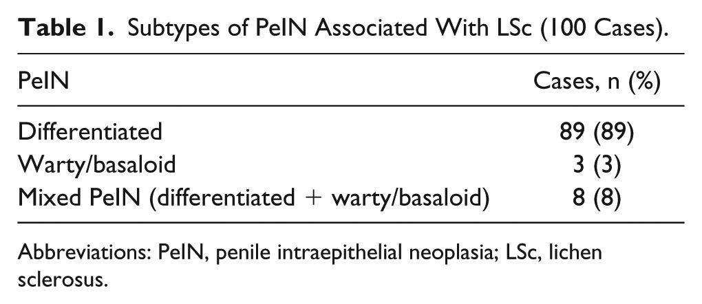
Abbreviations: PeIN, penile intraepithelial neoplasia; LSc, lichen sclerosus.
Subtypes of Squamous Cell Carcinoma Associated With LSc (109 Cases).
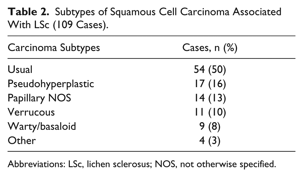
Abbreviations: LSc, lichen sclerosus; NOS, not otherwise specified.
Spatial Topography
There were 5 spatial patterns found (Table 3): (1) LSc was found adjacent to PeIN (23%; Figure 1), (2) LSc was adjacent and part of PeIN (42%; Figure 2), (3) LSc was in continuity to invasive carcinoma (8%), (4) LSc was advent to and part of PeIN and invasive carcinomas (24%; Figure 3), and (5) LSc was a separated, independent lesion, with normal tissue in between PeIN and carcinoma (3%). LSc, in continuity to PeIN and/or invasive carcinoma, was found in more than half of the cases (74%; patterns 2, 3, and 4).
Relationship of Lichen Sclerosus (LSc), Penile Intraepithelial Neoplasia (PeIN), and Invasive Carcinomas.
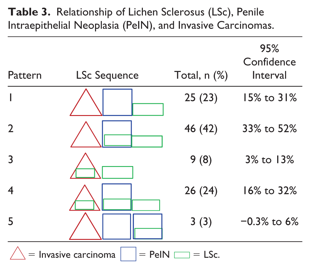
 = Invasive carcinoma
  = PeIN
  = LSc.

(A) Representation of invasive carcinoma, penile intraepithelial neoplasia (PeIN), and lichen sclerosus (LSc) as triangle, square, and rectangle, respectively. (B) LSc with hyperkeratois squamous hyperplasia, and dermal perivascular and linear hyalinization (see *). (C) PeIN without LSc. (D) Squamous carcinoma usual type without LSc. This pattern as observed in 23% of the cases.
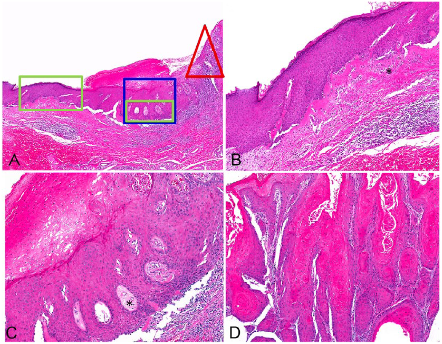
(A) Lichen sclerosus (LSc)-penile intraepithelial neoplasia (PeIN) sequence. (B) LSc in non-atypical squamous epithelium. Note changes of squamous hyperplasia and linear dermal hyalinization. Focal separation at basal layer. (C) Differentiated PeIN with hyper and parakeratosis, squamous hyperplasia, basal layers atypia, and perivascular hyalinization with interphase separation. (D) Invasive carcinoma without presence of LSc. This pattern was observed in 42% of the cases.
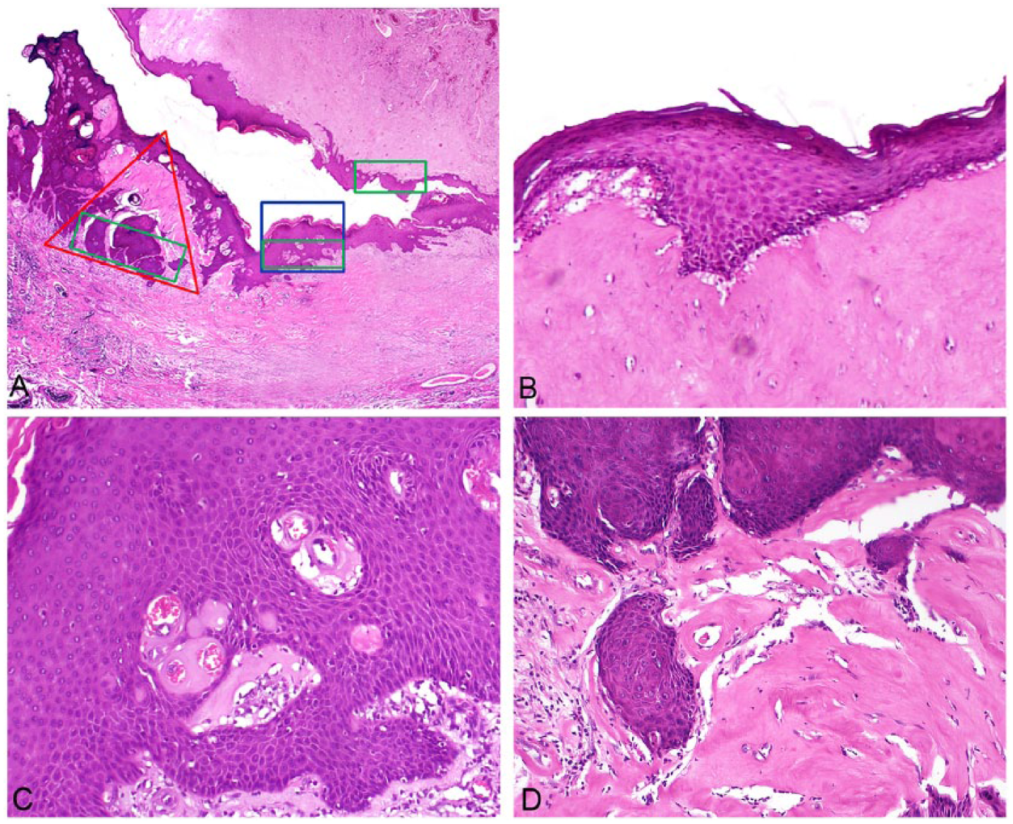
(A) Lichen sclerosus (LSc) in continuity with neoplasia (penile intraepithelial neoplasia [PeIN] and invasive carcinoma). (B). LSc and non-atypical squamous epithelium. (C). LSc changes (globular hyalinization) in relation to PeIN. (D) Presence of LSc within invasive carcinoma. This pattern was noted in 24% of the cases.
Discussion
We are presenting data supporting the spatial relation of LSc with intraepithelial and invasive penile neoplasia for the first time in this study. The morphological boundaries of penile precancerous lesions are not well defined, and they are usually determined by a constantly evolving consensus of experts. LSc is not registered in current classifications as a precancerous condition,7,9,14,15 but some studies in the penis and other areas point in that direction.16-19 There are some general criteria usually present considered useful to define a lesion as precancerous: (1) There should be a temporal relationship with premalignant lesion occurring on a population basis at an earlier age than invasive cancer. (2) They should also occur at a greater frequency, severity, and extent in organs harboring carcinoma. (3) Precancerous lesions should have similar morphologic features to invasive carcinoma. They should be similar or homogeneous at any hierarchical level of anatomical study, from microscopic to molecular. (4) Most important, there should be a spatial association with invasive cancer arising from the lesion. (5) Last, and most definitively, progression from the premalignant lesion into invasive carcinoma should be observed over time. Several of these criteria are present in penile LSc. When not associated with atypia or cancer, LSc is diagnosed about 10 years earlier than invasive carcinomas. 5 If not identical, the usual hyperplastic epithelium of LSc associated with neoplasia is morphologically not very different from differentiated PeIN. Hyperplasia and non-atrophy are the prevalent LSc epithelial lesions when associated with carcinomas. 11 There are not sufficient molecular studies to evaluate epithelial changes at the subclinical level.11,20,21 However, studies in other organs have demonstrated molecular alterations compatible with neoplastic changes in subclinical epithelial hyperplastic lesions with no or minimal atypia,22,23 and there are some studies indicating a conversion to in situ or invasive carcinoma in about 5% to 10% of patients with long-duration chronic LSc.24-26 The continuous sequence of LSc, precancerous, and invasive carcinomas found in this study suggest a spatial and probably causal relationship among these lesions. There were various combinations depicted in Table 3, most of which was of non-atypical LSc adjacent and as part of PeIN present in 42% of the cases. In one third of the cases, there was a continuous and contiguous relationship of LSc with invasive carcinomas, with (24%) or without PeIN (8%) in between. The presence of LSc within invasive tumors was a novel finding probably indicating tumor destruction of adjacent LSc/PeIN or tumor overgrowth. The continuity of the lesions also suggests a causal link. LSc was very rarely found separated from either PeIN or invasive SCC (3%). Our interpretation for the latter is that they may represent a coincidental multicentric or a collision phenomenon. The boundaries of normal epithelia/squamous hyperplasia and differentiated PeIN were gradual and subtle, and only the extremes or periphery of both lesions were well-defined.
In this series, the majority of the histological subtypes of SCCs present in association with LSc were of the non-HPV-related type. In other studies, we also found that if a sufficient number of sections is submitted, most cases of pseudohyperplastic, verrucous, and papillary NOS carcinomas will show foci of LSc in adjacent mucosae, 4 suggesting a link with these not too unusual well-differentiated subtypes of penile carcinomas. On the contrary, warty and basaloid SCC, HPV-related tumors were present in only 8% of the cases.
In summary, LSc was sequentially and continuously located (1) next to PeIN, (2) next to PeIN and affecting PeIN itself, (3) LSc in continuity and within invasive carcinomas, and (4) LSc throughout the sequence PeIN-invasive penile carcinoma. In a minority of cases, LSc was separate or discontinuous from the sequence LSc-PeIN-invasive carcinoma. The striking spatial relationship among LSc, PeIN, and invasive carcinoma supports the role of LSc as a penile precancerous lesion. The presence and spatial relation of LSc within invasive carcinoma is a novel finding.
Footnotes
Declaration of Conflicting Interests
The author(s) declared no potential conflicts of interest with respect to the research, authorship, and/or publication of this article.
Funding
The author(s) received no financial support for the research, authorship, and/or publication of this article.
Ethical Approval
Not applicable, because this article does not contain any studies with human or animal subjects.
Informed Consent
Not applicable, because this article does not contain any studies with human or animal subjects.
Trial Registration
Not applicable, because this article does not contain any clinical trials.
