
Abstract
Metastatic breast cancer resembling ductal carcinoma in situ (DCIS) is a rare phenomenon. In this article, we present a unique case of metastatic lobular carcinoma with DCIS-like morphology in the left axillary lymph nodes of a 52-year-old female. She presented with 2 lesions in the left breast on mammography, and a mastectomy with axillary lymph node dissection was performed. Gross examination showed a 3.5 × 2.5 × 1.0 cm indistinct tumor in the lower outer quadrant and a 2.5 × 2.5 × 1.8 cm tumor in the upper outer quadrant. Microscopic assessment revealed a pleomorphic lobular carcinoma in the lower outer quadrant and a grade 2 invasive ductal carcinoma in the upper outer quadrant. Sixteen of the 17 axillary lymph nodes showed metastatic lobular carcinoma with foci of solid and comedo-type DCIS-like features. Immunohistochemical analysis of the primary and metastatic lobular carcinoma showed no expression of E-cadherin and p63 antibodies. To our knowledge, metastatic lobular carcinoma exhibiting this pattern has not been reported. The case suggests that lobular carcinoma can morphologically recreate a primary microenvironment at a distant site and simulate in situ growth. Recognition of this pattern is important to avoid misdiagnosis.
Introduction
Metastatic invasive ductal carcinoma simulating various morphological forms of ductal carcinoma in situ (DCIS) in the lymph nodes has been described in the literature.1-8 Recent reports have also shown lobular carcinoma in situ with comedo-type necrosis mimicking DCIS. 9
Case Report
A 52-year-old female presented with 2 lesions in the left breast on mammography, and a mastectomy with axillary lymph node dissection was performed. Gross pathological examination showed a 3.5 × 2.5 × 1.0 cm indistinct tumor in the lower outer quadrant and a 2.5 × 2.5 × 1.8 cm circumscribed tumor in the upper outer quadrant. Microscopic assessment of the tumor in the lower outer quadrant revealed an invasive pleomorphic lobular carcinoma (PLC) and immunohistochemical analysis showed no expression of E-cadherin (Figure 1A and B). Sixteen of 17 axillary lymph nodes showed metastatic lobular carcinoma with foci of solid and comedo-type DCIS-like features (Figure 1C). Immunohistochemistry of the metastasis was negative for p63 and E-cadherin (Figure 1D). Microscopy of the second tumor in the upper outer quadrant revealed a grade 2 invasive ductal carcinoma that demonstrated strong E-cadherin expression on immunohistochemistry (Figure 1E and F). No ectopic breast tissue was identified.
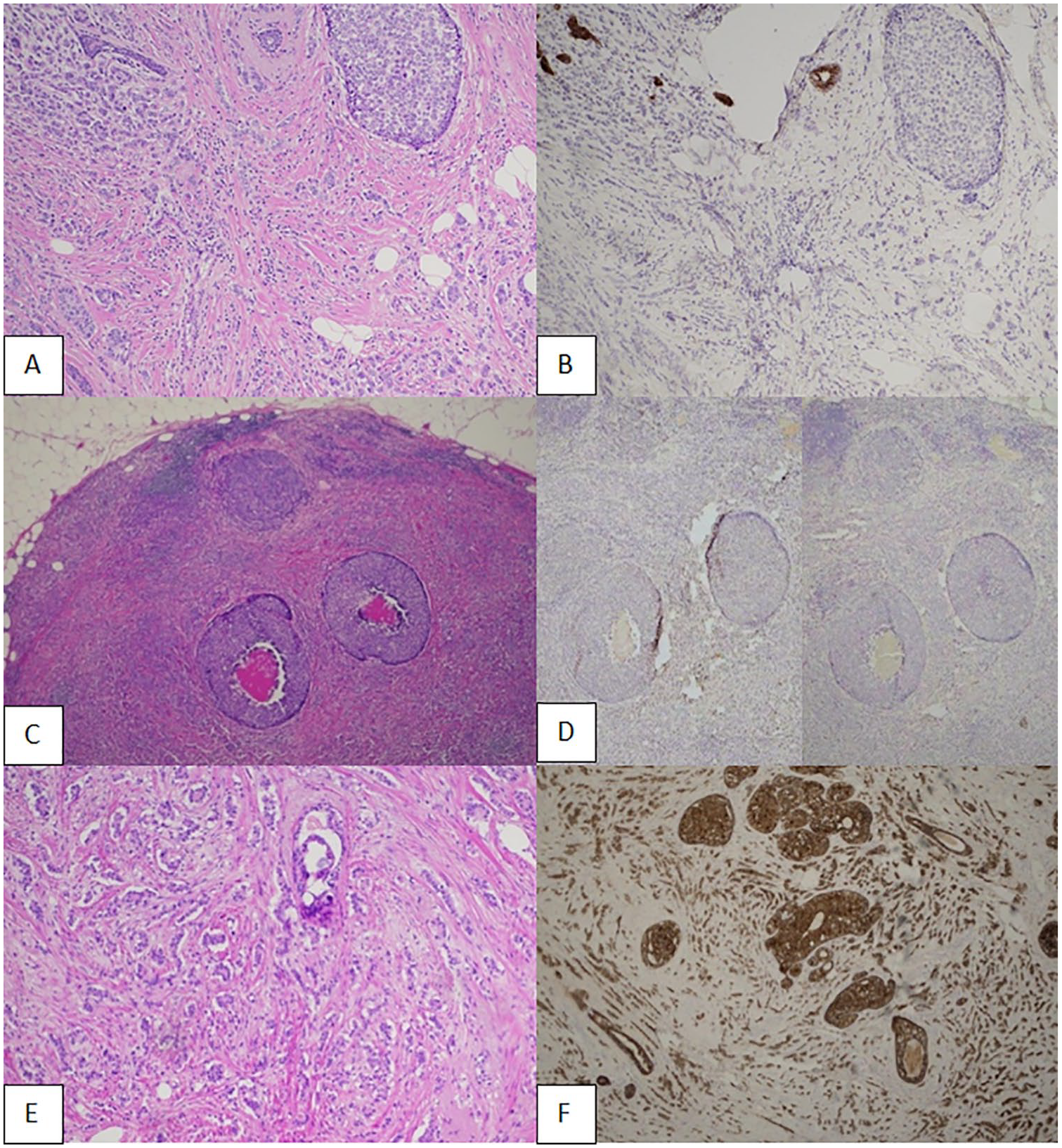
(A) Histological sections of the invasive lobular carcinoma (hematoxylin and eosin stain [H&E]). (B) The tumor cells are negative for E-cadherin. (C) Histological section of a positive lymph node (H&E). The sharply circumscribed oval tumor deposits show comedo-type necrosis, morphologically indistinguishable from high grade DCIS. (D) The tumor cells are negative for E-cadherin and p63, respectively, on immunostaining, confirming metastatic lobular carcinoma. (E) Invasive ductal carcinoma (H&E). (F) The tumor cells are positive for E-cadherin.
Discussion
Metastatic breast cancer resembling DCIS is a rare phenomenon that has been described in studies1-8 in the context of metastatic ductal carcinoma. To our knowledge, metastatic lobular carcinoma exhibiting this pattern has not been reported.
Reviewing 200 cases of metastatic human breast cancer using histopathological, digital image, and immunocytochemical analyses, Barky et al 2 revealed a 21% incidence of DCIS growth pattern within axillary nodal metastases. The authors concluded that the features represented cellular populations epigenetically derived from the primary tumor, capable of reverting back to their in situ growth state, and referred to the phenomenon as “revertant” DCIS. Earlier observations of this process were published by Cowen 1 in 1980 and Lee et al 2 in 1995. A summary of the published literature on DCIS-like metastasis is in Table 1.
Summary of Literature on DCIS-Like Metastasis.
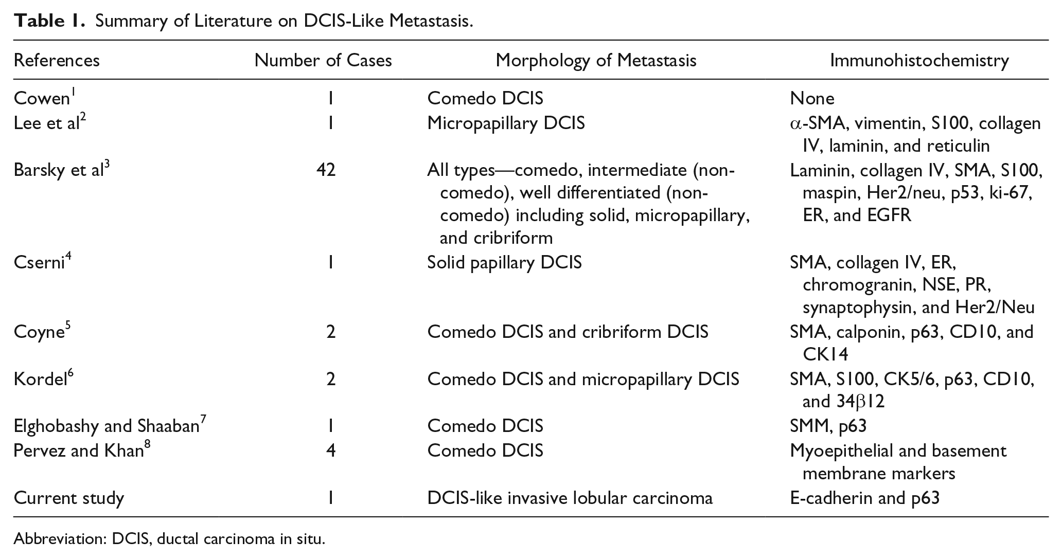
Abbreviation: DCIS, ductal carcinoma in situ.
While the “revertant” DCIS patterns in the lymph nodes simulated DCIS in the breast, they, however, lack a myoepithelial layer on immunohistochemical analysis. The phenomenon should be differentiated from primary DCIS arising in accessory 10 or ectopic breast tissue 11 in which a myoepithelial layer should be demonstrated by immunohistochemistry. Evidence of residual normal breast tissue is another clue.
Invasive lobular carcinoma, which represents 5% to 15% of invasive breast tumors, has different clinical and pathological characteristics from ductal carcinoma. 12 PLC is a rare form of lobular carcinoma with aggressive clinical features and worse prognostic factors. Morphologically, PLC has a growth pattern typical of lobular carcinoma, but has a higher degree of cellular atypia and a higher mitotic activity than classical lobular carcinoma. It is often associated with pleomorphic carcinoma in situ with comedo-type necrosis, 9 and unlike invasive ductal carcinoma, there is loss of E-cadherin on immunohistochemistry.
Our observation supports the hypothesis that metastatic lobular carcinomas can morphologically recreate their primary microenvironment at a distant site and simulate in situ growth (revertant form). However, without performing E-cadherin immunohistochemistry, the lesion could be misdiagnosed as DCIS with comedo-type necrosis or invasive ductal carcinoma.
Footnotes
Acknowledgements
Mr M. Youssef, Consultant Surgeon, is gratefully acknowledged for obtaining patient consent to publish this article.
Declaration of Conflicting Interests
The author(s) declared no potential conflicts of interest with respect to the research, authorship, and/or publication of this article.
Funding
The author(s) received no financial support for the research, authorship, and/or publication of this article.
Ethical Approval
Not applicable, because this article does not contain any studies with human or animal subjects.
Informed Consent
Patient consent was obtained to publish this article.
Trial Registration
Not applicable, because this article does not contain any clinical trials.