
Abstract
Glomus tumor can rarely arise in the central nervous system as a sella turcica mass. In this article, we report a case of sellar glomus tumor in a female patient who presented at the age of 8 years with visual impairment. The tumor recurred at 4 years and 26 years after initial excision and gamma knife therapy. Histologic examination showed a monotonous population of oval cells accompanied by delicate blood vessels, features mimicking pituitary adenoma. The tumor showed histologic progression at the second recurrence. Synaptophysin staining was positive, but chromogranin and CD56 were negative. The tumor cells were negative for epithelial markers but expressed actin and SMA. Awareness of the rare occurrence of glomus tumor at this region, careful analysis of morphology, and appropriate immunohistochemical workup are essential to solve this diagnostic challenge. The clinicopathologic features of all previously reported cases are reviewed.
Glomus tumor is a tumor of perivascular modified smooth muscle cells that mainly affects young adults with no gender predilection. It most commonly affects the distal extremities, such as the subungual region, hand, wrist, and foot, where the glomus body, a thermoregulatory structure, is normally found. Rare cases of glomus tumors have been reported in visceral sites such as gastrointestinal tract, 1 genitourinary system,2-4 thyroid gland, 5 mediastinum, and lung. 6 Occurrence of glomus tumor in the central nervous system is extremely rare, and it has only been reported in the sella turcica, presumably arising from the gomitoli, a physiological analog of glomus body responsible for regulating portal blood flow in the posterior lobe of the pituitary gland. 7 In this article, we report a case of sellar glomus tumor in an 8-year-old female, which recurred 26 years later with histologic progression; all cases reported in the literature are also reviewed.7-9
Case Report
The female patient presented at the age of 8 years in 1992 with visual impairment. Imaging and biochemical investigations showed a 2-cm pituitary tumor with suprasellar extension and complicated by panhypopituitarism. Craniotomy revealed a richly vascularized tumor in the pituitary fossa. Subtotal excision was done, and a diagnosis of glomus tumor was made. Postoperative gamma knife therapy was given for residual disease. Four years later, magnetic resonance imaging revealed an increase in tumor size, and another course of gamma knife was given. In 2018, 26 years after the initial presentation, she noticed progressive visual impairment of the left eye. Magnetic resonance imaging showed a recurrent 5-cm tumor at the suprasellar region with a large cystic component and strong contrast enhancement (Figure 1). Craniotomy revealed a large tumor in the pituitary fossa with extension to the frontal lobe. Excision was performed, and the patient was well at 1 year after the surgery and had regained full vision.
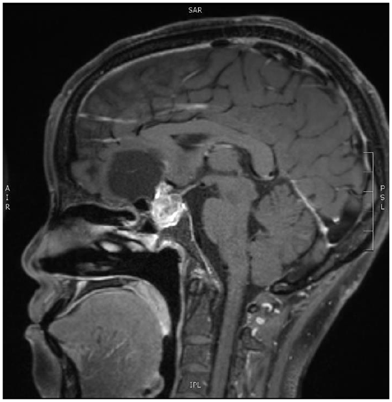
Magnetic resonance imaging shows a large contrast-enhancing mass in the sella turcica with a cystic component extending to the frontal lobe.
The initial excision specimen (1992) was not available for review. Histologic examination of the recurrent tumor showed tumor fragments of moderate cellularity, consisting of monotonous round cells with delicate intervening small blood vessels (Figure 2A). The tumor cells were arranged in sheets, packets, and small nests, and possessed central round to oval nuclei, pale chromatin, indistinct nucleoli, and moderate amount of eosinophilic cytoplasm. Pericellular basement membrane was demonstrated by diastase–periodic acid–Schiff and reticulin stains (Figure 2B). The intervening blood vessels had narrow or ectatic lumens, and the endothelial cells often appeared to directly abut on the surrounding tumor cells without intervening pericytes (Figure 2C). The stroma showed focal hyalinization, fibrosis, and hemosiderin deposition. In one tumor fragment, the cellularity was markedly increased compared with the rest of the tumor. The tumor cells showed increased nuclear cytoplasmic ratio, moderately pleomorphic nuclei, and occasional mitotic activity (Figure 2D). On immunostaining, the tumor cells were negative for cytokeratin MNF116, cytokeratin CAM5.2, growth hormone, prolactin, ACTH, S100, glial fibrillary acidic protein, and CD34. Synaptophysin was positive but not chromogranin and CD56 (Figure 3). The tumor cells were diffusely positive for muscle-specific actin, smooth muscle actin, and calponin. Actin immunostain also revealed absence of a layer of spindly pericytes between the endothelial cells and tumor cells in some blood vessels (Figure 4). The Ki-67 proliferation index was around 1%, but up to 15% in the fragment with histologic progression (Figure 2E). BRAF V600E staining was negative. The overall features were consistent with recurrent glomus tumor with focal histologic progression.
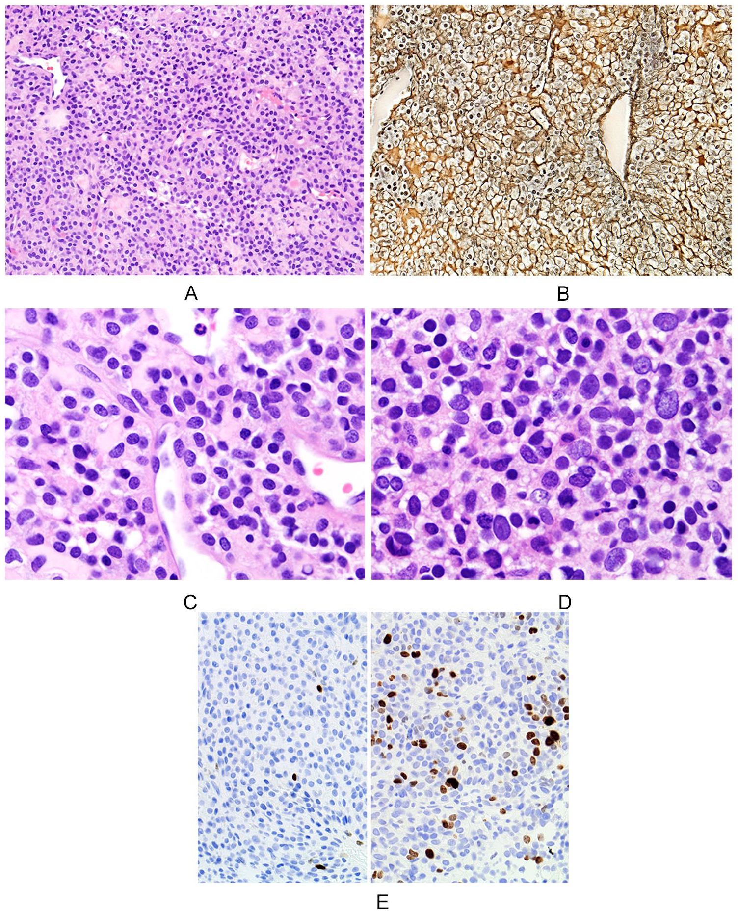
(A) Low-power magnification shows packets and nests of monotonous round cells with delicate intervening small blood vessels. (B) Reticulin stain highlights collagen fibers encircling individual tumor cells. (C) The spindly endothelial cells of small blood vessels abut directly on the surrounding tumor cells. (D) Histologic progression of glomus tumor is characterized by increased cellularity, raised nuclear cytoplasmic ratio, and moderate nuclear pleomorphism. (E) The Ki67 proliferative index is 1% in the glomus tumor (left), but has increased to 15% in the area with histologic progression (right).
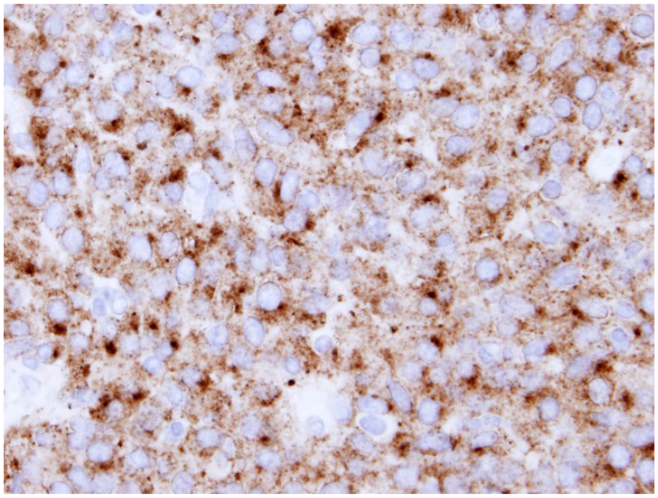
The tumor cells show diffuse granular cytoplasmic staining for synaptophysin on immunostaining.
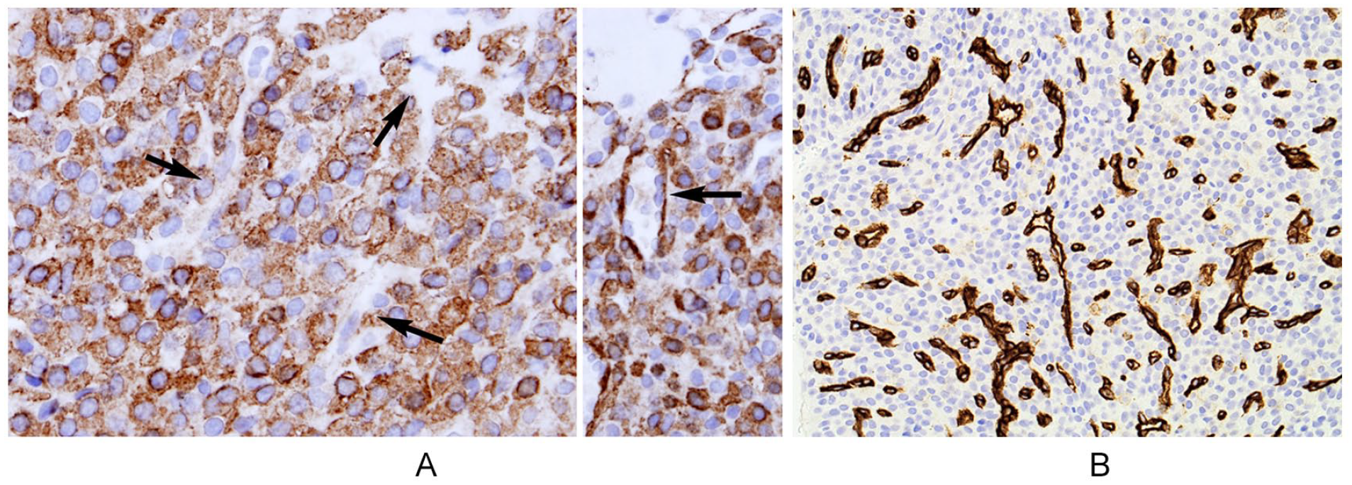
(A) Immunostaining for smooth muscle actin shows absence of pericytes in some blood vessels on the left (arrows). Pericytes are highlighted by smooth muscle actin in normal blood vessels on the right for comparison (arrow). (B) CD34 immunostaining highlights the endothelial cells, whereas the tumor cells are negative.
Discussion
Glomus tumor of the central nervous system is exceedingly rare, with only 3 cases (2 glomangiomas and 1 glomus tumor) reported in the English language literature. The current case, except for the unusual localization of sella turcica, shows the typical morphology and immunophenotype of glomus tumor. However, the packeting of bland-looking cells accompanied by a rich vascularity and expression of synaptophysin can easily lead to an erroneous diagnosis of pituitary adenoma, a much more common tumor at this anatomic location. Microscopically, the eosinophilic cytoplasm of the glomus tumor cells lacks the usually granular quality of cytoplasm in pituitary adenoma. The finding of vessels with spindly endothelial cells abutting directly on the surrounding tumor cells without an intervening layer of pericytes (better highlighted by actin and CD34 immunostain; Figure 4A and B) is a characteristic feature of glomus tumor, a growth pattern recapitulating the perivascular origin of the neoplastic cells in the normal glomus body. 10 Some normal blood vessels within the tumor should show strongly actin-positive cells in the vessel walls for comparison. Glomus tumors of soft tissues usually do not express synaptophysin, but focal to diffuse synaptophysin expression has been well documented in glomus tumors arising in visceral organs such as stomach, esophagus, duodenum, bronchus, kidney, liver, and nose.1,11-13 In all these cases, other neuroendocrine markers such as CD56 and chromogranin are negative, as in the current case. Other tumors of the sellar region, such as craniopharyngioma, germ cell tumor, pituicytoma, meningioma, hematolymphoid tumor, and metastatic tumor, should pose no problems in the differential diagnosis.
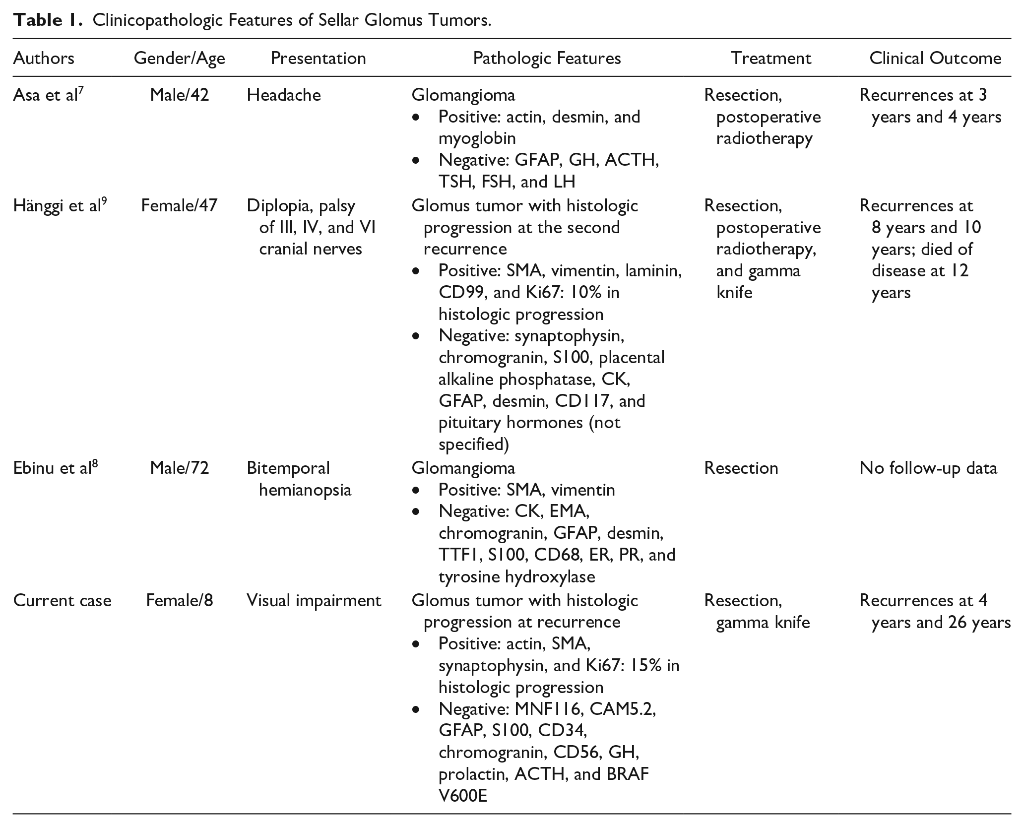
Literature review shows that the age of patients with sellar glomus tumors ranged from 8 to 72 years (median = 44.5 years), affecting 2 males and 2 females (Table 1). The most common presentation was visual impairment (3), followed by headache (1) and cranial nerves palsy (1). All patients developed local recurrences with long-term follow-up, and the mean duration from tumor excision to the first recurrence was 5 years. This is most probably due to the strategic site and high vascularity of the tumors precluding complete excision.
Clinicopathologic Features of Sellar Glomus Tumors.
In the current case, histologic progression with increased cellularity, nuclear pleomorphism, and mitotic activity were found focally, most consistent with glomus tumor with uncertain malignant potential. BRAF V600E mutation has been found in a subset of malignant glomus tumor, and this case is negative for BRAF V600E on immunostaining. 14 Histologic progression has also been reported in one previously reported sellar tumor, 9 and that patient died of disease at 12 years with no metastasis.
In conclusion, sellar glomus tumor is a rare tumor that can be potentially mistaken for pituitary adenoma. Awareness of this rare occurrence, careful analysis of morphologic features, and appropriate immunohistochemical workup would be essential to solve this diagnostic challenge.
Footnotes
Declaration of Conflicting Interests
The author(s) declared no potential conflicts of interest with respect to the research, authorship, and/or publication of this article.
Funding
The author(s) received no financial support for the research, authorship, and/or publication of this article.
Ethical Approval
Not applicable, because this article does not contain any interventional studies with human or animal subjects.
Informed Consent
Verbal informed consent has been obtained from the patient.
Trial Registration
Not applicable, because this article does not contain any clinical trials.