






Abstract
Papillary renal neoplasm with reverse polarity is a form of recently described tumor. These tumors are defined by GATA3 positivity, negative vimentin staining, and the presence of both papillary structures and a layer of eosinophilic cells with apical nuclei and a granular cytoplasm. In the present report, we review 7 cases of papillary renal neoplasm with reverse polarity that were GATA3+ and vimentin−, consistent with past reports. In all 7 of these cases, we found that these tumors were additionally positive for 34βE12. All 7 of these tumors were categorized as stage pT1. On histological examination, these tumors exhibited branching papillae with apical nuclei. All 7 of these patients were alive on most recent follow-up, with 6 being disease free and one having developed prostate cancer. Together, this overview of 7 additional cases of papillary renal neoplasm with reverse polarity offers further insight into this rare and poorly understood disease.
Introduction
Papillary renal neoplasm with reverse polarity is a form of recently described tumor that was initially defined by Al-Obaidy et al in 2019 following their review of 18 clinical cases of this proposed disease. 1 This tumor type has been described as being characterized by GATA3 and L1CAM positivity, negative vimentin staining, and the presence of both papillary structures and a layer of eosinophilic cells with apical nuclei and a granular cytoplasm. In a separate study, these same authors identified RAS missense mutations in 8/10 tested papillary renal neoplasm with reverse polarity tumors using a next-generation sequencing approach. 2
Papillary renal cell carcinoma (PRCC) was first defined as a distinct cancer in 1976, 3 and it is now recognized as the second most common form of renal cancer following clear cell renal cell carcinoma (ccRCC), making up between 15% and 20% of all kidney cancer diagnoses.4,5 Delahunt et al have previously proposed the further subclassification of PRCC into PRCC type 1 and PRCC type 2. 6 Under their proposed classification system, PRCC type 1 was defined by the presence of tubules and papillae that were covered by small cells with small nuclei, with the presence of psammoma bodies, glomeruloid papillae, papillary edema, and foam cells often being detected in these tumors. In contrast, PRCC type 2 tumors were defined as being composed of papillae that were covered by larger pseudostratified cells with large nuclei and clearly defined nucleoli, with the presence of psammoma bodies, glomeruloid papillae, papillary edema, and foam cells being significantly less common. Unfortunately, this classification system proved insufficient to capture the true diversity of PRCC tumors, with up to half of these tumors failing to correspond to the expected morphologic criteria for either of these tumor subtypes. This led Saleeb et al to propose a 4-tiered PRCC classification system (PRCC1-4) based on histological and immunohistochemical analyses of these tumor types, with PRCC4 being defined as a form of GATA3-positive PRCC in which tumor cells were arranged with an apical orientation in a linear manner with no evidence of nuclear pseudostratifcation. 7 Analyses of PRCC data sets present within the Cancer Genome Atlas indicate that type 1 PRCC tumors frequently exhibit MET alterations, while type 2 PRCC tumors often exhibit silencing of CDKN2A, mutations in SETD2, upregulation of genes involved in the NRF2-antioxidant response element (ARE) pathway, and fumarate hydratase (FH) gene mutations. 5
Given its very recent designation as a distinct form of cancer, there are few published articles discussing findings associated with cases of papillary renal neoplasm with reverse polarity. As such, in the present report, we describe 7 such cases that were identified on reexamination of previously diagnosed PRCC cases. These papillary renal neoplasm with reverse polarity tumors exhibited expected papillary structures and the presence of a layer of apically oriented tumor cells. In this article, we describe the clinical, pathological, and immunophenotypic findings pertaining to these cases, in addition to examining available patient follow-up data. Importantly, as has been previously reported, we found that all 7 of these tumors were GATA3+ and vimentin−, and we were additionally able to determine that these tumors were also all positive for 34β4E12, which has not been documented in previous studies. Our results thus offer new insights into papillary renal neoplasm with reverse polarity, thereby aiding future efforts to diagnose and potentially even to treat this disease.
Materials and Methods
Case Identification and Microscopic Examination
Findings from 190 PRCC cases from the Ruijin Hospital Pathology Department and Ningbo Diagnostic Pathology Center were reexamined, revealing 7 of these cases to exhibit tumor morphology consistent with papillary renal neoplasm with reverse polarity. This was then further validated by 2 independent pathologists on their examination of 4-µm-thick hematoxylin and eosin–stained tumor tissue sections that had been formalin fixed and paraffin embedded using standard protocols.
Immunohistochemistry and Fluorescence In Situ Hybridization (FISH)
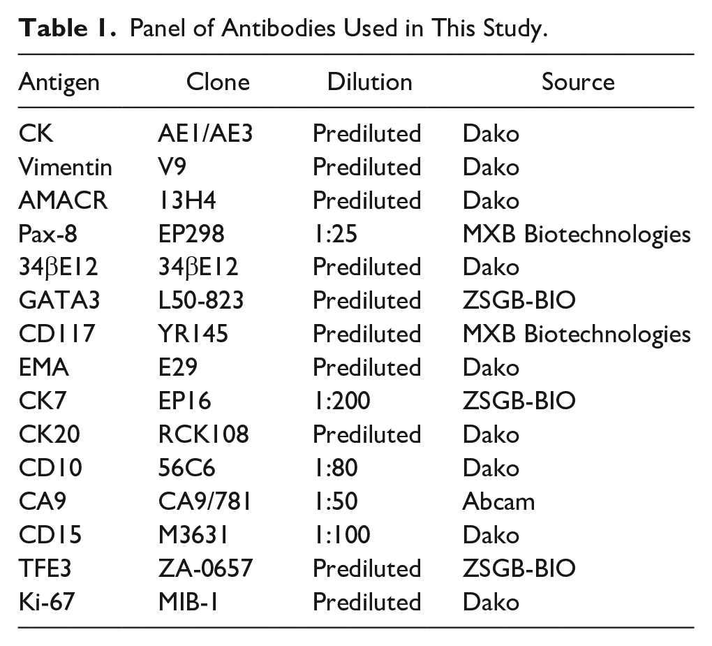
Tissue microarrays were prepared using core samples (2 mm in diameter) from patient formalin-fixed and paraffin-embedded samples, with 3 representative samples being selected per tumor. A Dako Omnis automated staining platform was then used for immunohistochemical staining of these tissue sections with antibodies specific for vimentin, GATA3, 34βE12, CD15, CA9, CK7, EMA, PAX-8, TFE-3, AMACR, CD10, and CD117 (see Table 1). In addition, FISH assays were performed as in previous studies. 8
Panel of Antibodies Used in This Study.
Results
Clinical Findings
Reexamination of tumor sections from 190 diagnosed cases of PRCC revealed 7 cases, wherein the tumors exhibited the apical nuclear localization and granular eosinophilic cytoplasm characteristic of papillary renal neoplasm with reverse polarity. Clinicopathological findings for these 7 patients are compiled in Table 2. These patients (3 female, 4 male) had both a mean and median age of 71 years, with no clear gender bias in diagnosis rates. Five of these patients originally presented with pain in their back or sides, whereas the tumors were originally identified incidentally during routine health care visits. Tumors were localized to the left kidney in 2 patients and to the right kidney in 5 patients. Computed tomography (CT) scans from these patients revealed the presence of a variable density pass (Figure 1). All 7 of these tumors were TNM stage I, were confined to the kidney, and were 1.2 to 2.8 cm in diameter (mean: 2.2 cm). Patient 4 had a 15-year history of hypertension. Two of these patients were treated via radical nephrectomy, while the remaining 5 were treated via partial nephrectomy. Patients were followed for 3 to 65 months, with all 7 patients being alive and free of tumor recurrence or metastasis as of their most recent follow-up. Patient 3 was diagnosed with prostate adenocarcinoma 3 months following nephrectomy.
Clinicopathologic Data.
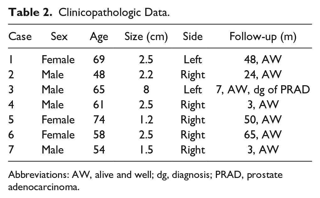
Abbreviations: AW, alive and well; dg, diagnosis; PRAD, prostate adenocarcinoma.
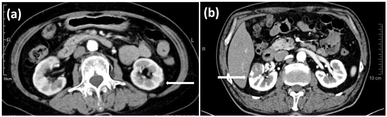
Imaging findings. (a) A mass (2.5 cm in diameter) of variable density was detected in the left kidney on computed tomography (CT) examination of case 1. (b) A mass (2.2 cm in diameter) of variable density was detected in the right kidney on CT examination of case 2.
Pathological Findings
Gross morphological examination revealed all 7 of these tumors to be round or ovoid in shape and to be well circumscribed with a prominent pseudocapsule. Tumor surfaces were red-to-gray in color, with a soft or rubbery consistency and no evidence of tissue necrosis or fibrosis. Three of these tumors had evidence of focal hemorrhage. A well-defined architecture of thin papillae was evident in all 7 tumors on microscopic examination, with arborizing proliferation of delicate fibrovascular cores. Tumor cells contained round nuclei and eosinophilic cytoplasm. Notably, nuclei localized to the apical surface of these cells consistent with reverse polarity, nucleoli were largely undetectable, and the majority of tumor cells were arranged in a monolayer on the papillary basement membrane. These tumors were all classified as World Health Organization/International Society of Urologic Pathologists (WHO/ISUP) grade 1 or 2. None of these tumors contained psammoma bodies, intracellular hemosiderin, mitotic figures, foam cell clusters, or necrotic sections (Figure 2).
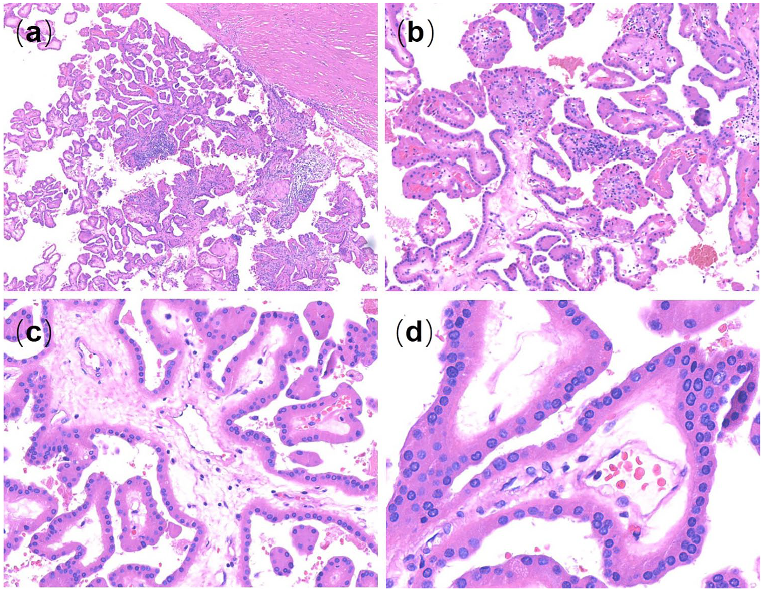
Histological features of papillary renal neoplasm with reverse polarity. (a) Tumors were well circumscribed (40×). (b-d) Tightly packed branching papillae were evident in all analyzed tumors. Cells contained small, hyperchromatic, wrinkled nuclei with primarily apical localization (b, 100×; c, 200×; d, 400×).
Immunohistochemical and FISH Findings
On immunohistochemical analysis, we found that all 7 of these tumors stained positive for GATA3, 34βE12, CK, CK7, EMA, PAX-8, and AMACR, and negative for vimentin, CD117, CK20, CD15, CD10, CA9, and TFE3. These tumors also had a low (1% to 2%) Ki-67 index (Figure 3). In addition to the previously reported diffuse strong GATA3 staining in these tumors, they were also strongly positive for 34βE12, which has not been reported previously. When we then further analyzed 34βE12 in the remaining 183 PRCC patient samples in this sample cohort, we found that only 12 exhibited positive 34βE12 expression. AMACR were all moderate positive in the 7 cases. EMA-positive staining was observed in the outer margin and reversed apical membrane pattern of neoplastic nests. All 7 of these samples were CD15 negative. FISH analyses further revealed that 3 of these exhibited trisomy of chromosome 7, 2 exhibited trisomy of chromosome 17, and 1 exhibited a Y chromosome deletion (Table 3).
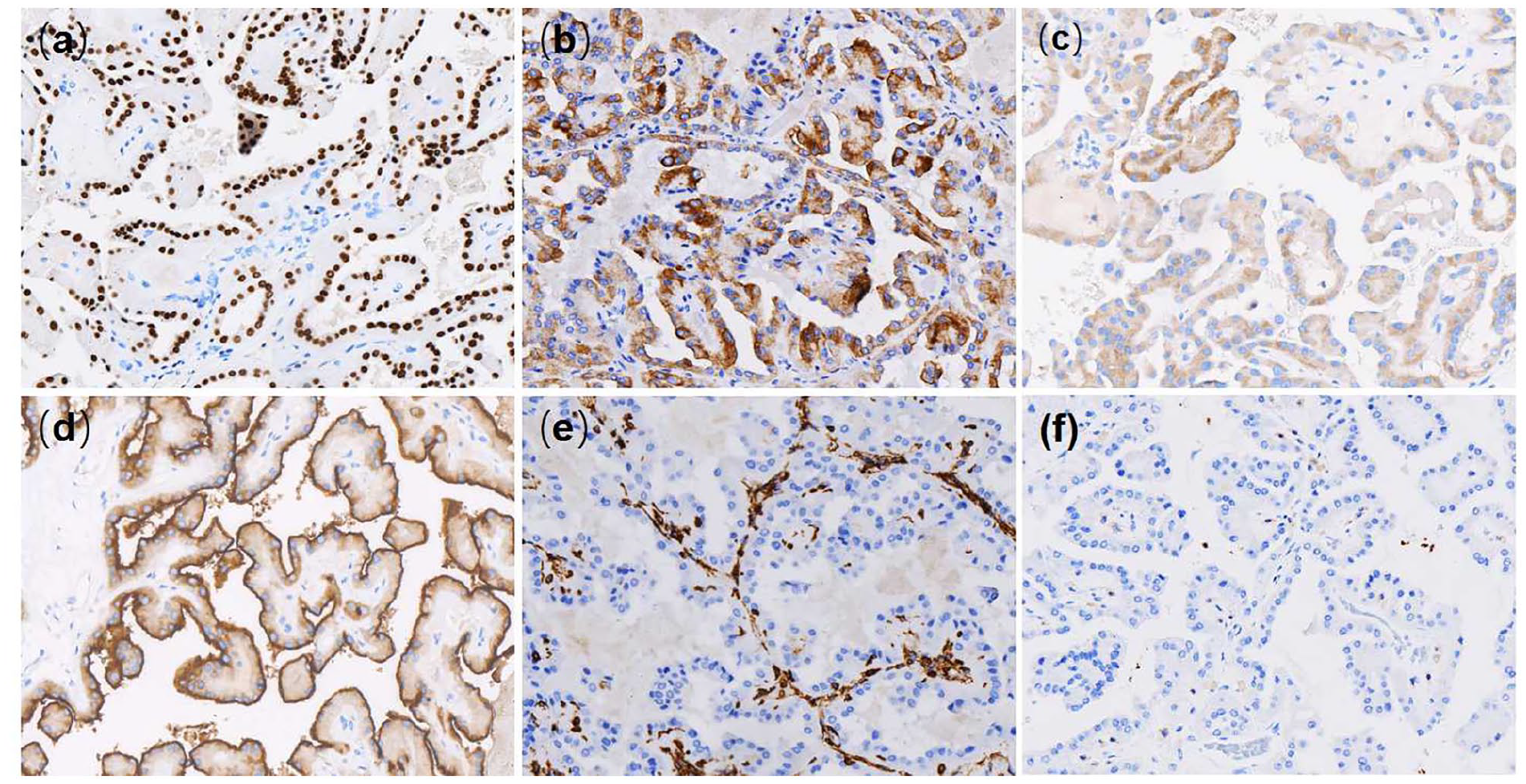
Immunohistochemical features of papillary renal neoplasm with reverse polarity. (a) Tumors exhibited strong GATA3 expression (200×). (b) Tumors exhibited strong 34βE12 expression (200×). (c) Tumors exhibited variable moderate AMACR expression (200×). (d) Tumors exhibited strong EMA expression (200×). (e) Tumors were negative for vimentin (200×). (f) Tumors were negative for CD15 expression (200×).
Immunohistochemical and FISH Results of Papillary Renal Neoplasm With Reverse Polarity.
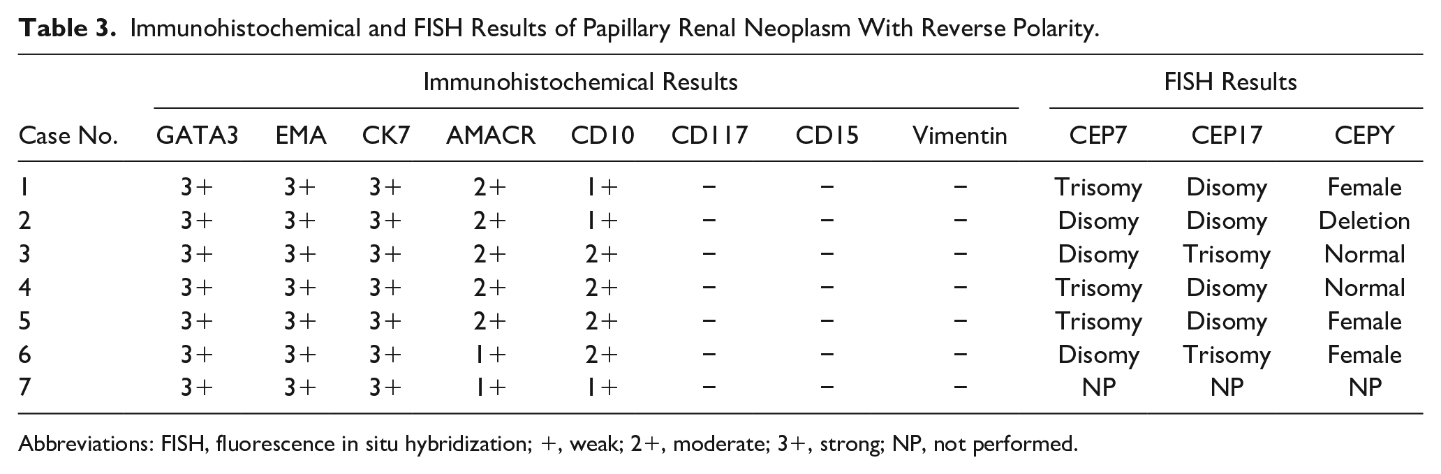
Abbreviations: FISH, fluorescence in situ hybridization; +, weak; 2+, moderate; 3+, strong; NP, not performed.
Discussion
In this report, we were able to identify 7 cases of papillary renal neoplasm with reverse polarity on reexamination of 190 previously diagnosed PRCC cases, consistent with the proposal that papillary renal neoplasm with reverse polarity is a rare and morphologically distinct papillary neoplasm. Few studies, to date, have studied papillary renal neoplasm with reverse polarity in depth, with the best-defined immunohistochemical features of these tumors being that they are typically GATA3+ and vimentin−. In addition, ~80% of papillary renal neoplasm with reverse polarity tumors have been shown to harbor KRAS missense mutations. Building on these previous findings, we further found that all papillary renal neoplasm with reverse polarity cases identified in this study were strongly 34βE12+, thus offering an additional immunohistochemical marker of value for the differential diagnosis of this PRCC subtype.
In previous reports, tumors exhibiting papillary renal neoplasm with reverse polarity-like morphology were classified as oncocytic or oncocytoid PRCC. In their study of 43 PRCC cases in 2003, Allory et al had identified 4 such oncocytic-type PRCCs and found them to have favorable outcomes. 9 Lefèvre et al also reported on 10 oncocytic renal papillary tumors that had favorable outcomes, a low proliferation index, and strong positive staining for CD10, AMACR, and vimentin, with the latter marker being inconsistent with papillary renal neoplasm with reverse polarity tumor staining profiles. 10 Park et al reported on a series of 7 cases of oncocytic PRCC that exhibited a pattern of nuclear reverse polarity consistent with that observed in papillary renal neoplasm with reverse polarity, although they found these tumors to stain positive for CD15, P504S, and vimentin. 11 These oncocytic or oncocytoid PRCCs reported were not completely consistent with papillary renal neoplasm with reverse polarity. Saleeb et al classified cases of some oncocytic eosinophilic PRCC using the PRCC4 designation, and first identified these tumors as being GATA3+. 7 In 2019, Al-Obaidy et al conducted a systematic analysis of 18 tumors, which they then went on to define as prototypical cases of papillary renal neoplasm with reverse polarity, summarizing the histologic and molecular characteristics of these tumors.1,2 In this article, we summarized our findings from our analyses of 7 additional papillary renal neoplasm with reverse polarity cases, with our most notable finding being that all of these tumors were positive for 34βE12 expression in addition to being positive for GATA3.
GATA3 is a transcription factor that has been shown to play key roles in regulating the differentiation and activity of a number of cells and tissue types.12,13 GATA3 is encoded on chromosome 10p14, 14 and in cases of carcinoma of unknown origin, GATA3 staining is often used as a marker that can confirm that a tumor originates from breast or urothelial tissue.15,16 The specific function and expression of GATA3 in cases of renal cell carcinoma is not as well documented. Mantilla et al found in a 2017 study that clear cell PRCC (CCPRCC) tumors frequently express GATA3, making it a valuable marker for distinguishing between these and morphologically similar tumor types. 17 Gonzalez-Roibon et al also reported in a 2013 study that a minority of oncocytomas, Xp11.2 renal cell carcinomas, and collecting duct carcinomas are positive for GATA3 expression. 18 As discussed above, Saleeb et al were the first to identify a subset of PRCC cases, which were GATA3 positive and which they classified using the PRCC4 subtype. In their recent study, Al-Obaidy et al found that their prototypical papillary renal neoplasm with reverse polarity cases were GATA3 positive, and we similarly found all 7 papillary renal neoplasm with reverse polarity tumors identified in the present study to be positive for the expression of this transcription factor. GATA3 has been demonstrated to be expressed in the distal convoluted tubules and collecting system, which suggested the possibility that papillary renal neoplasm with reverse polarity may originate in the distal convoluted tubule or collecting system rather than the proximal tubules. 17
The 34βE12 antibody is known to recognize certain forms of high-molecular-weight cytokeratin within the cytoplasm of cells, and positive 34βE12 staining has been explored as a diagnostic marker in prostate cancer, breast cancer, and collecting duct carcinomas. In the present study, we found that all 7 papillary renal neoplasm with reverse polarity cases stained strongly positive for 34βE12, suggesting that this may, therefore, be a valuable diagnostic biomarker when differentiating between papillary renal neoplasm with reverse polarity and morphologically similar tumor types. 34βE12 was a sensitive marker of renal collecting duct carcinoma, which also points to a distal nephron origin of papillary renal neoplasm with reverse polarity. Martignoni et al have previously shown that CCPRCC tumors often stain strongly positive for 34βE12, with 12/14 cases being 34βE12 positive, whereas no tested ccRCC tumors stained positive with this antibody. 19
In this study, we found that papillary renal neoplasm with reverse polarity tumors stained positive for both GATA3 and 34βE12, as do CCPRCC tumors. As such, it is essential that histological or immunohistochemical differences between these 2 tumor types be clearly defined so as to guide proper patient diagnosis and treatment. On morphological examination, the nuclei of these 2 tumor types align to the apical surface of cells and are primarily of low grade (ISUP nucleolar grade 1 or 2). CCPRCC tumor cells, however, typically have a clear cytoplasm and circumferentially aligned nuclei, whereas the cytoplasm of papillary renal neoplasm with reverse polarity cells is eosinophilic. Tumor necrosis, perirenal invasion, and lymphovascular invasion are absent in CCPRCC tumors, and CCPRCC tumor cells exhibit diffuse CK7 positivity, as well as carbonic anhydrase IX positivity in a cup-like distribution pattern. Our results also suggest that trisomy of chromosomes 7 or 17, as identified via FISH, may be an additional means of differentiating between CCPRCC and papillary renal neoplasm with reverse polarity.
All 7 of the tested papillary renal neoplasm with reverse polarity cases in the present study were found to be negative for CD15 expression. CD15 is the key enzyme in Lewis-Y molecules carried by glycoproteins and glycolipids, and Lewis-Y molecules are involved in glycosylation reactions that are common in the context of carcinogenesis.20,21 CD15 expression status is frequently associated with cancer prognosis in cases of breast cancer, thyroid cancer, and Hodgkin’s lymphoma.22,23 Notably, a loss of CD15 expression in ccRCC has been shown to correspond to a poorer patient prognosis. 24 The importance of CD15 as a prognostic or diagnostic biomarker in PRCC, however, is not as well understood. The fact that the papillary renal neoplasm with reverse polarity tumors analyzed in the present study were all negative for CD15 expression may suggest that a loss of CD15 is associated with a good prognosis in this cancer type, as all 7 of these tumors were TNM stage I and all patients remained alive and free of recurrence or metastasis over the 3- to 65-month follow-up period.
Papillary renal neoplasm with reverse polarity tumors must also be distinguished from papillary adenomas. Papillary adenomas also exhibit papillary, tubular, or tubulopapillary architecture with a low ISUP grade and trisomy 7 and 17, as well as loss of the Y chromosome. However, papillary adenomas are nonencapsulated tumors with a diameter ≤15 mm, and they lack apically localized nuclei.
Papillary renal neoplasm with reverse polarity tumors appears to exhibit an indolent clinical course. All the 7 patients in these study were alive and free of tumor recurrence or metastasis as of their most recent follow-up. Al-Obaidy et al also showed no metastasis of the 18 patients, which should be distinguished from PRCC type 2, and the size of these lesions were usually smaller than the other PRCC subtypes. The mean diameter of the 7 cases were only 2.2 cm.
In summary, through our identification of 7 additional cases of papillary renal neoplasm with reverse polarity, we have confirmed previous reports indicating that these tumors represent a rare and distinct form of PRCC characterized by apically localized nuclei and eosinophilic cytoplasm. We found these tumors to stain positive for both GATA3 and 34βE12, while they were negative for the expression of both vimentin and CD15. Based on presently available data, these papillary renal neoplasm with reverse polarity tumors appear to exhibit an indolent clinical course.
Footnotes
Declaration of Conflicting Interests
The author(s) declared no potential conflicts of interest with respect to the research, authorship, and/or publication of this article.
Funding
The author(s) disclosed receipt of the following financial support for the research, authorship, and/or publication of this article: This study was supported in funding by the Shanghai Science and Technology Development Fund (19MC1911000).
Ethical Approval
Not applicable, because this article does not contain any studies with human or animal subjects.
Informed Consent
Not applicable, because this article does not contain any studies with human or animal subjects.
Trial Registration
Not applicable, because this article does not contain any clinical trials.
