
Abstract
Background. Diagnosis of dedifferentiated liposarcoma (DDL) can sometimes be challenging due to a wide variety of histological features. “Meningothelial-like” whorl is an uncommon histological feature of DDL, which is also observed in neural tumors and follicular dendritic cell sarcoma. This feature is frequently associated with metaplastic bone formation. We conducted this study to describe the clinicopathological features of DDL with meningothelial-like whorls that would aid in establishing accurate diagnosis. Material and Methods. Microscopic glass slides of 5 cases of DDL with meningothelial-like whorls, diagnosed between January 2010 and December 2019, were reviewed. Results. Paratesticular region was the most common site. Whorls occupied 10% to 75% of tumor area and ranged in size from <0.1 cm to >2 cm. In 1 case, these whorls coalesced to form large areas of dedifferentiation. The cells forming whorls were spindle to epithelioid shaped and lacked significant nuclear pleomorphism and increased mitoses. Metaplastic bone formation was observed in 4 cases and cartilage formation in 3 cases. p16 and α-smooth muscle actin (α-SMA) immunohistochemical stains were positive in 2 cases, when performed. MDM2 gene amplification was observed in all cases by fluorescence in situ hybridization technique. These tumors showed aggressive behavior, similar to that of DDL without meningothelial-like whorls. Two patients died, 1 developed recurrence, 1 presented as recurrent tumor, and 1 developed metastasis. Conclusion. Meningothelial-like whorls in DDL most likely represent an early stage of dedifferentiation. Presence of well-differentiated liposarcoma areas, metaplastic bone formation, positive expressions for p16 and α-SMA immunohistochemical stains, and MDM2 gene amplification are useful diagnostic clues. These tumors have the potential to behave aggressively.
Introduction
Dedifferentiated liposarcoma (DDL) is a distinct form of liposarcoma that most commonly occurs in retroperitoneum of middle-aged individuals.1,2 Majority of the DDLs are primary tumors, and only 10% arise in recurrent cases of atypical lipomatous tumor/well-differentiated liposarcoma (ALT/WDL). 1 These locally aggressive tumors have high recurrence rate and possess metastatic potential.1,2 According to the World Health Organization classification of bone and soft tissue tumors, DDL is defined by the presence of non-lipogenic (or sometimes lipogenic) sarcomatous component along with WDL. 1 DDLs usually exhibit MDM gene amplification and ring chromosomes, which are also the molecular signature of WDL.1-3 The dedifferentiated component exhibits wide spectrum of morphological features encompassing variety of low-grade and high-grade sarcomas.1,2 The most common form is undifferentiated pleomorphic sarcoma or myxofibrosarcoma of intermediate to high grade.1,4 “Heterologous dedifferentiation” is in the form of myogenic, osteogenic, chondrogenic, or angiogenic differentiation. “Homologous (lipoblastic) dedifferentiation” takes the form of pleomorphic liposarcoma.1,2,5 “Meningothelial-like” or “neural-like” concentric whorls is a rare histological feature among variety of histological features seen in the dedifferentiated areas of DDL.1,5-8 These whorls are frequently associated with metaplastic bone formation.5,6,8 Some of the studies suggest these structures to be neural in nature, while others suggest myofibroblastic or pericytic differentiation. These meningothelial-like whorls are also seen in other tumors such as meningioma, perineurioma, and follicular dendritic cell sarcoma (FDCS).5,6,8 Owing to the diversity of histological features and rarity of meningothelial-like whorls, a general histopathologist might face challenge in reaching a correct diagnosis. Few studies attempting to ascertain the behavior of DDLs with this feature have observed variable outcomes.5-8
The aim of this study is to describe the clinicopathological features of DDL with meningothelial-like whorls that will be helpful in making their correct diagnosis and appropriate management.
Materials and Methods
The Surgical Pathology database of Aga Khan University Hospital was searched through Integrated Laboratory Management System (ILMS) software for keyword “dedifferentiated liposarcoma with meningothelial-like whorls” reported between January 1, 2010, and December 31, 2019. Demographic data were recorded from patient’s reports available on ILMS. Microscopic glass slides of these cases were retrieved and reviewed for histologic features such as histologic differentiation and grade of DL component; presence, proportion, and type of ALT/WDL component; percentage, maximum size, and distribution of meningothelial-like whorls; histological features of cells forming meningothelial-like whorls; and presence of metaplastic bone and its relationship with whorls.
Information regarding treatment and follow-up was obtained from the patients or their attendants through telephonic communication.
Results
Five cases of DDL with meningothelial-like whorls were included in the study. All patients were male. Patients’ age ranged from 28 to 66 years, and tumor size ranged from 10 to 31.2 cm with median of 16.5 cm. Paratesticular region was the most common site. Resection margins were positive or close in almost all cases (Table 1). Cut surface of all these cases was multinodular, firm, and gray white. Areas of gross calcifications were observed in 4 cases, myxoid areas in 2 cases, and fleshy appearance in a single case.
Summary of Clinicopathological Features and Follow-up Information of DDL Cases With Meningothelial-Like Whorls in This Study (N = 5).
Abbreviation: DDL, dedifferentiated liposarcoma.
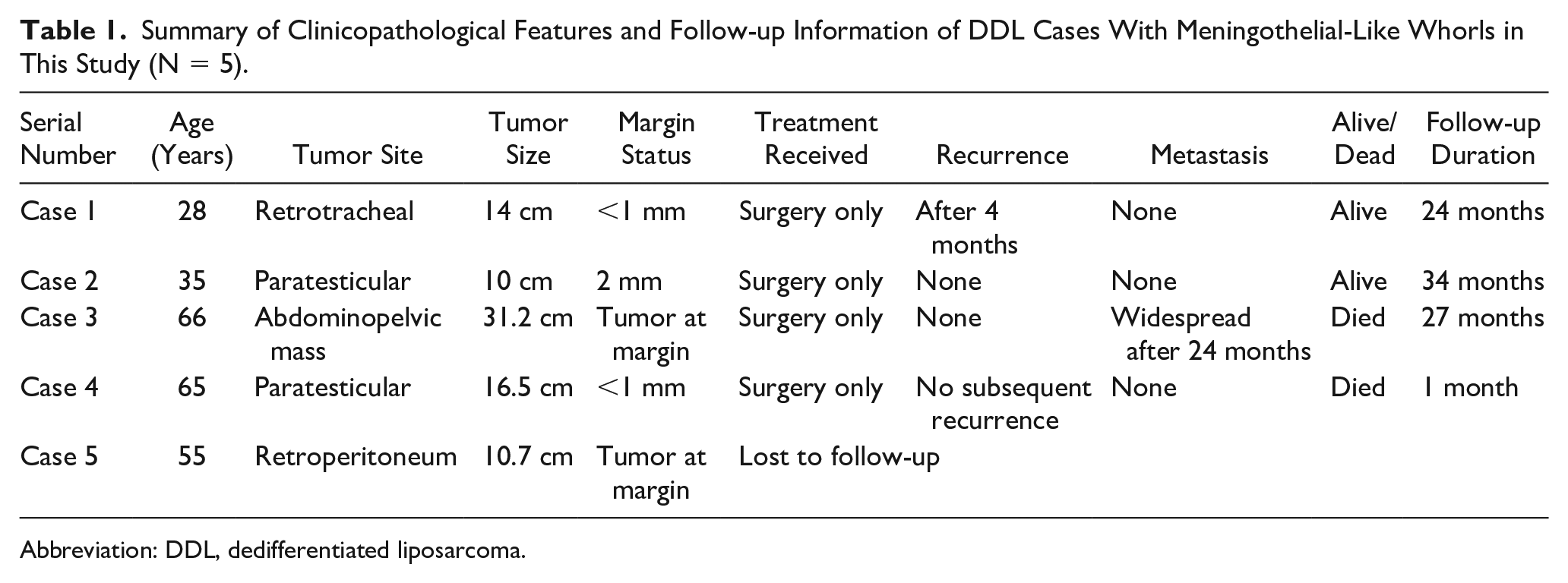
Microscopically, all cases showed areas of WDL and areas of dedifferentiation in varying proportions. Percentage of WDL component ranged from 5% to 80% of total tumor area. In 3 cases, WDL subtype was combination of sclerosing and lipoma-like, while WDL subtype was exclusively sclerosing in 2 cases (Table 2; Figure 1A). In 4 cases, the DDL component was in the form of low to intermediate spindle cells sarcoma that lacked any definitive differentiation (Figure 1B). In 1 case, it was in the form of intermediate-grade leiomyosarcoma and myxofibrosarcoma (Figure 1C and D). Tumor necrosis and multinucleated tumor giant cells were seen in 2 cases. Lymphocytic infiltrate and lymphoid aggregates were observed in 4 cases.
Summary of Histological Features of DDL and WDL Components Described in Different Studies.
Abbreviations: DDL, dedifferentiated liposarcoma; WDL, well-differentiated liposarcoma; MFH, malignant fibrous histiocytoma.
(A) Well-differentiated liposarcoma component, sclerosing subtype. Dedifferentiated liposarcoma component in the form of (B) intermediate-grade unclassifiable spindle cell sarcoma, (C) myxofibrosarcoma, and (D) leiomyosarcoma.
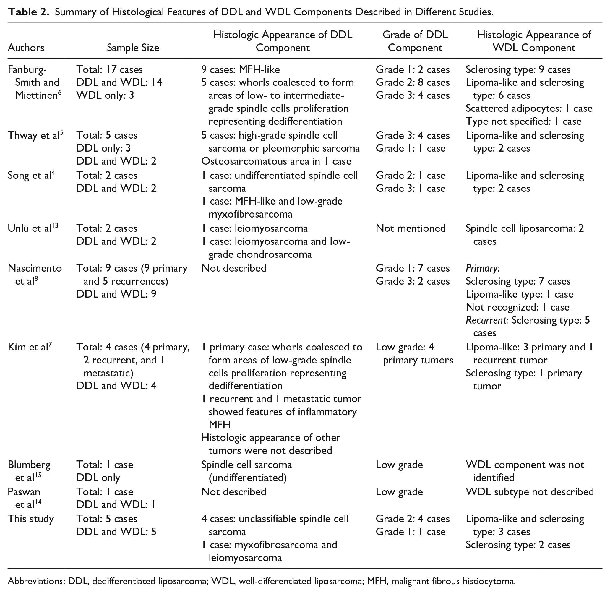
Concentric meningothelial-like whorls comprised 10% to 75% of the total tumor area. The size of these whorls ranged from <1 mm to >2 cm (Figure 2A). These whorls were present singly or in clusters in areas of WDL and areas of dedifferentiation (Figure 2B). These whorls coalesced in 3 cases. In 1 case, the whorls coalesced to form large areas of dedifferentiation (Figure 2C). The cells that formed the whorls were spindle to epithelioid shaped, had indistinct cellular outlines, and moderate amount of pale to eosinophilic cytoplasm. The nuclei were oval, vesicular, had finely dispersed chromatin, and inconspicuous nucleoli. Nuclear pleomorphism and mitotic activity was not appreciated in these cells (Figure 2D). The background stroma was variably myxoid and collagenous in 3 cases and exclusively collagenous in 2 cases. Psammomatous calcification was not observed.
(A) Whorls (arrows) in areas of well-differentiated liposarcoma component. (B) Whorls coalescing to form areas of low-grade spindle cells sarcoma (arrows). (C) High-power view of the whorl showing concentrically arranged spindle- to epithelioid-shaped cells lacking pleomorphism. Thin-walled blood vessels and lymphocytes are appreciable in the background. (D) Whorl with collagenous background.
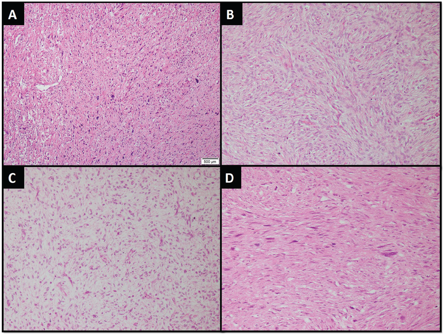
Metaplastic bone formation merging with the whorls was observed in 4 cases, and it was present in the form of rounded nodules and trabeculae of mature woven bone (Figure 3A and B). These trabeculae were lined by osteoblasts in all cases and osteoclasts in a single case. Cartilage formation was observed in 3 cases, and endochondral ossification in 2 cases (Figure 3C and D).
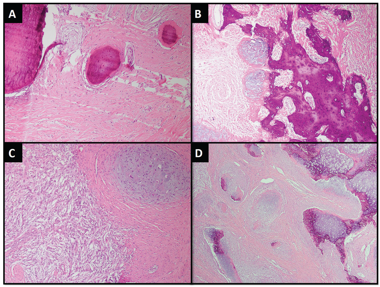
Metaplastic bone in the form of (A) nodules and (B) trabeculae of mature woven bone. (C) Cartilage formation. (D) Cartilage exhibiting endochondral ossification.
The whorl-forming cells were positive for α-smooth muscle actin (α-SMA) and p16 in 2 cases, when performed (Figure 4A and B). Epithelial membrane antigen (EMA), cytokeratin AE1/AE3, desmin, S100, SOX-10, HMB45, CD34, CD31, CD117, DOG-1, and Alk-protein immunohistochemical (IHC) stains were negative, when performed (Table 3).
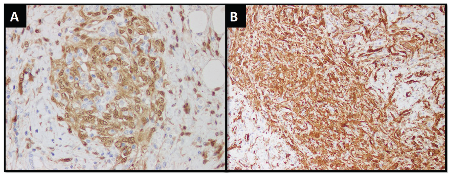
Whorl-forming cells demonstrating positive expression for (A) p16 and (B) α-smooth muscle actin immunohistochemical stain.
Summary of Immunohistochemical Features of Neoplastic Cells Forming Meningothelial-Like Whorls Described in Different Studies.
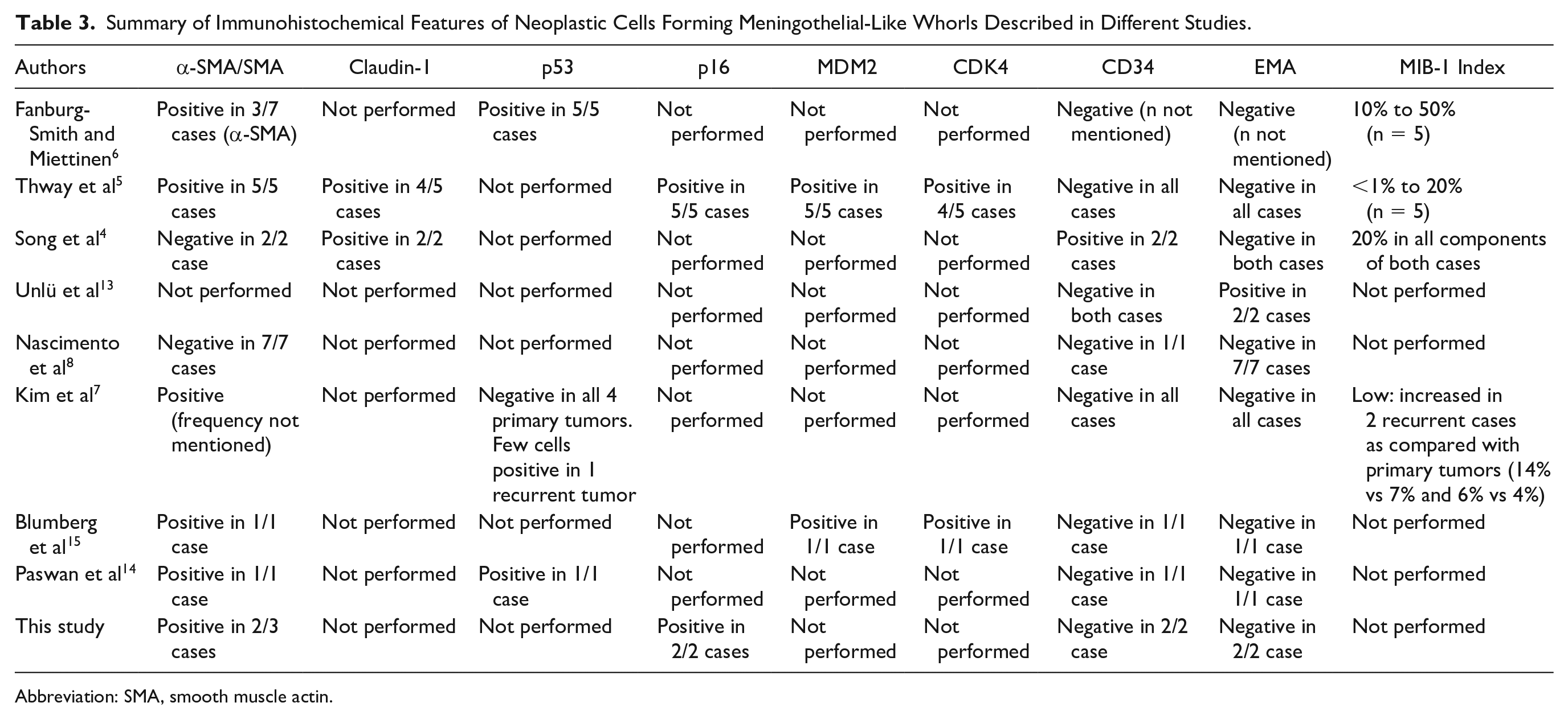
Abbreviation: SMA, smooth muscle actin.
MDM-2 amplification was identified in all cases by fluorescence in situ hybridization (FISH) method (Figure 5).
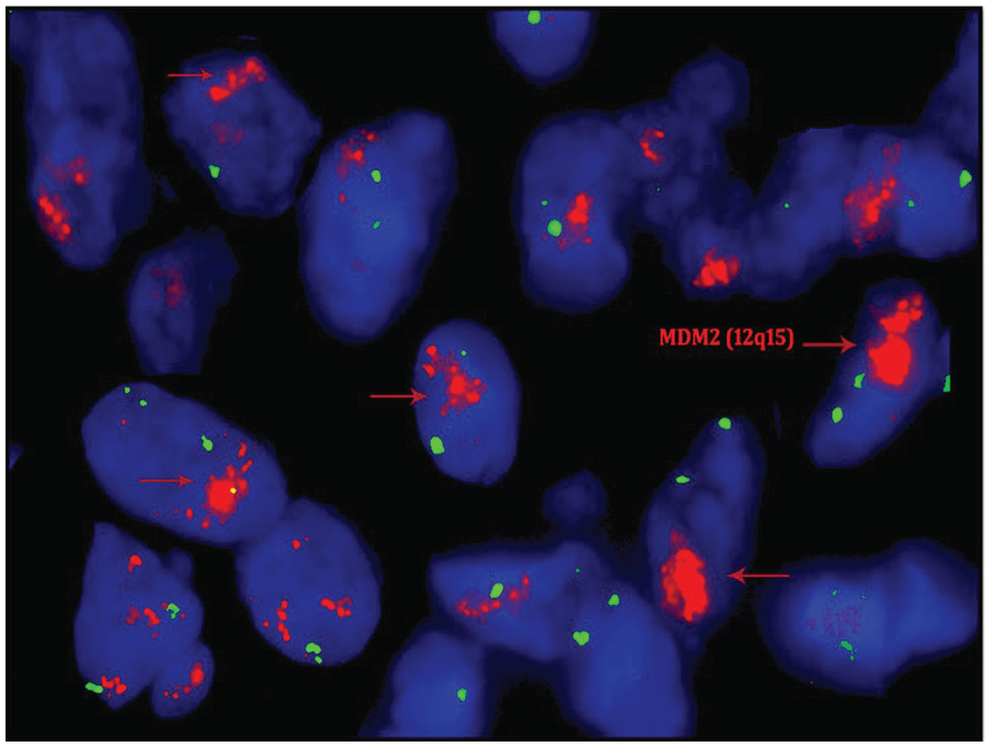
Fluorescence in situ hybridization test for MDM2 gene amplification is positive. The amplified gene signals are increased red signals (arrows).
Follow-up information was available for 4 patients. All of these patients were treated with surgery alone. No adjuvant chemotherapy or radiotherapy was given. Follow-up duration ranged from 1 to 34 months. Two patients were alive, and 2 died of disease. Local recurrence and widespread metastasis were observed in single case each. Case 4 presented to us for excision of recurrent tumor. He had an initial surgery 4 years ago, the record of which was not available. Patient was advised radiotherapy, but he refused any additional treatment (Table 1).
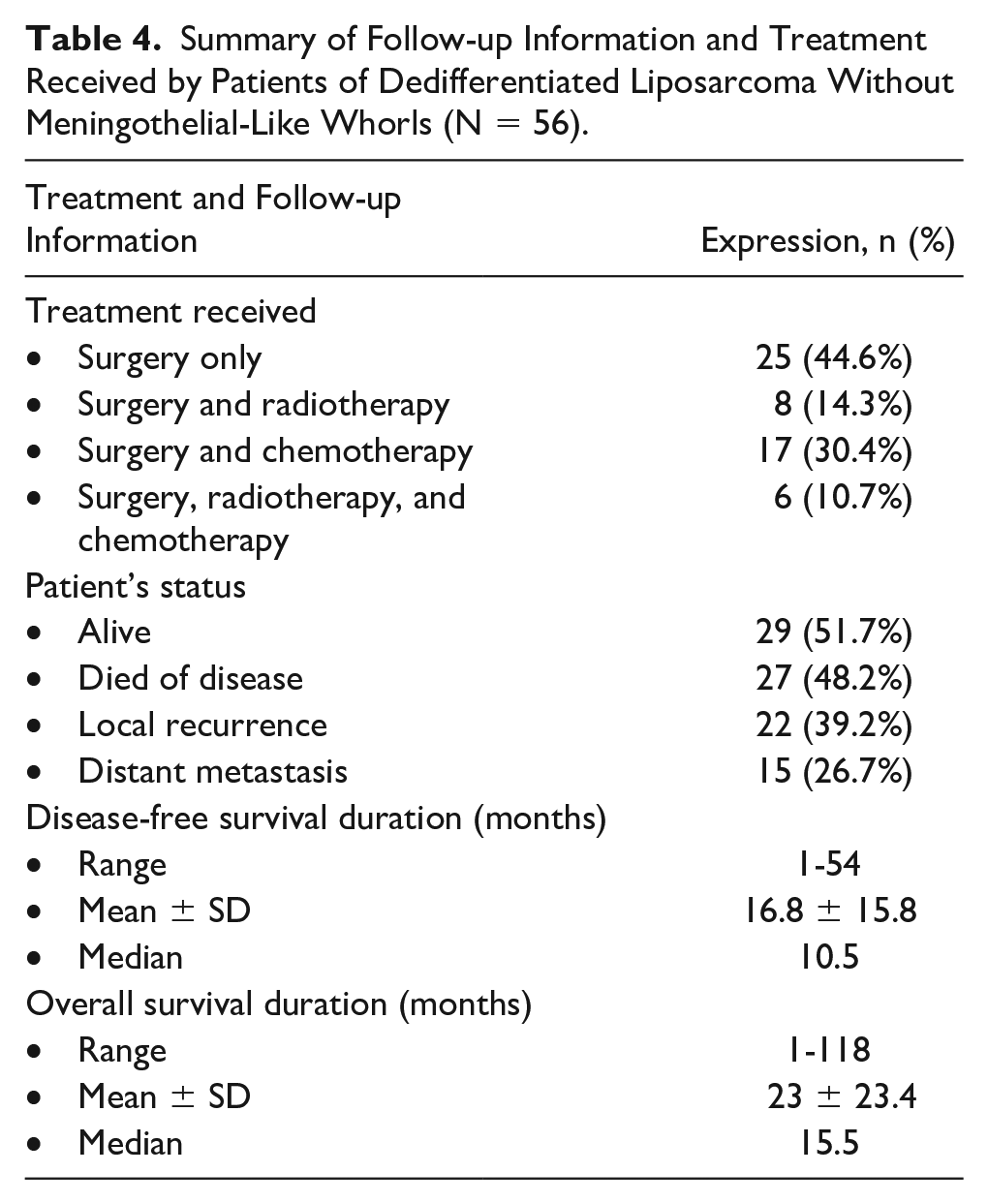
Between January 2010 and December 2019, 104 cases of DDL were diagnosed at our institution. Follow-up information was available for 56 patients (Table 4). Follow-up duration ranged from 1 to 118 months. These patients were treated with combination of surgery, radiotherapy, and chemotherapy. Almost half of the patients died of disease. Thirty-two (57.1%) cases either developed recurrence or metastasis. Among 22 patients who developed recurrence, 9 developed recurrences within 1 year of surgery and 3 patients developed multiple recurrences. Among 15 patients who developed distant metastasis, 9 developed metastases within 1 year of initial presentation. Lung was the most common metastatic site, and 4 developed metastases in multiple organs.
Summary of Follow-up Information and Treatment Received by Patients of Dedifferentiated Liposarcoma Without Meningothelial-Like Whorls (N = 56).
Discussion
Dedifferentiation is an uncommon phenomenon observed in few malignant soft tissue and bone tumors such as liposarcoma, chondrosarcoma, parosteal osteosarcoma, chordoma gastrointestinal stromal tumor, and solitary fibrous tumor. It is defined as the presence of sarcomatous component with divergent differentiation along with tumor component exhibiting actual/original differentiation. 9 Dedifferentiation is observed in 10% liposarcomas, and it is most commonly observed in retroperitoneal tumors.1,4-6 Meningothelial-like whorls and metaplastic bone formation are rare features of DDL, and majority of the studies reporting these features have not estimated their incidence. However, few studies have reported its frequency to range from 6% to 12% of cases.10-12 To the best of our knowledge, there are around 50 reported cases of WDL and DDL with meningothelial-like whorls and metaplastic bone formation in English literature.4,5,7,13-16 Song et al comprehensively summarized the findings of 34 such cases reported in the literature and 2 new cases of his study. 4 These tumors show male predilection, wide age range (24-78 years, mean 58.2 years), and predominantly involve retroperitoneum. Less common sites include extremities, spermatic cord, scrotum (paratesticular tissue), anterior mediastinum, and head and neck region. Tumor size ranged from 3 to 48 cm, and the mean tumor size was 12 cm. Interestingly, all patients of our study were male. Patients’ age also had wide range, and median was 55 years. Median tumor size was 16.5 cm, and only a single case of our cohort involved retroperitoneum (Table 1). Radiological examination revealed adipocytic and non-adipocytic areas. Similarly, the dedifferentiated areas appeared solid, gray white, and sometimes calcified. These areas may have diffuse appearance or form intratumoral nodules.4,7,8,15 Similarly, on gross examination, we also observed gray areas of dedifferentiation and calcification.
These features can be seen in areas of WDL and/or DDL.6-8,13 Histological features of DDL and WDL components described in different studies have been summarized in Table 2. Meningothelial-like whorls can comprise 5% to 60% of the total tumor volume, and these have been reported to range from <0.1 mm to greater in 1 cm in the areas of coalescence.4-7,15 The whorls may form a discrete mass or may be intermingled with WDL areas. 7 In our series, whorls occupied 10% to 75% of total tumor area. Their size ranged from <0.1 cm to more than 2 cm. In 1 case, these whorls coalesced to form areas of low- to intermediate-grade spindle cell sarcoma lacking any definitive differentiation. This phenomenon had also been described by Fanburg-Smith and Miettinen in 5 out of 17 cases. 6 These whorls were formed by spindle to epithelioid cells arranged in concentric lamellae around small-sized, thin-walled blood vessels against myxoid to sclerotic stroma. The cells are plump with indistinct cell borders, pale fibrillary cytoplasm, distinct nuclear membranes, vesicular nuclei, and small indistinct nucleoli.5,6,8,14,15 Mitotic activity is usually low, and up to 5 mitoses/10 high-power fields have been observed. Marked atypia can be occasionally seen in whorl-forming cells. 5 Occasional atypical mitoses may also be observed. 6 These whorls were seen in clusters and scattered throughout the tumor. Similarly, we also observed spindle to epithelioid morphology of whorl-forming cells. Increased mitotic activity and significant nuclear pleomorphism was not seen. The background in the whorls may show mast cell population.5,7 Diffuse lymphocytic infiltrate intermixed with neoplastic cells or lymphoid follicles at the periphery of whorls may be seen in all cases reported by Nascimento et al. 8 Lymphoid aggregates and diffuse lymphocytic infiltrate was also seen in 4 cases of our study.
Multinucleated tumor giant cells may also be seen in dedifferentiated areas. 5 Focal “paraganglioma-like” pattern has also been reported in 1 case. 4
Metaplastic bone formation has been observed in 59% to 100% cases where it is seen either in the center or adjacent (merging) to the whorls. Mostly, the bone is in the form of spicules and trabeculae of mature woven bone, sometimes lined by osteoblasts and osteoclasts. The bone-forming cell may occasionally exhibit atypia.4-8,15 The close association of whorls with metaplastic bone raises the possibility of production of osteogenic promoter substance by whorl-forming cells. 8 Cartilage formation has been reported in up to 60% cases. 5 In our study, metaplastic bone formation was also observed in 4 cases, and it was in the form of nodules and trabeculae of mature woven bone. Whorls were seen in the close proximity of metaplastic bone. Cartilage formation was also observed in 3 of our cases.
The cells forming whorls shared positivity for MDM2, CDK4, and p16 IHC stains with neoplastic cells in dedifferentiated areas. 5 Positive expression is also noted for vimentin, α-SMA, claudin-1, and p53 IHC stains. In our study, 2 cases were stained with p16 and α-SMA IHC stains, and the whorl-forming cells were positive for these markers. P16 expression was also observed in the adjacent DDL component. Some nonspecific markers that can be occasionally positive in these cells include CD56, CD57, CD10, and CD99. These cells are consistently negative for cytokeratin AE1/AE3, synaptophysin, chromogranin A, neurofilament, NSE, CD31, CD34, CD21, CD35, CD117, CD68, β-catenin, S100 protein, h-caldesmon, and desmin IHC stains (Table 3).4-8,14,15 EMA has also been reported negative in majority of the studies except for a single study that reports positive expression. 16 Positive expression for SMA favors the possibility of myofibroblastic differentiation, and lack of h-caldesmon and desmin IHC stains’ expression does not support the possibility of myopericytic differentiation. Claudin-1 IHC expression in these cells favors perineural differentiation, but negative expression for EMA and CD34 IHC stains (in majority of the studies) reduces the likelihood of this possibility. 4 However, the only study reporting positive EMA expression did not evaluate claudin-1 expression. 13 Mib-1 index was higher than the surrounding WDL areas. Expression of p53 was also observed in dedifferentiated areas but not in WDL areas.6,14
Two differential diagnoses related to the meningothelial-like whorls, that is, malignant meningioma and FDCS, can also rarely occur in retroperitoneal region.8,17,18 Presence of WDL and association of these whorls with metaplastic bone formation are useful diagnostic clues for DDL. 6 The presence of prominent myxoid background stroma of the whorls brings nerve sheath myxoma into the differential diagnoses. When the background stroma of the whorls is hyalinized, fibrotic, and hypocellular, it resembles neurothekeoma. 8 Immunohistochemistry can also be helpful as the whorls seen in neural tumors (meningioma and perineuroma) demonstrate positive expression for EMA IHC stain, while the whorls in DDL are demonstrate negative expression.18,19 Prominent lymphocytic infiltrate is commonly observed in FDCS. However, this feature was also frequently observed in DDLs with meningothelial-like whorls. 8 The ultrastructural features of the whorl-forming cells have been assessed in a single case. The features favored FDC differentiation 8 ; however, these cells do not demonstrate positive expression for IHC markers of FDC differentiation, that is, CD21 and CD35 IHC stains.8,17 According to Fanburg-Smith and Miettinen, the cells forming whorls in DDL show pericytic and myofibroblastic differentiation as these cells not only resemble pericytes and myofibroblasts morphologically, but these are also positive for α-SMA. In addition, the cells of whorls around the metaplastic bone show osteoblastic differentiation as these cells are positive for osteocalcin IHC stain. 6
The cells forming the metaplastic bone are cytologically bland, while the cells in areas of heterologous osteogenic (osteosarcomatous) dedifferentiation show malignant features. 6 According to Fanburg-Smith and Miettinen, based on positive p53 IHC expression, increased MIB-1 index in contrast to the surrounding WDL areas, and formation of dedifferentiation-like areas by coalescence of whorls, these structures are considered to represent an early stage of dedifferentiation. 6 Nascimento et al consider these whorls as a sign of dedifferentiation because of increased cellularity and different appearance from background WDL. 8 These cases also exhibit complex karyotype comprising several structural and numeric chromosomal abnormalities including ring and giant chromosomes. 3
The coalescence of whorls to form areas of low- to intermediate-grade spindle cell sarcoma, as observed in our study and the study by Fanburg-Smith and Miettinen, provides important evidence in favor of whorls representing initial stage of dedifferentiation. 6
Surgical resection is the mainstay of treatment for liposarcomas. High-grade tumors may benefit from adjuvant chemotherapy and radiotherapy. Patient’s age, primary tumor site, presentation status, histologic subtype, margin status, and tumor burden independently predict disease-free survival. 20 The behavior of tumors with these whorls has been quite variable. In a large study of 155 DDL cases, behavior was not related to the histologic grade. 21 However, in a series of 17 DDL cases with meningothelial-like whorls, 7 patients developed metastasis and died of disease. In one of these cases who died of disease, whorls were present in the background of WDL and areas of dedifferentiation were not observed. The observation points toward malignant potential of tumors and the possibility of dedifferentiated areas which remained unnoticed. The authors suggested that the variation in behavior was attributed to the variation in histologic grades. 6 In another study, none out of 4 patients developed recurrence, metastasis, or died of disease. 5 In 9 cases reported by Nascimento et al, recurrence was observed in 5 cases. None of the patients developed metastasis or died of disease. 8 Another study of 4 cases reported recurrence in 2 cases and metastasis to cervical spine in 1 case after 32 months. 7 In our study, follow-up information was available for 4 cases and the tumor behaved aggressively in these cases. One of the patients developed local recurrence after 4 months and was alive with disease. One of the patients presented as a recurrent tumor after 4 years of initial surgery, and he died of disease 1 month after second surgery. Another patient developed distant metastasis after 2 years of surgery and died 3 months later. The behavior of these tumors was similar to the behavior of DDL cases without meningothelial-like whorls, diagnosed during this period. More than half of these cases either developed recurrence or metastasis, and almost half of them died of disease. A substantial number of recurrences and metastasis were observed within 1 year.
Hence, DDL with meningothelial-like whorls demonstrate similar aggressive behavior for which DDL are generally known for.
Conclusion
Meningothelial-like whorls and metaplastic bone formation are uncommon features of DDLs. Areas of WDL in the vicinity, formation of metaplastic bone, positivity for p16, and α-SMA IHC stains, negativity for EMA, CD21, CD23, and CD35 IHC stains, and MDM2 gene amplification by FISH technique are helpful features for ruling out differential diagnoses and reaching the correct diagnosis. These whorling structures most likely represent an early stage of dedifferentiation. Like all DDLs, tumors with whorls also bear the potential to behave aggressively.
Footnotes
Declaration of Conflicting Interests
The author(s) declared no potential conflicts of interest with respect to the research, authorship, and/or publication of this article.
Funding
The author(s) received no financial support for the research, authorship, and/or publication of this article.
Ethical Approval
Since this study was conducted by reviewing the patient’s records and reviewing the archived material, exemption from the ethical approval was granted by the Hospital’s Ethical Review Committee (2020-3346-7362).
Informed Consent
Not applicable, because this article does not contain any studies with human or animal subjects.
Trial Registration
Not applicable, because this article does not contain any clinical trials.