






Abstract
Hereditary pancreatitis is a rare form of recurrent acute pancreatitis that typically has an onset in early adulthood. We report a rare case of hereditary pancreatitis in an individual with a serine protease inhibitor Kazal type 1 (SPINK1) mutation. Histologically the pancreas showed features of chronic pancreatitis with variable fibrosis, acinar destruction, and prominent inspissated secretions within the pancreatic ducts. Additionally, focal areas of low-grade pancreatic intraepithelial neoplasia (PanIN) were present. Although the histopathology of common gene mutations resulting in hereditary pancreatitis have been well described, the histopathology of pancreatitis secondary to SPINK1 mutation has been described in only one previous study.
Introduction
Hereditary pancreatitis is an incredibly rare cause of recurrent bouts of acute pancreatitis. 1 Patients with hereditary pancreatitis ultimately progress to develop the sequelae of chronic pancreatitis at a young age and are at an increased risk for development of pancreatic adenocarcinoma. 2 Many genes have been implicated in the development of hereditary pancreatitis, most notably the cationic trypsinogen gene serine protease 1 (PRSS1), as well as the cystic fibrosis transmembrane conductance regulator (CFTR) and the serine protease inhibitor Kazal type 1 (SPINK1). 3 The histopathology of hereditary pancreatitis due to mutations in PRSS1 has previously been described. 4 In this article, we are presenting the clinical features and histopathology of a patient with recurrent pancreatitis due to SPINK1 mutation.
Case Report
A 24-year-old female presented 2 years prior to an outside hospital with recurrent bouts of abdominal pain with elevated lipase levels of 5674 U/L, consistent with acute pancreatitis. Her past medical history was notable for multiple episodes of pancreatitis over the past 5 years with numerous admissions for pain management. She denied tobacco or alcohol use. She denied any family history of acute or chronic pancreatitis. She endorsed frequent abdominal discomfort but denied steatorrhea or symptoms of vitamin deficiencies seen in exocrine insufficiency. Workup at the time, including computed tomography of the abdomen and pelvis and an endoscopic ultrasound demonstrated mild peripancreatic inflammatory changes surrounding the pancreas, a peripancreatic fluid collection, and absence of cholelithiasis and choledocholithiasis. Pancreatobiliary anatomic alterations were not identified.
Given the recurrent nature of the patient’s pancreatitis with unclear etiology, she underwent genetic testing with a pancreatitis panel, which assayed mutations in 6 genes (PRSS1, CPA1, SPINK1, CTRC, CASR, and CFTR) commonly associated with hereditary pancreatitis. Next-generation sequencing and Sanger sequencing revealed that she harbored a mutation in the serine protease inhibitor Kazal type 1 (SPINK1) gene. She was heterozygous for a substitution mutation (c101A>G), which changed the nucleotide position 101 from A to G, which results in an amino acid substitution at amino acid 34 from asparagine to serine (p.Asn34>Ser). No mutations in PRSS1, CPA1, CTRC, CASR, or CFTR were identified.
The patient subsequently transferred care to Northwestern Memorial Hospital for clinical and surgical management. Repeat computed tomography imaging demonstrated diffuse interstitial pancreatic edema with moderate amount of peripancreatic fluid (Figure 1). One year later, she underwent total pancreatectomy with autologous islet cell transplantation, splenectomy, cholecystectomy, and duodenectomy. The head of the pancreas was submitted for examination and consisted of a 9.5 g, 4.5 × 3.2 × 1.5 cm pancreas specimen. On gross examination, the pancreas head was surrounded by a moderate amount of peripancreatic fat (Figure 2A and B). The pancreatic parenchyma was white, tan, and lobulated with small areas of firmness. There were no gross masses or lesions present (Figure 2C). The head of the pancreas was serially sectioned and entirely submitted.
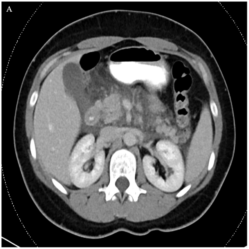
Computed tomography (CT) imaging of pancreas demonstrating diffuse pancreatic edema, consistent with pancreatitis.
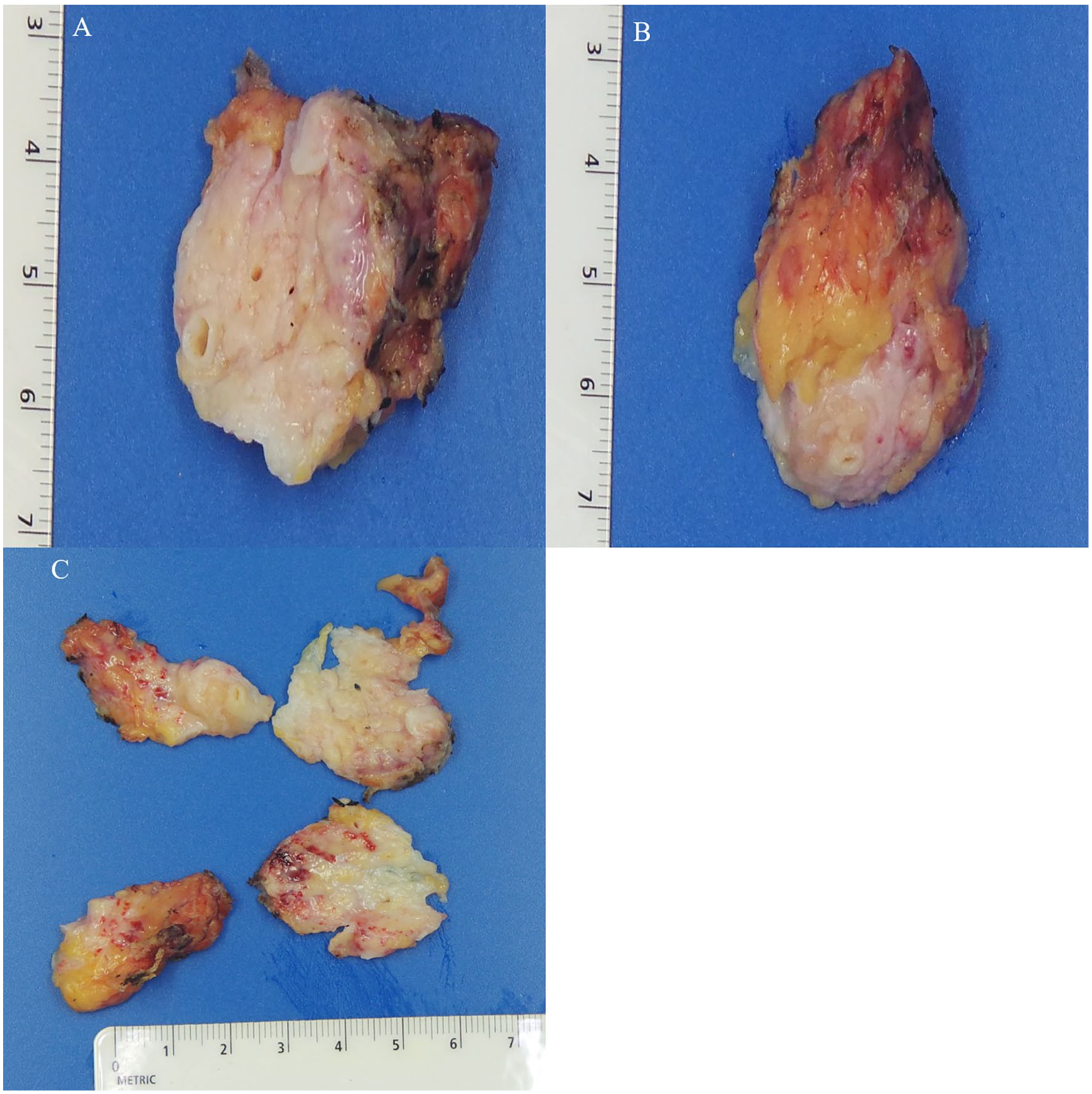
Gross examination of the pancreas. (A) The head of the pancreas is firm and tan-white. (B) There is extensive peripancreatic fat surrounding the head of the pancreas. (C) Cut surface shows multilobulated parenchyma with no definitive masses or lesions.
Microscopic examination showed pancreatic lobules were varying degrees of interlobular and perilobular fibrosis and acinar destruction (Figure 3A and B). Additionally, there were scattered areas of ducts lined by single layer of epithelium with abundant intracellular mucin without nuclear atypia, consistent with low-grade pancreatic intraepithelial neoplasia (PanIN-1A; Figure 3C).There were multiple areas of dilated ducts filled with inspissated, lamellated eosinophilic secretions (Figure 3D and E), which stained negative for Congo red (Figure 3F).
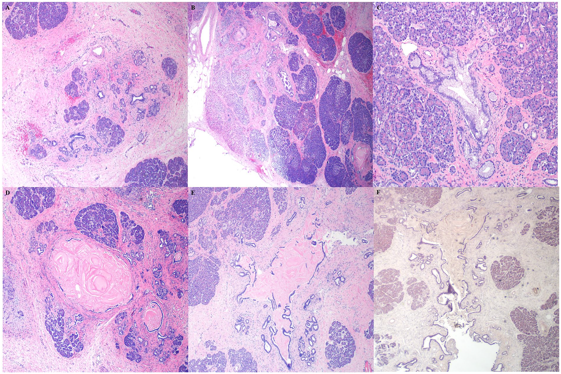
Histologic examination of the pancreas. (A) The pancreatic parenchyma consists of areas of perilobular fibrosis. (B) There are areas of sharp demarcation between atrophic lobules and viable parenchyma. (C) Scant areas of the pancreas demonstrated low-grade pancreatic intraepithelial neoplasia 1 (PanIN 1A). (D-E) Large and small ducts were filled with eosinophilic lamellated secretions. (F) The secretions stained negative for Congo red.
The patient’s postoperative course was unremarkable. She was discharged on supplemental pancreatic enzyme therapy.
Discussion
Hereditary pancreatitis was first described in the 1950s through a case series of 4 family members who had recurrent occurrences of pancreatitis. 5 Although alcohol and gallstones are the prevailing etiologic causes of a majority of cases of pancreatitis, up to 8.7% of cases of chronic pancreatitis were found to be caused by genetic factors. 6 Hereditary pancreatitis tends to have extremely high penetrance. Multiple genes have implicated in the pathogenesis, including PRSS1, SPINK1, CFTR, and CTRC. 1 In a survey of patients with hereditary pancreatitis in Japan, 41% were caused by PRSS1 mutations and 36% were caused by SPINK1 mutations. 7
SPINK1 plays a pivotal role in the inhibition of trypsinogen activation to trypsin in the pancreas. SPINK1 mutants create a disproportion of proteases and inhibitors in the pancreas, leading to unchecked activation of trypsin with resulting enzymatic cascades leading to autodigestion of the pancreas and pancreatitis. 8 In a murine model with deficient SPINK3, the homologue to human SPINK1, elevated trypsin activity in pancreatic acinar cells was detected within 0.5 days of birth compared with wild-type expression. 9 In a similar SPINK3 murine model, pancreatic acinar cells underwent autophagy with impaired ability to regenerate, highlighting the essential role for the balance of proteases and inhibitors in the pancreas. 10
Here, we described the unique histopathology of SPINK1 hereditary pancreatitis in a 24-year-old female who underwent pancreatectomy for recurrent episodes of acute pancreatitis leading to chronic pancreatitis. Her first episodes of pancreatitis dated back to 19 years of age. Individuals with hereditary pancreatitis tend to develop symptoms by the age of 10 to 12 years old; however, malabsorption, diabetes mellitus, and pancreatic cancer tend to occur at much older age. 11
Although a grading system for chronic pancreatitis does not exist, there is consensus on the histological aspects. Notably, a triad exists of fibrosis, duct changes, and loss of acinar tissue. 12 However, histologic analysis alone is not sufficient to make an exact diagnosis of the cause of chronic pancreatitis, and genetic testing is paramount to uncovering the etiology.
The histopathology of this case was quite unique in comparison to pancreatitis caused by PRSS1 and CFTR mutations. Our case showed varying amounts of perilobular and interlobular fibrosis. There were areas of sharp demarcation from viable pancreatic acini from the fibrotic, destroyed ducts. PRSS1 mutations tend to develop similar appearing pancreatic fibrosis. 4 Unlike patients with PRSS1, our case showed extensive involvement of ducts clogged with inspissated secretions. Pancreatic plugged ducts are very commonly observed in patients with CFTR mutations. 13 Amyloid deposition was not observed in either the ducts or the islets of Langerhans cells confirmed by negative Congo red staining. Additionally, our case showed variable peripheral adipose replacement, which was observed in PRSS1 cases. The patient had mild symptoms of pancreatic insufficiency, including bloating. Although striking on histology, the inspissated, lamellated eosinophilic secretions contained within the ducts did not portend a worse degree of pancreatic insufficiency, as the patient did not have steatorrhea or vitamin deficiencies.
A recent study of 28 cases of SPINK1 chronic pancreatitis demonstrated unique characteristics within the cohort, namely, the co-occurrence of additional germline alterations in pancreatitis-associated genes and histology demonstrating parenchymal fibrosis. 14 In our case, the patient was heterozygous for the SPINK1 N34S mutation and did not harbor additional germline mutations in other pancreatitis genes. Additionally, while parenchymal fibrosis was identified in our case, the presence of inpissated ducts filled with lamellated secretions was not identified in the study.
Increased risk of pancreatic carcinoma is well documented in patients with hereditary pancreatitis, with patients developing a cumulative risk of roughly 40% by the age of 70 years. 15 Low-grade PanIN was observed in our case. Although low-grade PanIN is typically an incidental finding, high-grade PanIN does carry an increased risk for the development of pancreatic adenocarcinoma. 16 Thus, close surveillance is warranted in patients with hereditary pancreatitis.
Our case elucidates the histopathology of SPINK1 hereditary occurring in one patient. The histology of the pancreas had characteristics similar to those seen in both PRSS1 and CFTR hereditary pancreatitis, and similar findings to the recent published study by Jones and colleagues. 14 Although we elucidate the histopathology of SPINK1 pancreatitis, further studies with an expanded patient cohort are needed to develop a consensus on the features that are common in these patient populations. Additionally, our patient was heterozygous for the N34S mutation, and further studies in homozygous patient populations should be undertaken.
Footnotes
Declaration of Conflicting Interests
The author(s) declared no potential conflicts of interest with respect to the research, authorship, and/or publication of this article.
Funding
The author(s) received no financial support for the research, authorship, and/or publication of this article.
Ethical Approval
Not applicable, because this article does not contain any studies with human or animal subjects.
Informed Consent
Not applicable, because this article does not contain any studies with human or animal subjects.
Trial Registration
Not applicable, because this article does not contain any clinical trials.
