
Abstract
Only four cases of colorectal adenocarcinoma with a yolk sac tumor (YST) component have been reported in the English literature. No genetic investigation has been performed in these cases. We report a case of colorectal adenocarcinoma in which the recurrent tumor had a YST component. A 49-year-old woman presented with a pelvic tumor three years after endoscopic mucosal resection of sigmoid colon adenocarcinoma. The pelvic tumor consisted of an undifferentiated carcinoma component and a YST component. The serum alpha-fetoprotein level was elevated to 42 ng/mL. Treatment as conventional colorectal carcinoma produced some anticancer effects, but the patient died 14 months after the recurrence and 49 months after the EMR. With the help of the next-generation sequencing results of the recurrent tumor, APC c.835 − 8A > G and TP53 c.524G > A (p.R175H) mutations were identified by direct sequencing in both the primary and the recurrent tumors, confirming the relationship between the two metachronous tumors.
Introduction
Cases of conventional carcinoma with germ cell tumor components such as yolk sac tumor and choriocarcinoma have been reported in multiple organs. Serum markers such as alpha-fetoprotein (AFP) and human chorionic gonadotropin (hCG) may be elevated in these cases. In the large intestine, a small number of adenocarcinoma cases with choriocarcinoma component have been reported, 1 but cases with yolk sac tumor component are exceedingly rare.2–5 Herein, we report a case of recurrent colon cancer with a yolk sac tumor component and evidence of shared TP53 and APC mutations in the primary and recurrent lesions.
Case Report
A 49-year-old female patient presented to our hospital for the management of a pelvic tumor with metastasis to lymph nodes, lungs, liver and bone and peritoneal dissemination. She had undergone endoscopic resection of a polypoid carcinoma of the sigmoid colon three and a half years prior to this presentation. Six months before presentation to our hospital, a pelvic tumor involving the sigmoid colon and the left ureter was detected. Under the presumptive diagnosis of ovarian cancer, total hysterectomy, bilateral adnexectomy, omentectomy, and sigmoid colectomy were performed. The involved left ureter was spared. Shortly after the surgery, lung metastasis developed. The clinical decision was to treat this tumor, which was difficult to diagnose histopathologically, as ovarian cancer. After treatment failure with paclitaxel and carboplatin, and then pegylated liposomal doxorubicin and bevacizumab, the patient came to our institution for a second opinion.
A review of the histological specimens and the radiological images at our hospital raised the possibility of recurrence of colonic cancer, though the histology was not typical for colorectal cancer recurrence and had a yolk sac tumor component. The serum AFP level measured at our hospital was elevated at 42 ng/mL. Next-generation sequencing (NGS) of the pelvic tumor using FoundationOne Companion Diagnostic panel (Foundation Medicine) revealed APC c.835 − 8A > G and TP53 c.524G > A (p.R175H) mutations. No mutations were found in BRAF, KRAS, or NRAS genes and the tumor was microsatellite-stable.
With the presumptive diagnosis of recurrent colorectal cancer, the patient was treated with chemotherapy with FOLFOXIRI (fluorouracil, leucovorin, oxaliplatin, and irinotecan) plus bevacizumab. She was treated with this regimen for four months, followed by trifluridine/tipiracil plus bevacizumab for a month. She died 8 months after presenting to our institution, 14 months after the discovery of the pelvic tumor, and 49 months after the endoscopic resection of the colon cancer.
The histology of the pelvic tumor was reviewed. The pelvic tumor was composed of a solid component and a yolk sac tumor (YST) component, with the solid component accounting for the majority of the tumor. The solid component consisted of solid proliferation of pleomorphic neoplastic cells. Geographic necrosis of the solid tumor cells resulted in a pseudopapillary, vaguely transitional cell carcinoma–like appearance, which was reminiscent of high-grade serous carcinoma. Syncytiotrophoblast-like giant cells, which were positive for hCG by immunohistochemistry, were scattered in the vicinity of the YST component, but there was not enough morphological or immunohistochemical evidence to diagnose choriocarcinoma (Figure 1). The YST component consisted of similar pleomorphic cells. They exhibited a variety of patterns characteristic of a YST: reticular, microcystic, glandular, and papillary patterns, in some places associated with loose myxoid stroma (Figure 2). A few Schiller–Duval bodies were observed (Figure 2A). The immunohistochemistry performed at the previous institution and ours showed that the YST component was positive for SALL4, glypican 3, AFP, and CDX2 and negative for keratin 7 and keratin 5/6. The solid component was positive for SALL4, CDX2, and keratin 7, partially positive for keratin 5/6, and negative for AFP and glypican 3. Both components were positive for p53 (Figure 3A) and AE1/AE3, negative for OCT3/4, KIT, D2-40, CD30, PAX8, WT1, estrogen receptor, calretinin, and vimentin, and partially positive for chromogranin A, synaptophysin, and CD56. β-catenin nuclear accumulation was positive in the solid component and negative in the YST component (Figure 3B). During the process of histopathological diagnosis, our thinking was that if we interpreted the solid component of this pelvic tumor as undifferentiated carcinoma of the colon and the YST component as an abnormal but not impossible differentiation, the diagnostic possibility of recurrent colonic cancer could be sustained.
Undifferentiated carcinoma area of the recurrent colon cancer. Geographic necrosis gives the solid proliferation of neoplastic cells a pseudopapillary, vaguely transitional cell carcinoma–like appearance (A). A small number of syncytiotrophoblast-like giant cells were scattered (B), which were positive for hCG by immunohistochemistry (not shown). (Hematoxylin–eosin, original magnification × 10, bar = 1 mm [A] and × 200, bar = 100 µm [B]).

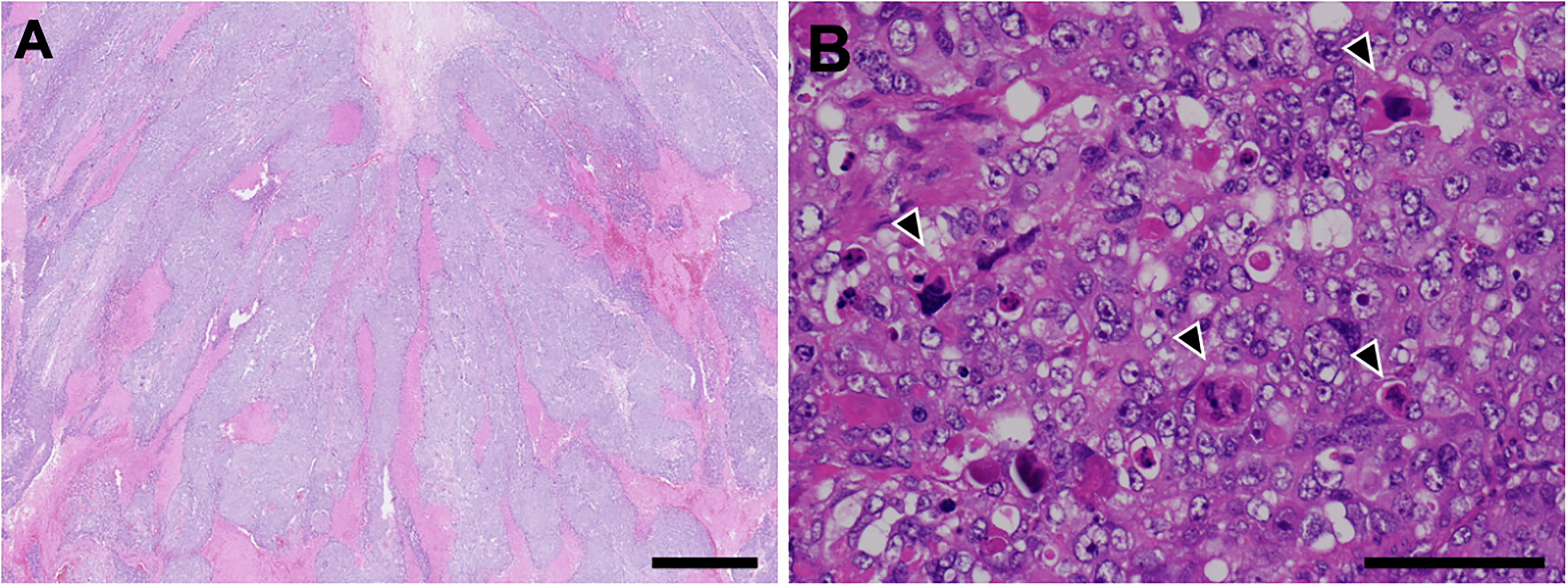
Yolk sac tumor area of the recurrent colon cancer. Neoplastic cells exhibited reticular (A&B), microcystic (B&C), glandular (B&D), and papillary (D) pattern, in some places associated with loose myxoid stroma (C). A few Schiller–Duval bodies were observed (asterisks in A). (Hematoxylin–eosin, original magnification × 20, bar = 1 mm [A] and × 100, bar = 200 µm [B-D]).
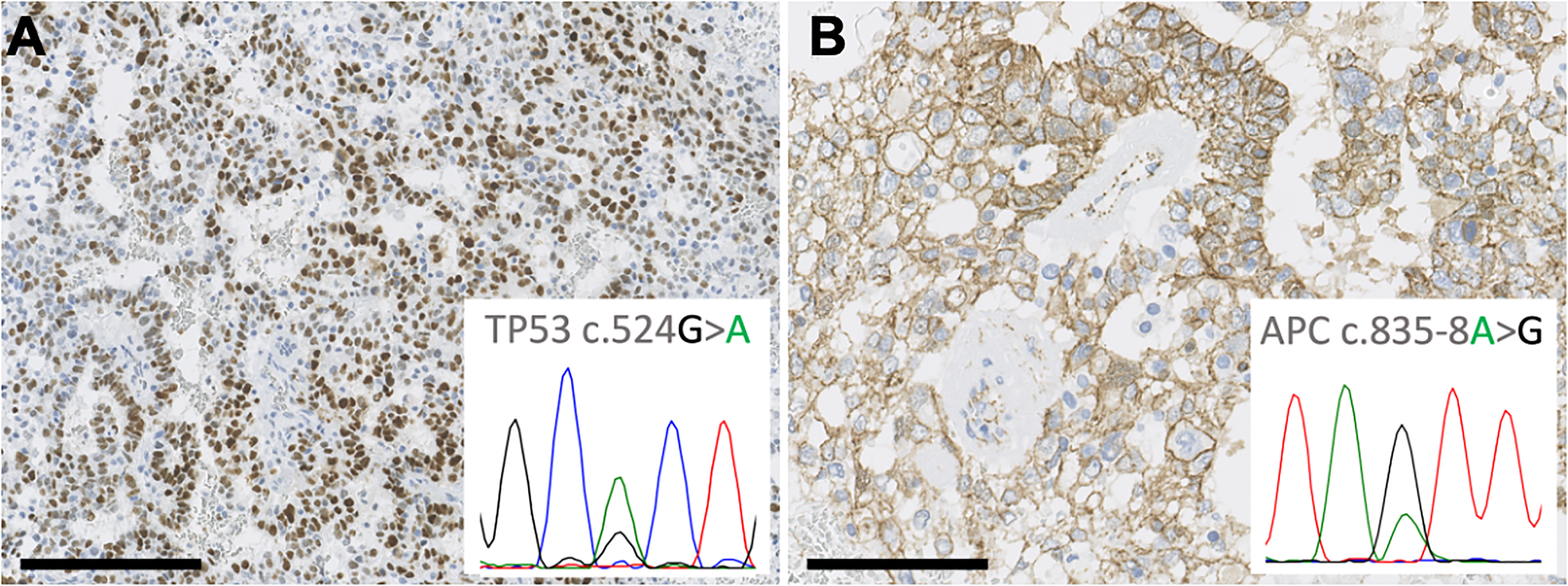
Immunohistochemical and molecular analysis of the recurrent colon cancer. TP53 c.524G > A (p.R175H) mutation and APC c.835-8A > G mutation detected by next-generation sequencing was confirmed by direct sequencing (A&B, inset). In yolk sac tumor area, immunohistochemistry was positive for p53 (A) in accordance with TP53 mutation, but β-catenin did not show nuclear accumulation (B) in spite of APC mutation (see text). (Original magnification × 100, bar = 200 µm [A] and × 200, bar = 100 µm [B]).
To further investigate the possibility of recurrence, the histological specimen from the endoscopic resection of sigmoid colon cancer performed four years earlier was reexamined. It showed morphologically conventional tubular adenocarcinoma, but with marked invasiveness and lymphatic invasion (Figure 4). Additional immunohistochemistry at our institution revealed β-catenin nuclear positivity and p53 positivity (Figure 5).
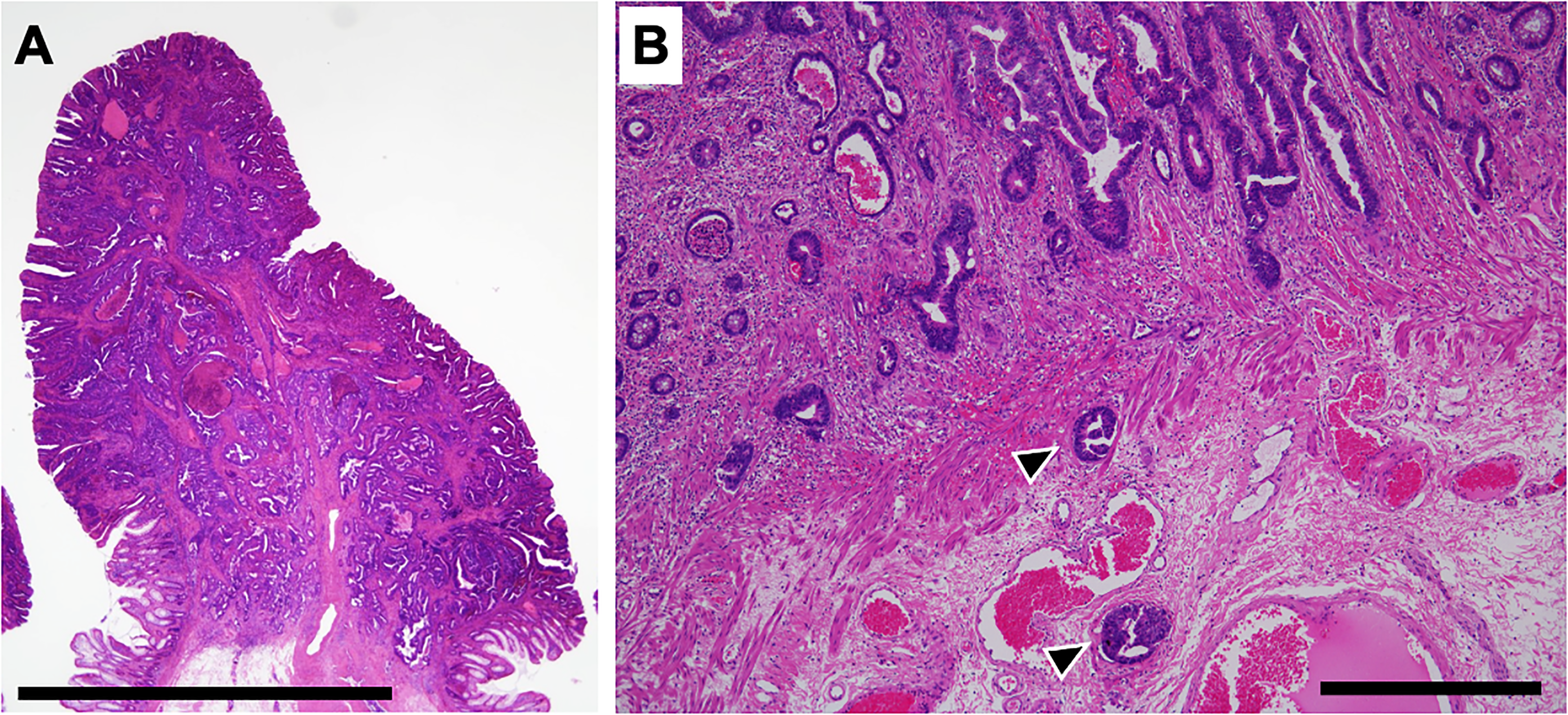
Primary sigmoid colon adenocarcinoma. The tumor resected endoscopically three years prior to the discovery of the pelvic tumor was a morphologically conventional adenocarcinoma with marked invasiveness and lymphatic infiltration. Arrowheads in B indicate tumor cell nests within lymphatics. A and B show different sections. (Hematoxylin–eosin, original magnification × 10, bar = 5 mm [A] and × 40, bar = 500 µm [B]).
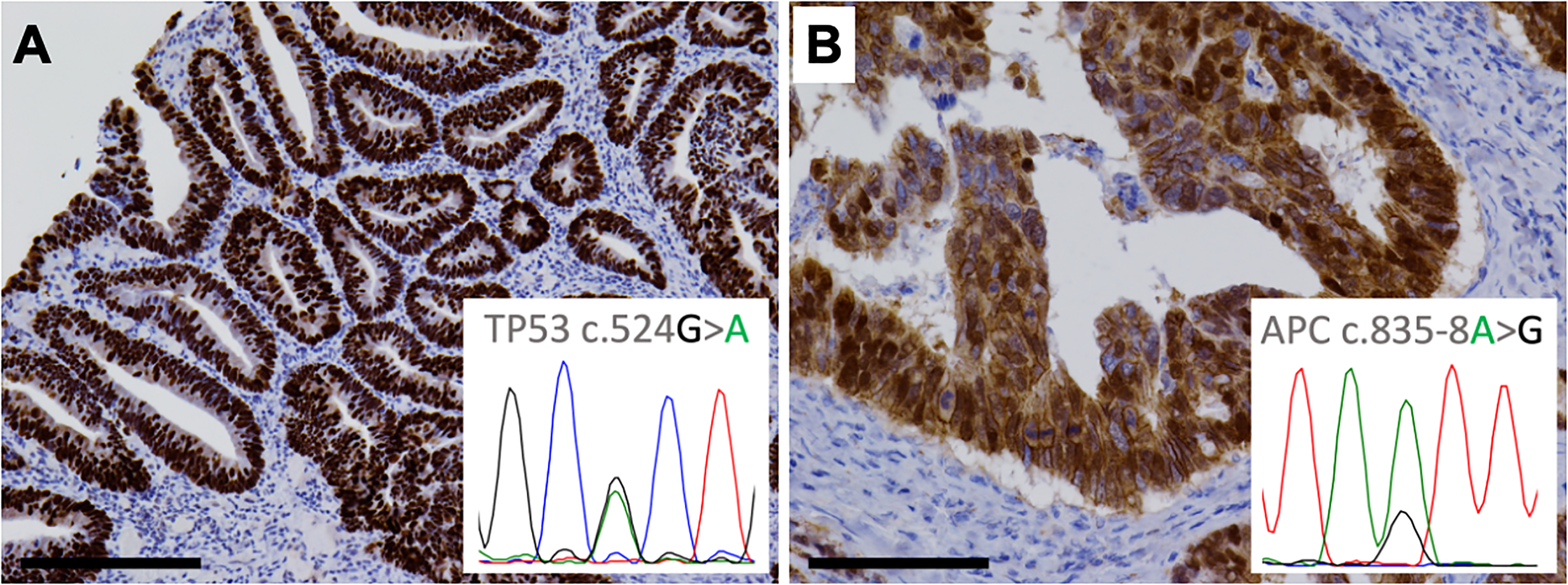
Immunohistochemical and molecular analysis of the primary colon cancer. Immunohistochemistry was positive for p53 (A) and nuclear accumulation of β-catenin (B), in accordance with the TP53 c.524G > A (p.R175H) mutation (A, inset) and APC c.835-8A > G mutation (B, inset) detected by direct sequencing. (Original magnification × 100, bar = 200 µm [A] and × 200, bar = 100 µm [B]).
In preparing this report, we performed direct sequencing of the genomic regions harboring the two mutations revealed by next-generation sequencing in both the colon and the pelvic tumor tissues. The APC c.835 − 8A > G and TP53 c.524G > A (p.R175H) mutations were detected in both tumors and confirmed the monoclonal nature of these tumors (Figures 3&5).
Discussion
A small number of cases of carcinoma with a germ cell tumor component, such as choriocarcinoma and YST, have been reported in various organs, including, but not limited to, the stomach 6 , gallbladder 7 , ovary and endometrium8–10, and urinary bladder. 11 In the colon, although both are rare, choriocarcinoma 1 is more frequently reported than YST.2–5 Seemingly more common is colorectal adenocarcinoma with enteroblastic differentiation,12,13 recently reported in case series.14,15 This is an AFP-producing adenocarcinoma demonstrating differentiation to the fetal gut, analogous to the better known gastric adenocarcinoma with enteroblastic differentiation.
In the English literature, only four cases of colorectal adenocarcinoma with a YST component have been reported.2–5 Two4,5 of these cases were in young patients in their 20s or twenties or “third decade of life”; this raises the possibility that they may be “true” germ cell tumors with an endometrioid-like or mucinous carcinoid component, rather than adenocarcinoma differentiating into germ cell tumor.16–18 The remaining cases are those reported by Yu et al 2 and Kawahara et al, 3 who were 54 and 62 years old, respectively. The latter case also had a choriocarcinoma component.
Our report is the first to genetically prove that the YST component arises from colorectal adenocarcinoma. We detected genomic mutations common to both the primary colon adenocarcinoma and the recurrent tumor by direct sequencing of both tissues, demonstrating their monoclonality (Figures 3&5). Moreover, a mutation in the APC gene, which is strongly associated with colorectal carcinogenesis, was identified (Figures 3&5B). APC c.835 − 8A > G has been reported as a causative mutation of colorectal adenocarcinoma 19 and colon polyposis. 20 This mutation leads to a frameshift in the protein by introducing a new splice acceptor site and is associated with nuclear accumulation of β-catenin on immunostaining. 19
The pathogenesis of the germ cell tumor component in adenocarcinoma remains unknown. Testicular post-pubertal germ cell tumors and ovarian non-teratomatous germ cell tumors (Type II germ cell tumors according to Oosterhuis and Looijenga 21 ) are strongly associated with abnormalities of chromosome 12p, usually isochromosome 12p and/or 12p overrepresentation. But studies of cases of germ cell tumors associated with conventional carcinoma of the ovary and the endometrium, which are better recognized than the colorectal example, did not find characteristic genomic abnormalities.8,9 We were unable to perform fluorescene in situ hybridization study in this case, but FoundationOne did not reveal evidence of copy number alterations of the genes located on this chromosomal segment, including KRAS, KDM5A, CCND2, FGF6, and FGF23, although, admittedly, the sensitivity of NGS-based approach to infer the presence or absence of chromosomal abnormalities is not high. The negative β-catenin nuclear accumulation in the YST component in our case is interesting. This might suggest a “switching off” of the WNT pathway activated in adenocarcinoma and dependance on another yet unidentified pathway of the YST component (Figures 3B&5B).
The differential diagnoses of a pelvic tumor with a YST component in an older adult population includes ovarian and endometrial carcinoma with a YST component,8,9 which are more common, as stated above. It is also not entirely unheard of for metastatic cancer from outside the pelvis to be accompanied by a YST component. 22 Pathologists often do not include YSTs in the differential diagnosis when diagnosing tumors in patients outside the predilected age group, and therefore do not readily recognize them. A low threshold to perform the relevant immunochemistry, such as SALL4 and AFP, is required. In our case, the paradoxically weaker staining of several keratins (keratin 7 and keratin 5/6) in the component that appeared to be better differentiated (ie, papillary and glandular architecture) compared to the solid component was one of the clues to the correct diagnosis. The possibility of a collision tumor was an important consideration in our case, although the combination of undifferentiated colorectal carcinoma and perimenopausal pelvic YST would be no less unusual than colorectal carcinoma with YST component. In our case, the YST component was embedded in the much larger component of undifferentiated carcinoma and one component exhibited gradual transition to another. Moreover, syncytiotrophoblast-like giant cells in the undifferentiated component seemed to suggest a relationship of this component to germ cell tumors. These findings supported the monoclonality of the recurrent tumor in our case.
Although we are tempted to think that our thorough reevaluation of the case led to the appropriate choice of chemotherapy, the number of cases of undifferentiated carcinoma with or without a YST component of the colon is very small, and more studies are needed to determine whether the treatment for ordinary adenocarcinoma of the colon is optimal for these patients. In any case, the first step is to recognize the phenomenon of YST differentiation in colorectal adenocarcinoma.
Footnotes
Acknowledgments
The authors thank Naoki Shiraishi, Genome Medical Center, Kindai University Hospital for providing advice on interpretation of genetic data. The authors also thank Life Science Research Institute, Kindai University for technical assistance.
Author Contributions
TO drafted the manuscript. TO, MK, and TS performed the histopathological diagnosis. TO and AI performed the molecular analysis. HK, SM, MT, MM, NM, and KN cared for the patient. All authors revised the manuscript.
Declaration of Conflicting Interests
The authors declared no potential conflicts of interest with respect to the research, authorship, and/or publication of this article.
Funding
The authors received no financial support for the research, authorship and/or publication of this article.
Ethical Approval
This article is a report of a single case.
Informed Consent
The patient provided written informed consent for the publication of this case.
Trial Registration
Not applicable, because this article does not contain any clinical trials.