
Abstract

Keywords
Previously we studied the expression of insulinoma-associated protein 1 (INSM1) in two series of breast tumors, thereby demonstrating that INSM1 immunohistochemistry is a useful addition to traditional biomarkers for neuroendocrine differentiation, such as chromogranin A and synaptophysin. 1 INSM1 is a nuclear marker, which is often easier to interpret than cytoplasmic proteins. Kawasaki et al recently reported a series of three breast tumors harboring morphologic characteristics of neuroendocrine differentiation, without expression of chromogranin A and synaptophysin. 2 Nevertheless, these lesions presented with diffuse nuclear INSM1 immunoreactivity, thereby illustrating its added value in the work-up of breast tumors with morphological features hinting at neuroendocrine differentiation. 2 Based on their findings, Kawasaki et al requested further clarifications on the morphological features of the tumors included in our study, which we provide here.
Our first cohort comprised 22 breast tumors with varying degrees of neuroendocrine differentiation, including three invasive carcinomas of no special type (NST) with variable levels of neuroendocrine biomarker expression (ie INSM1, synaptophysin and chromogranin A). We did not observe any breast tumors with diffuse strong INSM1 expression in the absence of traditional neuroendocrine biomarker expression. The haematoxylin and eosin-stained slides of these three carcinomas were reviewed as per request. The presence of characteristic morphologic features of neuroendocrine differentiation was evaluated (Table 1). One tumor displayed a diffusely infiltrative growth pattern. Two tumors had a heterogeneous architecture with partly frankly infiltrative areas and partly expansile growth with pushing borders. Two out of three breast tumors showed a ductal carcinoma in situ (DCIS) component with predominant cribriform architecture. None of the lesions contained overt spindle cells or plasmacytoid cells. Extensive central necrosis was identified in one tumor. All three lesions contained solid nests of variable size, combined with other architectural patterns. None of these lesions showed mucinous differentiation (defined by the presence of signet ring cells and/or extracellular mucin lakes).
Detailed Morphologic and Immunohistochemical Features of Four Breast Tumours Designated as Invasive Carcinomas of no Special Type with Neuroendocrine Differentiation.
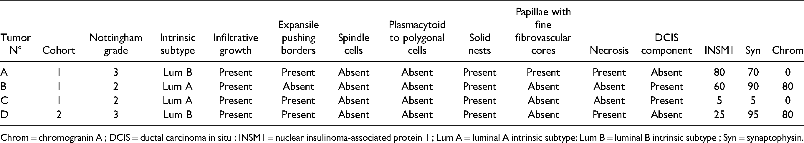
Chrom = chromogranin A ; DCIS = ductal carcinoma in situ ; INSM1 = nuclear insulinoma-associated protein 1 ; Lum A = luminal A intrinsic subtype; Lum B = luminal B intrinsic subtype ; Syn = synaptophysin.
Although it can be debated whether these infiltrative lesions should be designated as neuroendocrine tumors (NETs), large-cell neuroendocrine carcinomas (NEC) or invasive solid papillary carcinomas (SPC) because of the presence of solid nests, focally pushing borders and heterogeneous neuroendocrine biomarker expression, we felt that their global image fell short of such a diagnosis. Reasons for preferring the diagnosis of “invasive carcinoma of NST with neuroendocrine differentiation” over SPC or NET comprised the presence of substantial tumor necrosis, extensive destructive infiltrative growth, the lack of mucinous and spindle cell differentiation, and the presence of high mitotic activity and/or areas with high-grade nuclear atypia with prominent nucleoli and substantial pleomorphism (Figure 1). However, mitotic counts and nuclear atypia seemed insufficient to qualify for NEC, and diffuse hormone receptor expression did not support this diagnosis either. One tumor showed immunoreactivity for neuroendocrine biomarkers in only 5% of the cells, which seemed insufficient to qualify for a mammary NET, as the WHO classification of breast tumors requires both diffuse histological and immunohistochemical features of neuroendocrine differentiation. 3 There is currently no well-defined threshold for positivity versus negativity for neuroendocrine biomarkers. 1 For instance, the ASCO/CAP guidelines use a 1% cut-off for hormone receptor-positivity, as there is ample evidence for an association between hormone receptor status and response to adjuvant hormonal treatment in invasive breast cancer. 4 A strong and straightforward prognostic and/or predictive association with neuroendocrine biomarker expression levels in breast cancer is lacking. We and others therefore used an arbitrary 5% threshold to distinguish INSM1 positivity from negativity,1,5 but we would like to emphasize that there is currently no clinical outcome data to support this threshold. Others adopted a 1% cut-point. 6 Additional studies are required to explore which cut-point is clinically and biologically meaningful, especially since Razvi et al suggested that INSM1 expression in breast tumors is associated with a more favorable outcome. 7

Haematoxylin/eosin stained slides of three invasive breast carcinomas with neuroendocrine differentiation in the first cohort. Tumour A showed necrosis (black asterisks; A), diffuse infiltrative growth (B) and focal areas of trabecular and papillary architecture (C). Tumour B showed infiltrating solid nests (D) without spindle cells. Tumour C contained areas of small solid nests (E) as well as diffuse infiltrative growth and a cribriform ductal carcinoma in situ component (red asterisks; F). Original magnification 50x (A, D) and 100x (B, C, E, F).
We agree with Kawasaki et al that any breast lesion harboring the aforementioned “classic” morphological features suggestive of neuroendocrine differentiation should provoke a more detailed work-up by means of immunohistochemistry for synaptophysin, chromogranin A and INSM1, as these stains might aid to establish a final diagnosis. It should however be noted that the WHO classification of breast tumours does not provide clear information on the prevalence of neuroendocrine biomarker expression in special type lesions such as SPC or invasive mucinous carcinoma.
3
Neuroendocrine differentiation is described as “frequent”, without any reference to studies providing a more precise percentage. The WHO classification also states that neuroendocrine biomarker expression is “frequent” in low or intermediate grade breast tumors, but only tumors with both extensive neuroendocrine biomarker expression
In a second cohort, we screened 66 invasive breast carcinoma biopsies for nuclear INSM1 immunoreactivity. 1 Only one tumor was considered INSM1-positive, with nuclear INSM1 expression in 25% of the cells. This tumor had a luminal B subtype, characterized by hormone receptor-positivity, HER2-negativity, and a Ki-67 positivity rate of 60%. Mucinous differentiation, spindle cells and plasmacytoid cells were absent. Tumor necrosis was focally present, as well as high mitotic activity (20 mitoses per 10 high-power fields) and high-grade nuclear atypia (Figure 2; Table 1). Immunohistochemistry for synaptophysin and chromogranin showed immunoreactivity in 95% and 80% of tumor cells, respectively. Although one could consider a rare diagnosis of mammary large cell NEC or grade 3 NET, a diagnosis of invasive carcinoma with neuroendocrine differentiation was preferred, because of the heterogeneous morphology. Hormone receptor positivity and strong GATA3 immunoreactivity pleaded against a NEC. Again, the WHO classification lacks strict criteria for this differential diagnosis. 3 This lack of consensus and the evolving concept of mammary neuroendocrine neoplasms is reviewed in detail by Uccella et al, who emphasize that neuroendocrine biomarker expression in special type and NST carcinomas without evident or uniform neuroendocrine morphology is more common than the pure neuroendocrine phenotype. 10

Haematoxylin/eosin stain of a breast biopsy containing an invasive carcinoma of NST with neuroendocrine differentiation (A; original magnification 50x), with presence of focal areas of necrosis (asterisks) and small solid nests (B; original magnification 100x). The tumor showed immunoreactivity for chromogranin (C; original magnification 50x) and synaptophysin (D; original magnification 50x).
In conclusion, the comments of Kawasaki et al are valuable as they highlight the difficulties in distinguishing invasive carcinoma with neuroendocrine differentiation from mammary NEC and NET. According to the WHO classification, this distinction is based on the presence and extent of the histological features characteristic of neuroendocrine differentiation in invasive carcinomas. 3 Nevertheless, a precise cut-point for neuroendocrine biomarker expression is lacking at present. It is therefore to be expected that this differential diagnosis is prone to some degree of subjectivity, and thus interobserver variability. Future studies should identify a clinically meaningful cut-point for neuroendocrine biomarkers, as well as clarify the clinical value of the distinction between invasive carcinoma with neuroendocrine differentiation versus mammary NET and NEC. Additional molecular studies, in line with the work of Pareja et al, might support these morphologic and immunohistochemical findings. 9
Footnotes
Author Contributions
MRVB: data collection; writing - original draft. CG: writing - review & editing.
Declaration of Conflicting Interests
The author(s) declared no potential conflicts of interest with respect to the research, authorship, and/or publication of this article.
Funding
M.R. Van Bockstal received a postdoctoral clinical mandate (2019-089) from the not-for-profit organisation “Foundation Against Cancer” (Brussels, Belgium).
Ethical Approval
This is a reply to a letter to the editor. Approval by the local ethics committee has been obtained for the original study (published as Seijnhaeve et al. IJSP 2021).
Informed Consent
Not applicable, because this article does not contain any studies with human or animal subjects.
Trial Registration
Not applicable, because this article does not contain any clinical trials.