
Abstract
The distinction of mesenchymal tumors of the uterus is a frequent diagnostic challenge in gynecologic pathology. Especially, distinguishing low-grade endometrial stromal sarcoma (ESS) from leiomyoma or distinguishing low-grade ESS from high-grade ESS can be difficult. Epithelial-mesenchymal transition (EMT) is a physiological and pathological process in which epithelial cells lose their morphological features, become elongated and acquire mesenchymal traits. The signaling pathway of Zinc finger E-box binding homeobox 1 (ZEB1) is one of the most significant pathways involved in the EMT process and it has a crucial role in cancer progression, metastasis, and therapy resistance. We studied a series of 69 uterine mesenchymal neoplasms including 18 endometrial stromal sarcomas (10 cases of low grade and 8 cases of high grade endometrial stromal sarcomas), 26 leiomyosarcomas (8 cases of grade 1 and 19 cases of grade 2-3 leiomyosarcomas), 15 leiomyomas, and 10 rhabdomyosarcomas, using an antibody ZEB1. We graded the leiomyosarcomas depending on the FNCLCC grading system. It was observed that leiomyosarcoma was more intensely stained with ZEB1 than leiomyoma (P < 0.001) and high-grade ESS was significantly more intensely stained with ZEB1 protein than low-grade ESS (P < 0.004). It also was observed that high-grade leiomyosarcoma was significantly more intensely stained with ZEB1 protein than low-grade leiomyosarcoma (P < 0.000). Our data suggest that Zeb1 can be used to differentiate high-grade sarcomas from their low-grade counterparts as well as benign and malignant smooth muscle tumors of the uterus.
Introduction
Leiomyomas are the most common neoplasms of the female genital tract. Other types of benign and malignant mesenchymal tumors of the uterus are less frequent. Malignant mesenchymal tumors constitute less than 3% of uterine malignancies. 1 The distinction of mesenchymal tumors of the uterus is a frequent diagnostic challenge in gynecologic pathology. Especially, distinguishing low-grade endometrial stromal sarcoma (ESS) from leiomyoma or distinguishing low-grade ESS from high-grade ESS can be difficult. Accurate diagnosis of uterine mesenchymal tumors can usually be made histomorphologically. However, for challenging cases, immunohistochemical auxiliary techniques can be used for confirmation of the diagnosis. 2
Epithelial-mesenchymal transition (EMT) is a physiological and pathological process in which epithelial cells lose their morphological features, become elongated and acquire mesenchymal traits after many biochemical changes.3–6 EMT has physiologically important roles in certain parts of embryogenesis and it has been associated with various pathological processes, including inflammation, fibrosis, wound healing, and malignant progression.3,4,7,8 Several transcription factors that induce EMT have been described, including SNAI1/2, PRRX1, TWIST1/2, ZEB1, and ZEB2.3,9 The signaling pathway of Zinc finger E-box binding homeobox 1 (ZEB1) is one of the most significant pathways involved in the EMT process and it has a crucial role in cancer progression, metastasis, and therapy resistance.5,10 While EMT contributes to malignant progression, it also imparts resistance to cell death and thence to chemotherapy and immunotherapy.7,11
Even though many studies have shown the role of ZEB1 in various carcinomas11–16, there is no yet available data about the expression and function of ZEB1 in mesenchymal tumors of the uterus. In this study, we investigated the expression of the ZEB1 marker in mesenchymal tumors of the uterus by immunohistochemistry and analyzed the relationships among this marker and various histological tumor types.
Materials and methods
Case selection
A total of 69 patients with mesenchymal tumors of the uterus diagnosed between 2009 and 2021 were selected from the archives of the Department of Pathology at Selcuk University, Faculty of Medicine, including 18 endometrial stromal sarcomas (10 tumors of low grade and 8 tumors of high grade endometrial stromal sarcomas), 26 leiomyosarcomas (8 tumors of grade 1 and 19 tumors of grade 2-3 leiomyosarcomas), 15 leiomyomas, and 10 rhabdomyosarcomas. All of the patients were female. Patients were aged 18-94 years (mean 59; median 58). Histological diagnosis was made according to World Health Organization (WHO) 2020 criteria 17 . This study was conducted in accordance with the Declaration of Helsinki.
Immunohistochemistry
Immunohistochemical staining was performed on the formalin-fixed, paraffin-embedded, 4-μm-thick tissue sections. Heat-induced antigen retrieval was performed with 10 mM of sodium citrate buffer (pH 6.0). Endogenous peroxidase was quenched with 3% hydrogen peroxide for 15 min. Immunohistochemistry was then performed using the following antibody: ZEB1 rabbit polyclonal antibody (NPB1-05987; Novus Biologicals, 1:100 dilution; 4 °C overnight).
Immunohistochemical Evaluation:
Immunohistochemical examinations were conducted by two independent pathologists blinded to the type of tissues at different times. Nuclear staining was regarded as positive. Cytoplasmic positivity was interpreted as a non-specific reaction. Different staining intensities were observed in the same case. In cases with different staining intensities, the score with the highest percentage of staining was accepted.
The staining of immunohistochemical ZEB1 for tumor cells was evaluated with regard to intensity. Endothelial cells were used as internal positive controls. The ZEB1 staining intensity was scored as none (0), weak (1 + ), intermediate or strong (2 + ). In detail, none = no immunolabeling; weak = less intense than immunolabeling of in adjacent endothelial cells; intermediate = similar with endothelial cells; and strong = more intense than endothelial cells.
Statistical Analyses
All the data were analyzed by the software Statistical Package for the Social Sciences (SPSS) V.25.0. The chi-square test was applied to correlate the expression of the immunohistochemical ZEB1 expression with the histological subtypes and tumor grade. P < 0.05 was considered for statistical significance level.
Results
Data on the demographic characteristics, age, tumor size, count of mitosis, and presence of necrosis for each of the patients included in the study are presented in Table 1. The results of immunohistochemistry are summarized in Tables 2-6. ZEB1 expression was demonstrated by nuclear staining. All of the mesenchymal tumors of the uterus in our study showed positivity for ZEB1, but there was a consistent difference in staining intensity between benign and malignant tumors or between low-grade malign tumors and high-grade malignant tumors. It was observed that leiomyosarcoma was more intensely stained with ZEB1 than leiomyoma, and this was statistically significant (P < 0.001) (Figure 1). ESS was separated into two subgroups according to WHO 2020 classification 17 as low grade and high grade, and the relationship between these subgroups and ZEB1 immunohistochemical staining was examined. It was observed that high-grade ESS was significantly more intensely stained with ZEB1 protein than low-grade ESS (P < 0.004) (Figure 2). Leiomyosarcoma was separated according to the Fédération Nationale des Centers de Lutte Contre le Cancer (FNCLCC) grading system 18 as Group 1 (grade 1) and Group 2 (grade 2 and grade 3), and the relationship between these subgroups and ZEB1 protein staining was examined. It was observed that Group 2 was significantly more intensely stained with ZEB1 protein than Group 1 (P < 0.000) (Figure 3). In addition, strong (2 + ) nuclear staining was detected in all rhabdomyosarcomas (Figure 4). There was no difference in ZEB1 expression between ESS and leiomyosarcoma (P = 0.289). Similarly, no difference was detected in ZEB1 expression between ESS and leiomyoma (P = 0.289). All of the mesenchymal tumors of the uterus were separated according to the count of mitosis as group 1 (>10 / mm2) and group 2 (<10 / mm2), and the relationship between these subgroups and ZEB1 staining was examined. It was observed that group 1 was significantly more intensely stained with ZEB1 than group 2 (P < 0.000). Also, all of the mesenchymal tumors of the uterus were separated according to the presence of necrosis as group 1 (tumors with necrosis) and group 2 (tumors without necrosis), and the relationship between these subgroups and ZEB1 staining was examined. It was observed that group 1 was significantly more intensely stained with ZEB1 than group 2 (P < 0.000). No significant relationship was observed between age (p = 0.312), tumor size (p = 0.294), and ZEB1 expression.

A. High-grade endometrial stromal sarcoma (HESS) (H&E, × 400). B. Diffuse and strong nuclear staining immunoreactivity with ZEB1 in HESS (immunohistochemistry (IHC), × 400). C. Low grade endometrial stromal sarcoma (LESS) (H&E, × 400). D. Diffuse but weak nuclear staining immunoreactivity with ZEB1 in LESS (IHC, × 400).

A. Leiomyosarcoma (LMS) (H&E, × 200). B. Diffuse and strong nuclear staining immunoreactivity with ZEB1 in LMS (IHC, × 200). C. Leiomyoma (H&E, × 200). D. Diffuse but weak nuclear staining immunoreactivity with ZEB1 in leiomyoma (IHC, × 400).

A. High-grade leiomyosarcoma (H&E, × 400). B. Diffuse and strong nuclear staining immunoreactivity with ZEB1 in high-grade leiomyosarcoma (IHC, × 400). C. Low-grade leiomyosarcoma (H&E, × 400). D. Diffuse but weak nuclear staining immunoreactivity with ZEB1 in low-grade leiomyosarcoma (IHC, × 400).

A. High-grade endometrial stromal sarcoma (HESS) (H&E, × 400). B. Diffuse and intermediate nuclear staining immunoreactivity with ZEB1 in HESS (IHC, × 400). C. Rhabdomyosarcoma (H&E, × 400). D. Diffuse and strong nuclear staining immunoreactivity with ZEB1 in rhabdomyosarcoma (IHC, × 400).
Demographics of Patients According to Tumor Subtypes.
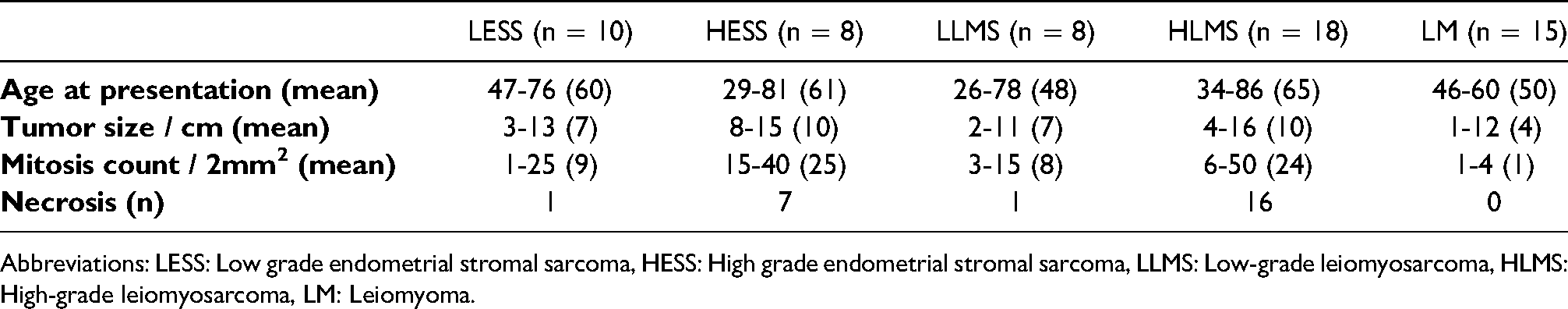
Abbreviations: LESS: Low grade endometrial stromal sarcoma, HESS: High grade endometrial stromal sarcoma, LLMS: Low-grade leiomyosarcoma, HLMS: High-grade leiomyosarcoma, LM: Leiomyoma.
ZEB1 Expression in Endometrial Stromal Neoplasms.
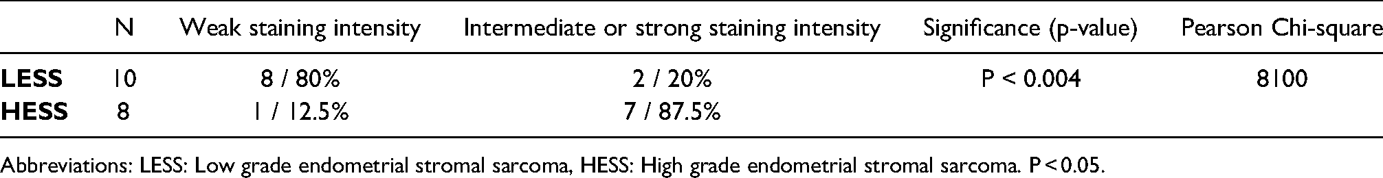
Abbreviations: LESS: Low grade endometrial stromal sarcoma, HESS: High grade endometrial stromal sarcoma. P < 0.05.
ZEB1 Expression in Uterine Smooth Muscle Tumors.
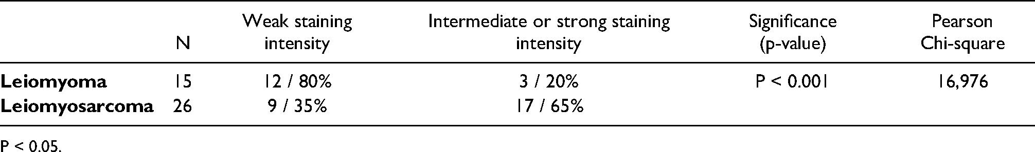
P < 0.05.
ZEB1 Expression in Leiomyosarcoma by the FNCLCC Grading System.
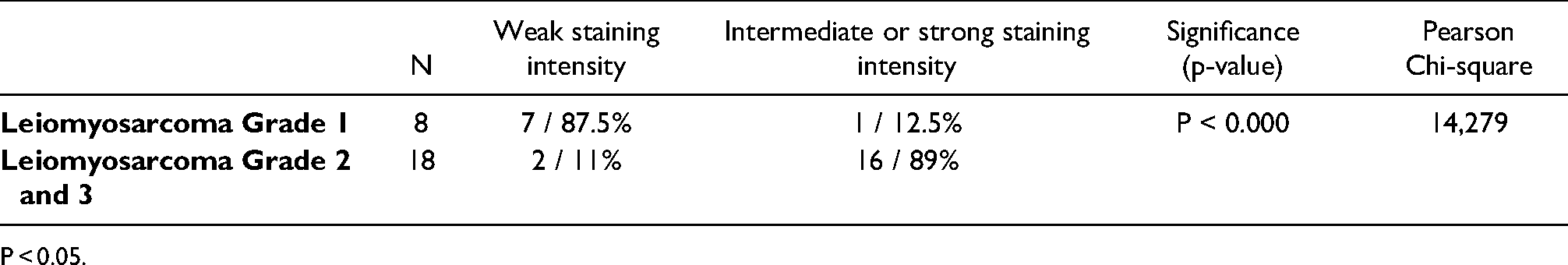
P < 0.05.
Relationship Between the Count of Mitosis and ZEB1 Expression at all Mesenchymal Tumors of the Uterus.
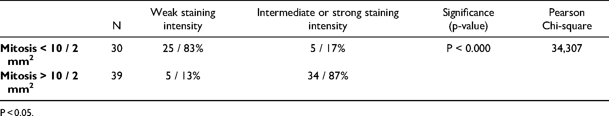
P < 0.05.
Relationship Between the Presence of Necrosis and ZEB1 Expression at all Mesenchymal Tumors of the Uterus.
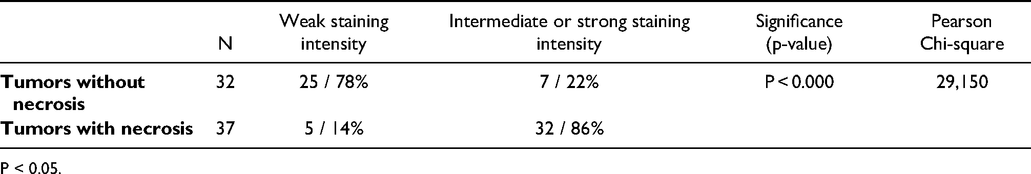
P < 0.05.
Discussion
Several reports have demonstrated that ZEB1 plays an important role in promoting EMT and a sarcomatoid phenotype.3–11 In the literature, there are no data on immunohistochemical ZEB1 expression in uterine mesenchymal neoplasms.
In the present study, we analyzed the immunohistochemical expression of ZEB1 in uterine mesenchymal neoplasms and revealed some significant differences in high-grade sarcomas from their low-grade counterparts as well as benign and malignant tumors.
Careful pathologic sampling and morphologic examination remain the mainstay in the diagnosis of uterine mesenchymal lesions. However, there are some areas in which immunohistochemistry may be useful.19,20
Uterine leiomyoma is a very common benign tumor that can be diagnosed clinically and pathologically without difficulty. However, because some leiomyomas may exhibit considerable cellularity and cellular atypia, a problem occurs in differentiating leiomyoma and leiomyosarcoma. 21 In our leiomyoma cases, there was not such a feature but ZEB1 expression was significantly more intense in leiomyosarcoma cases than leiomyomas. Of the leiomyomas, 3 tumors were stained moderately with ZEB1 and 2 tumors of high-grade leiomyosarcomas were stained weakly with ZEB1. And also a low-grade leiomyosarcoma with strong ZEB1 staining was present. Although there was an overlap, statistical results were significant. But as a result of these findings, we conclude that ZEB1 can not be used to distinguish leiomyoma from low-grade leiomyosarcoma. But we also suggest that ZEB1 can be used to differentiate high-grade leiomyosarcoma from leiomyomas.
In addition, we determined more intense staining of ZEB1 in grade 2 and 3 leiomyosarcomas than grade 1 as well as in high-grade ESS than low grade. Given some high-grade ESS are biphasic and have low-grade ESS-like component 22 , ZEB1 would be potentially very useful in cases of high-grade ESS with limited high-grade areas.
We also detected strong nuclear staining of ZEB1 in rhabdomyosarcomas. The value of ZEB1 in the differential diagnosis of rhabdomyosarcomas versus leiomyosarcomas is limited because both rhabdomyosarcomas and leiomyosarcomas are usually ZEB1 strong positive. These results may support that high ZEB1 expression is associated with higher tumor grade and is a poor prognostic parameter in uterine sarcomas.
The distinction of endometrial stromal neoplasms from cellular smooth muscle tumors of the uterus is sometimes difficult. In their study, Nucci et al 23 emphasized that h-Caldesmon appears to be a more sensitive and specific marker of smooth muscle differentiation than desmin and may distinguish uterine smooth muscle tumors from endometrial stromal lesions. In the present study, no significant difference was detected in terms of ZEB1 expression between uterine smooth muscle tumors and endometrial stromal neoplasms.
In the literature, in hepatic 12 , gastric 13 , and pancreatic tumors 24 , high ZEB1 expression was associated with prognosis as well as poorer tumor differentiation. In the present study, we analyzed ZEB1 expression in low-grade and high-grade uterine sarcomas and put out some significant differences in terms of tumor differentiation. Our findings provide a new contribution to the literature.
As we generally know, higher-grade tumors have a worse prognosis than low-grade tumors. Although we did not reveal the relation between ZEB1 expression and prognosis because of the limited number of patients, we can suggest that patients with high ZEB1 expression may have a worse prognosis than patients with low ZEB1 expression. Further studies with larger series are needed to support this hypothesis.
Conclusion
In conclusion; ZEB1 is a marker that can be used to differentiate high-grade sarcomas from their low-grade counterparts as well as benign and malignant smooth muscle tumors of the uterus. Further studies with larger series are needed to support these findings and reveal the clinical significance of this marker in uterine mesenchymal tumors.
Footnotes
Declaration of Conflicting Interests
The author(s) declared no potential conflicts of interest with respect to the research, authorship, and/or publication of this article.
Funding
The author(s) received no financial support for the research, authorship, and/or publication of this article.
Trial Registration
Not applicable, because this article does not contain any clinical trials.
Informed Consent
Not applicable, because this article does not contain any studies with human or animal subjects.
Ethical Approval
Not applicable, because this article does not contain any studies with human or animal subjects.