






Abstract
Dermatofibrosarcoma protuberans (DFSP) is a rare, CD34+ mesenchymal neoplasm that classically involves the dermis. A COL1A1::PDGFB t(17;22) translocation is present in 91.4% to 96% of cases, resulting in aberrant proliferation due to tyrosine kinase hyperactivity. Here, we present a postmenopausal woman with a CD34-positive spindle cell neoplasm of the breast without cutaneous involvement, lacking muscle marker expression, STAT6 expression, and 13q14 deletion by fluorescence in situ hybridization (FISH). Although the classic PDGFB translocation was not detected by FISH, the overall features were highly suspicious for DFSP. Subsequent RNA-based next-generation sequencing revealed an EMILIN2::PDGFD fusion. A literature review showed that PDGFD fusions can be detected in up to 55% PDGFB FISH negative cases, with EMILIN2::PDGFD fusion highly associated with fibrosarcomatous transformation. This holds important diagnostic and prognostic information as fibrosarcomatous-DFSP is associated with higher recurrence and metastatic potential. The tumor was completely resected with clear margins, showed no fibrosarcomatous areas, and no evidence of recurrence is documented 2 years since resection. This review and case report adds to the literature regarding PDGFD-translocation positive DFSP as a differential diagnosis of CD34-positive spindle cell tumors of the breast, while emphasizing the prognostic importance of EMILIN2::PDGFD fusions.
Introduction
Dermatofibrosarcoma protuberan (DFSP) is the second most common primary cutaneous sarcoma (after Kaposi's sarcoma), comprising 18.4% of cases. 1 Survival data are limited; current reported 5- and 10-year mortality rates have been shown to be less than 1%.1,2 However, DFSP does have high morbidity due to a need for surgical excision with wide margins and a high rate of recurrence. Outcomes are significantly worse when DFSP undergoes fibrosarcomatous (FS) transformation (FS-DFSP), which is associated with an increased risk of metastasis, reduced CD34 staining, and a herringbone pattern with increased atypia and mitoses.3,4
A systematic review of outcomes for FS-DFSP and DFSP showed that the risk of local recurrence, metastasis, and death from disease in FS-DFSP was significantly higher as compared to DFSP (local recurrence 29.8% vs 13.7%, risk ratio 2.2, 95% confidence interval [CI] 1.7-2.9; metastasis 14.4% vs 1.1%, risk ratio 5.5, 95% CI 4.3-7.0; and death from disease 14.7% vs 0.8%, risk ratio 6.2, 95% CI 5.0-7.8). 4
In the United States, the overall annual incidence of DFSP was 4.1 persons per million person-years in 2000 to 2010. 2 DFSP cases in the US showed DFSP has a slight predilection for females with a male-to-female ratio of 0.9.1,2 DFSP is most often located on the trunk (31.7%) followed by the head (28.9%), lower limb (18.3%), and upper limb (16.9%). 2
A survey of patients conducted through a Facebook support group showed the tumor often presents macroscopically as a flat lesion in 44.8% (87/194) before becoming protuberant and polypoid in nature in 73.6% of flat lesions (64/87), while the lesion first presented as a bump in 55.2% of patients (107/194). 5 DFSP are most often mistaken for benign cysts, lipomas, scars, dermatofibromas, or keloids. 5 Although rarely metastasizing, DFSP does frequently infiltrate surrounding tissues, causing the classic “honeycombing” microscopic pattern when infiltrating into adipose. Histologically, DFSP also demonstrates spindle cells in a storiform pattern and generally monomorphic ovoid nuclei with minimal atypia.
In most cases (91.4%-96%), a COL1A1::PDGFB t(17;22) translocation leads to proliferation due to tyrosine kinase hyperactivity, caused by the translocated highly expressed collagen type 1 alpha 1 chain promoter (COL1A1, located on chromosome 17q21) driving unregulated autocrine platelet-derived growth factor subunit B signaling (PDGFB, located on chromosome 22q13).6–9 Less than 10% of DFSP may be negative for the COL1A1::PDGFB fusion, and instead have an atypical fusion such as COL6A3::PDGFD t(2;11), EMILIN2::PDGFD t(11;18), or TNC::PDGFD t(9;11).3,7–12 Similar to COL1A1::PDGFB, the COL6A3::PDGFD translocation brings together a different collagen gene (COL6A3, located on chromosome 2q37) and platelet-derived growth factor gene (PDGFD, located on chromosome 11q22) to drive unregulated autocrine expression of platelet-derived growth factor subunit D driven by the highly expressed collagen type IV alpha 3 chain. Unlike PDGFB, the native PDGFD protein has an N-terminal CUB domain, which must be proteolytically cleaved at Arg-247 or Arg-249 by urokinase plasminogen activator before it can stimulate platelet-derived growth factor receptor signaling. 13 The EMILIN2::PDGFD fusion brings the Elastin Microfibril Interfacer 2 (EMLIN2, located on chromosome 18p11) promoter and protein N-terminus in contact with the active domain of PDGFD. Similarly, the TNC::PDGFD fusion brings the tenascin C (TNC located on 9q33) promoter and protein N-terminus next to the active domain of PDGFD.
Methods
Immunohistochemical stains were performed on formalin-fixed paraffin-embedded (FFPE) blocks using an automated stainer (Ventana BenchMark Ultra systems, Ventana Medical Systems). All immunohistochemical stains are validated for clinical use.
Total nucleic acid was extracted from FFPE tissue sections with the Promega ReliaPrep FFPE Total RNA Miniprep System, omitting the RNase step to isolate total nucleic acid. RNA-targeted sequencing was performed using a custom Archer FusionPlex panel with anchored multiplex PCR, following the manufacturer's instructions. Nanodrop and PreSeq qPCR are used to assess the quantity and quality of RNA. Libraries were created using 200 ng of total nucleic acid and pooled in groups of 6 samples at 4 nM with 2% PhiX. Sequencing is performed on an Illumina MiSeq. The FASTQ files were loaded into the vendor-provided Archer Analysis 7.0.
The fusion transcript was confirmed using Sanger sequencing on the ABI 3500XL instrument. The cDNA was amplified and sequenced with the following M13-tagged primers: 5′EMILIN2 M13 chr18:2885137:+ TGTAAAACGACGGCCAGTGAATGGAGGTGCTGTCCTGG, 3′PDGFD M13 chr11:103797854:- GCTTGGCATCATCATTGAGC.
Case Report
A 60-year-old woman presented with a palpable growing breast mass, 1.7 cm in the greatest dimension. The lesion was first noticed 4 months prior. There were no overlying skin changes or nipple discharge. The patient denied pain, prior radiation, and weight loss but endorsed trauma. Bilateral diagnostic mammogram confirmed a BIRADS 4 mass with mildly irregular borders, 10 cm from the nipple near the inframammary fold. Histologically, the core needle biopsy showed a cellular spindled cell lesion exhibiting delicate thin-walled blood vessels with focal perivascular accentuation of tumor cells. The tumor cells had ill-defined cytoplasmic borders, round to oval to spindled nuclei lacking nuclear atypia or appreciable mitotic activity. Immunohistochemical stains showed the lesion to be strongly positive for CD34. STAT6, desmin, and CD99 were negative. A benign mammary myofibroblastoma was preliminarily suggested at the time of core needle biopsy with complete excision recommended for further diagnostic evaluation.
Upon excision, gross examination revealed a 1.5 × 1.4 × 1 cm firm to gelatinous irregular lesion with a well-defined border. Histological examination revealed uniform-appearing spindled cells with a storiform and focally whorled architecture (Figure 1). The spindled cells showed monomorphic fibroblastic cytomorphology with tapering nuclei and limited amounts of amphophilic cytoplasm. The tumor infiltrated adjacent fat cells with honeycomb-patterned fat entrapment (Figure 1). The tumor showed a mitotic rate of 1 in 10 high-power (40×) fields. The tumor was diffusely positive for CD34 and patchily positive for ER, and stained negative for desmin, PR, STAT6, keratin 5/6, p63, myeloperoxidase, c-kit, and keratin AE1/AE3. Rb staining was non-specific. The tumor was negative for the 13q14 (RB1) rearrangement as well as the t(17;22) COL1A1::PDGFB translocation, both by fluorescence in situ hybridization (FISH) at an outside laboratory. The differential diagnosis included myofibroblastoma and DFSP. Based on morphology, lack of muscle marker expression and lack of 13q14 gene rearrangement, myofibroblastoma was not favored. A diagnosis of DFSP without FS transformation was made. Subsequent nonclinical RNA-based next-generation sequencing (NGS) revealed an EMILIN2::PDGFD fusion between exon 3 of EMILIN2 (NM_032048.2) and exon 6 of PDGFD (NM_025208.4) (chr17:48271304 and chr22:39631879, hg19), which was confirmed with Sanger sequencing (Figure 2). The entire lesion was submitted for histologic evaluation and no FS transformation was seen.
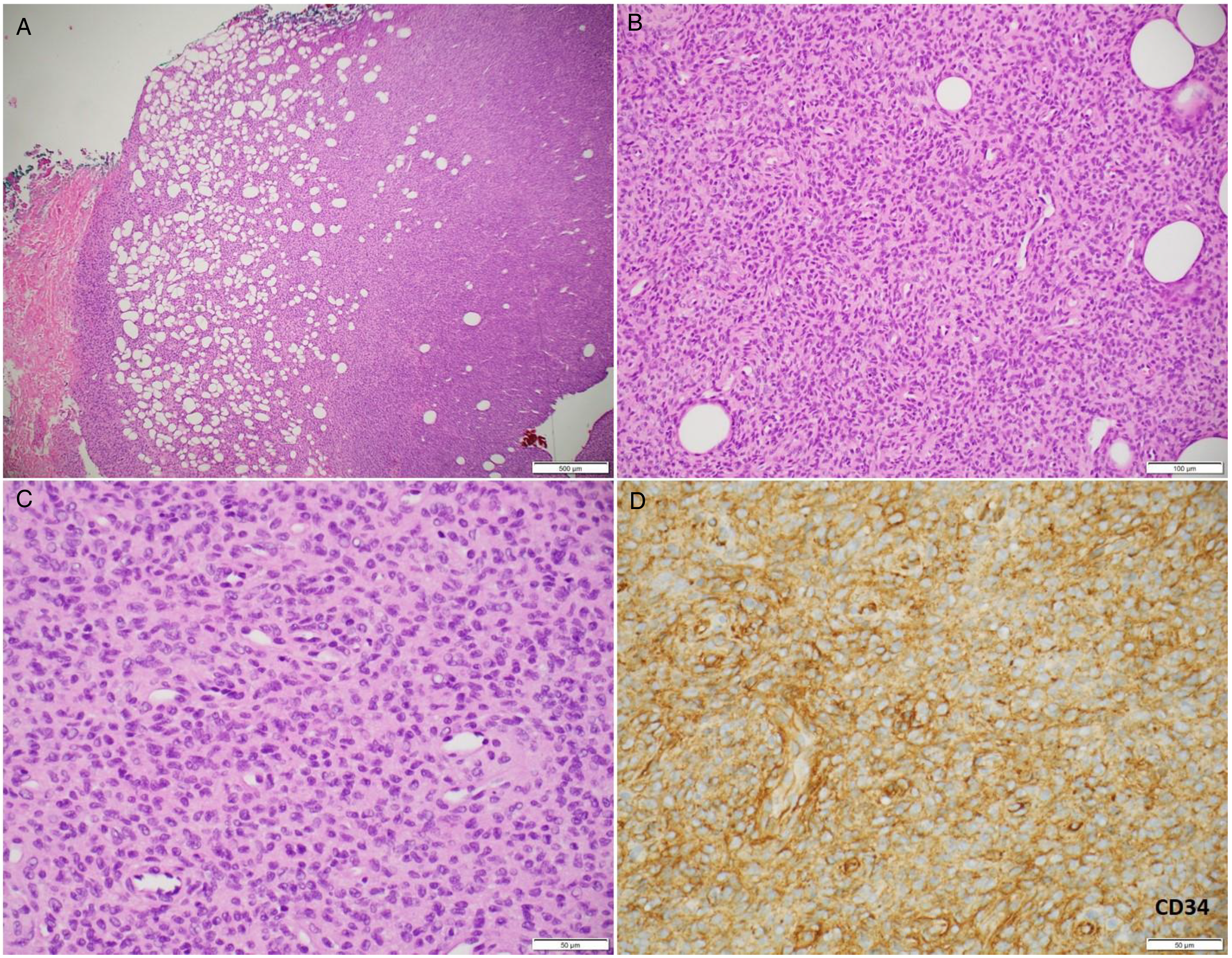
Histopathologic images of DFSP. (A) Low-power view of tumor with honeycomb entrapment of fat (4× objective, H&E stain); (B) Storiform growth pattern in tumor (20× objective, H&E stain); (C) High-power view of tumor cytomorphology with spindle cells showing minimal atypia (40× objective, H&E stain); (D) Lesional cells are diffusely positive for CD34 immunohistochemistry (40× objective).
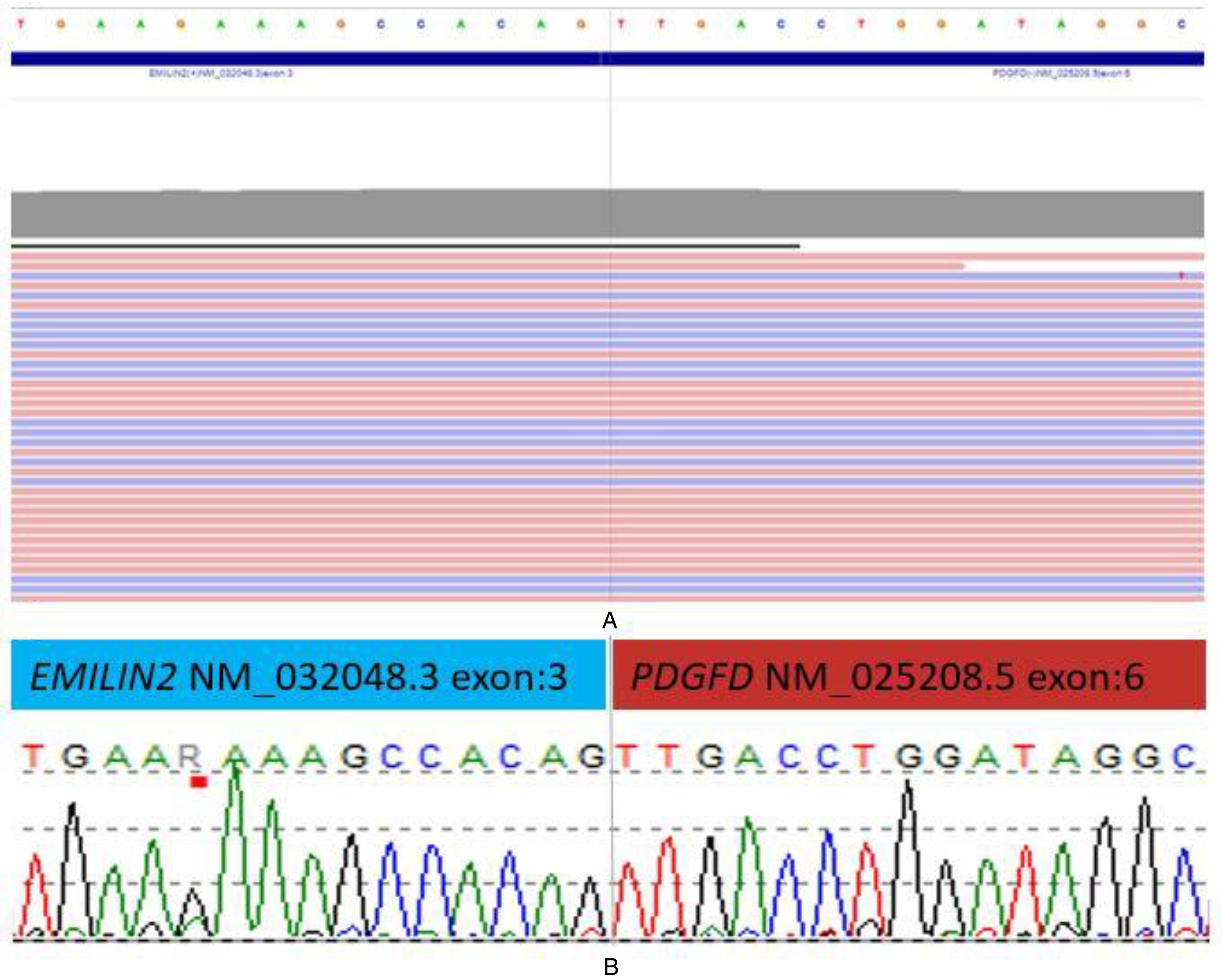
DFSP sequencing results with EMILIN2::PDGFD fusion. (A) Integrative Genomics Viewer (IGV) representation of RNA-targeted next-generation sequencing with anchored multiplex PCR demonstrating the EMILIN2::PDGFD fusion with exon breakpoints corresponding to chr17:48271304 and chr22:39631879 (hg19). (B) Sanger sequencing chromatogram demonstrating Sanger confirmation of the EMILIN2::PDGFD fusion.
Due to positive margins and a diagnosis of DFSPs, the surrounding tissue and overlying skin were surgically removed 2 months later. No residual tumor or dermal involvement was noted on re-excision. No recurrence has been documented for 2 years following resection.
Discussion
This case highlights challenges in the diagnosis of atypical CD34-positive spindle cell lesions of the breast. Spindle cell lesions of the breast that diffusely express CD34 include myofibroblastoma, solitary fibrous tumor, pseudoangiomatous stromal hyperplasia (PASH), and DFSP. Myofibroblastomas usually form a bland circumscribed spindle cell proliferation intersected by bundles of collagen, coexpressing muscle markers such as desmin, SMA, and h-caldesmon. They also show deletion of the 13q14 region detectable by FISH. Solitary fibrous tumors share some morphologic features of myofibroblastoma; in addition, they show prominent staghorn-like vasculature, lack muscle markers, and most importantly, express STAT6 by immunohistochemistry. PASH has immunohistochemical overlap with myofibroblastoma, but is morphologically distinct, forming slit-like clefts in the stroma. DFSP is a locally aggressive spindle cell tumor with a storiform pattern of growth, ill-defined margins, subcutaneous involvement; lacks muscle marker expression; and is often positive for PDGFB rearrangement by FISH. This case report highlights an atypical presentation of DFSP lacking dermal involvement and typical PDGFB rearrangement, with PDGFD fusion detected by RNA sequencing. A comprehensive literature review explores the diagnostic, prognostic, and therapeutic implications of PDGFD fusions in DFSP.
DFSP is usually confirmed with a t(17;22) COL1A1::PDGFB translocation via FISH in 91.4% to 96% of cases.6–9 This fusion results in aberrant proliferation due to autocrine tyrosine kinase hyperactivity through activation of PDGFB receptor (PDGFRB) signaling.6,14 In a study of 20 DFSP cases with a negative t(17;22) FISH, RNA-targeted NGS showed 40% (8/20) of cases were positive for a cryptic COL1A1::PDGFB fusion. 7 Of the remaining cases, 55% (11/20) had PDGFD fusions and one case did not demonstrate a PDGFD or PDGFB fusion. 7
Both platelet-derived growth factor D (PDGFD) and platelet-derived growth factor B (PDGFB) activate platelet-derived growth factor receptor beta (PDGFRB) signaling. 11 PDGFRB is a cell-surface tyrosine kinase, which can drive cellular proliferation when activated. 11 Due to the shared activation of PDGFBR, both PDGFB fusion-positive and PDGFD fusion-positive tumors may benefit from tyrosine kinase inhibitor therapy, such as the FDA-approved imatinib. 11
Thus far, COL6A3, EMILIN2, and TNC have been described as forming fusion products with PDGFD in DFSP.7–10,12,15 Table 1 enumerates the 30 PDGFD fusion-positive cases reported in the literature, as well as the case presented here. Two of the cases in Table 1 are found in a retrospective cohort of 16 breast and upper chest DFSPs, resulting in a slight selection bias for breast tumors. 8 PDGFD fusion-positive tumors were histopathologically indistinguishable from classic DFSP. The literature to date describes 16 COL6A3::PDGFD fusions, 7 EMILIN2::PDGFD fusions, one TNC::PDGFD fusion, and 6 cases where the PDGFD fusion partner was not identified. Strikingly, it has been reported that EMILIN2::PDGFD fusion-positive DFSP is strongly associated with FS transformation (5/7) relative to COL6A3::PDGFD (1/16). 9 This is especially clinically relevant, as FS-DFSP is associated with a higher recurrence rate and metastatic potential.4,5 The single TNC::PDGFD-fusion positive DFSP reported exhibited FS transformation, but more data on other cases with this fusion are required in order to draw conclusions about the likelihood of FS change with this fusion. 10 Atypically, our case and previously reported EMILIN2::PDGFD-fusion positive DFSP tend not to involve the dermis (6/7).7,9 COL6A3::PDGFD fusions are most often found in female patients (14/16), and most often located on the trunk (14/16) with 6/16 reported in the breast.6,8,9 Our literature review identified breast as a common location for PDGFD-rearranged tumors, with 36% (11 out of 30) of PDGFD-rearranged tumors occurring in the breast. However, 2 of these cases may have selection bias, as one component of Dickson et al's study examined fusions in breast DFSP. 8 Excluding those 2 cases still yields a high prevalence of PDGFD-fusion positive DFSP in the breast with 32% (9/28) of PDGFD-rearranged DFSP occurring in the breast.
Literature Review of PDGFD Rearranged DFSP.
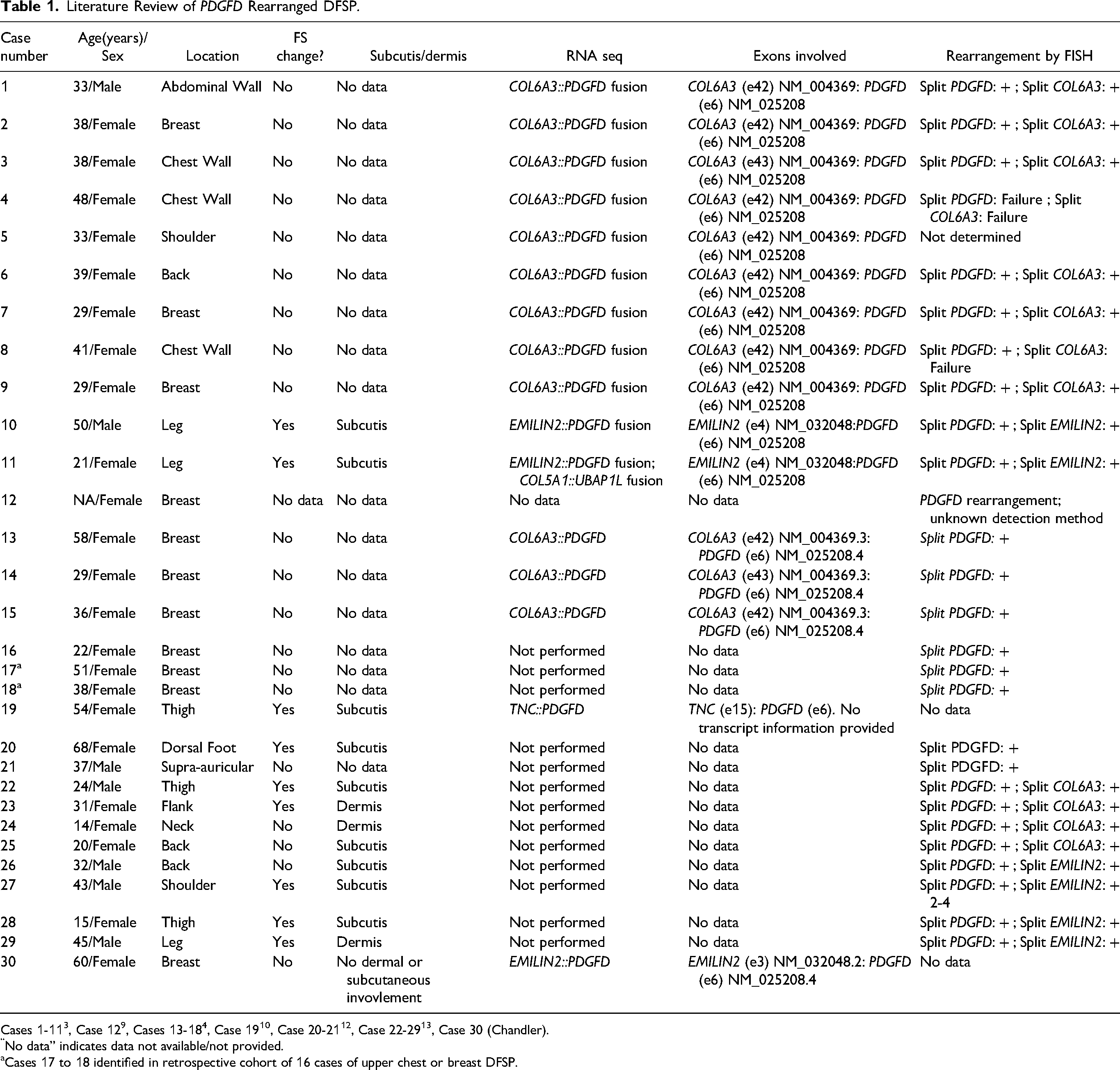
Cases 1-113, Case 129, Cases 13-184, Case 1910, Case 20-2112, Case 22-2913, Case 30 (Chandler).
No data” indicates data not available/not provided.
Cases 17 to 18 identified in retrospective cohort of 16 cases of upper chest or breast DFSP.
A recently published literature review of breast DFSP between 1988 and 2019 reported 36 cases of breast DFSP after excluding those with limited clinical data and those not reported in English. 3 Of the 5 cases with molecular testing results available, all 5 were positive for the more common PDGFB rearrangement. 3
Of note, all sequenced PDGFD-fusion positive DFSP cases have a consistent fusion junction of exon 6 of PDGFD, occurring within amino acid 258 and leaving exons 6 and 7, which code for the PDGFD, receptor binding form, according to UniProt annotation. 16 This is likely because the lack of exons 1 to 5 in PDGFD removes the CUB domain, which is normally proteolytically cleaved by urokinase plasminogen activator at Arg-247 or Arg-249 to create the active platelet-derive growth factor subunit B. 13 Within COL6A3, exon 42 is most often present at the fusion junction (10/12), with exon 43 involved less frequently (2/12). Among the 3 sequenced EMILIN2-fusion positive cases, exon 4 is present at the fusion junction in 2 cases (2/3), while exon 3 is present at the fusion junction in one case, which we present here (1/3).
Our case is the first EMILIN2::PDGFD-fusion positive DFSP reported in the breast. This review adds to the literature regarding PDGFD-fusion-positive DFSP while emphasizing the prognostic importance of EMILIN2::PDGFD fusions, which are associated with a notably higher rate of FS transformation. The case presented here was unusual due to the rarely reported EMILIN2::PDGFD fusion, dermal sparing, and presence in deep breast tissue.
Conclusion
This report expands upon the current literature describing the less common fusions found in DFSP, particularly in the breast. In this article, we report the seventh case of EMILIN2::PDGFD fusion-positive DFSP. This case and the review of the current literature on PDGFD-fusion positive DFSP demonstrate the need to include DFSP in the differential of CD34+ soft tissue tumors regardless of cutaneous involvement. When PDGFB FISH results are negative, alternative PDGFD fusions should be considered. For unknown reasons, EMILIN2::PDGFD-fusion positive DFSP has a markedly increased rate of FS transformation and is also associated with dermal sparing. In the future, it will be interesting to better understand the molecular basis of why EMLIN2-driven fusions tend to have FS transformation at a higher frequency. Due to the similar mechanism of PDGFBR activation between PDGFB and PDGFD, FDA-approved imatinib can likely be considered for patients with PDGFB- or PDGFD-fusion-positive DFSP when clinically indicated, particularly in cases where complete surgical excision is not feasible.8,11
Footnotes
Declaration of Conflicting Interests
The author(s) declared no potential conflicts of interest with respect to the research, authorship, and/or publication of this article.
Funding
The author(s) received no financial support for the research, authorship, and/or publication of this article.
