





Abstract
Background. Fibroadenoma (FA) and benign phyllodes tumor (PT) of the breast often have similar appearances on imaging. While an exact diagnosis of biopsy specimens is required to choose adequate treatment, including surgical procedures, it is sometimes difficult to pathologically differentiate these 2 tumors due to histological resemblances. To elucidate markers for distinguishing FA from benign PT, we analyzed clinical samples immunohistochemically. Methods. We retrospectively investigated 80 breast fibroepithelial lesions. As a discovery set, 60 surgical excision samples (30 FA and 30 benign PT) were examined. Twenty biopsy samples (10 FA and 10 benign PT) were examined as a validation set. To determine targets for immunohistochemistry, we first tested some proteins based on previous reports. As a result, Ki67 was chosen for differentiating FA and PT; thus further examinations were conducted with this protein. Results. Among the proteins examined, stromal Ki67 was significantly higher in PT than in FA. Benign PT had significantly higher stromal Ki67 expression both at random and at hotspots (p < .001 and <.001, respectively). The receiver operating characteristic curve analysis identified 3.5% and 8.5% (at random spots and hotspots, respectively) as the optimal cutoff values of stromal Ki67 for distinguishing between these 2 tumors. In the validation cohort employing needle biopsy specimens, we confirmed that these 2 cutoff values properly classified these 2 tumors (p = .043 and .029, respectively). Conclusion.We revealed that stromal Ki67 might be a potential marker for distinguishing FA from benign PT.
Introduction
Management of Fibroepithelial Lesions of the Breast
Breast fibroepithelial lesions can be divided into fibroadenoma (FA) and phyllodes tumor (PT) ranging from benign to malignant, representing a heterogeneous group of biphasic neoplasms.1,2 FAs generally need no treatment or are removed if they grow larger. In contrast, PTs need to be resected taking into consideration surgical margins because they have the potential to grow larger and can sometimes, albeit infrequently, be malignant. Even if the primary tumor is benign, it may become malignant in the case of local recurrence.3,4 Hence, an exact diagnosis of biopsy specimens is ideally required to choose adequate treatment, including surgical procedures.
Histological Diagnosis for Fibroepithelial Lesions by Core Needle Biopsy
Once the fibroepithelial lesion is detected clinically, a core needle biopsy is commonly taken for diagnosis before moving on to the next step. However, biopsy sections with small samples sometimes imply big challenges for the pathologist when diagnosing the subtype, because the histological features of the 2 subtypes are similar, both featuring a slit-shaped duct with a mild proliferation of the epithelium and cellular stroma with fewer or without mitoses.1,3,5-7 Commonly used auxiliary examinations for checking breast tumors clinically (ultrasound, mammography, and magnetic resonance imaging) display comparable difficulties in identifying FA and benign PT due to the visual similarities of the subtypes.7-10
Molecular Biological Features in FA and PT
Some studies have investigated molecular biological differences in FA and benign PT.4,11-13 Mediator complex subunit 12 (MED12) is one of the predominant popular genes with mutations in fibroepithelial lesions, regulating transcription of all RNA polymerase II-dependent genes. One study suggested that MED12 protein expression was slightly higher in benign PT than in FA. 14 However, in most reports there is little difference between the 2 tumors, with a high frequency of MED12 mutations observed in both FA and benign PT, up to ∼80%.15,16 TERT, which encodes the catalytic domain of the human telomerase reverse transcriptase, is found frequently in PT, especially borderline PT. However, the difference between FA and benign PT is not pronounced with 7% and 50% mutation frequency, respectively. 17 Despite this previous work, these genes might not be useful as diagnostic markers for differentiating FA and benign PT.
In this study, to elucidate potential markers to be utilized for distinguishing FA from benign PT in biopsy specimens, we retrospectively analyzed clinical samples of breast fibroepithelial lesions using immunohistochemistry. Morphology was also assessed.
Methods
Clinical Samples
We retrospectively examined 80 breast fibroepithelial lesions that were diagnosed and treated at our hospital. These samples comprised 60 surgical excision samples (30 FA, 30 benign PT) as a discovery set and 20 biopsy samples (10 FA, 10 benign PT) for validation. Samples for the discovery set were randomly chosen from cases from January 2015 to December 2020. For the validation set, which needed a biopsy and surgical specimen pairs from the same patients, the period was extended back to January 2008 to accumulate the number of cases required. In the current study, the final diagnoses of surgical specimens were used to classify all cases. Clinicopathological characteristics of the discovery set (n = 60) are shown in Supplemental Table 1. While there was no difference in age, tumor size was significantly larger in benign PT than in FA (p < .001). We observed the same trend in the validation set (n = 20, p = .044).
This study was carried out with approval from the Ethics Committee of our Hospital (No. E22-0008-H01) and complies with the 1964 Helsinki Declaration and its later amendments or comparable ethical standards. All participants were informed that the research policy was available on the homepage of the hospital and that they had the opportunity to opt out of the study at any time.
Analysis of Morphological Appearance of Nuclei
The morphological appearance was analyzed in hematoxylin and eosin-stained surgical sections. Three spots in a high-power field (400×) were randomly selected in each section and then captured with cellSens Standard software (OLYMPUS). KS 400 software (Carl Zeiss, Bioz) was used to measure the length and width of nuclei by recognizing the nuclear membrane (Figure 1), and the aspect ratio of each nucleus was automatically calculated.
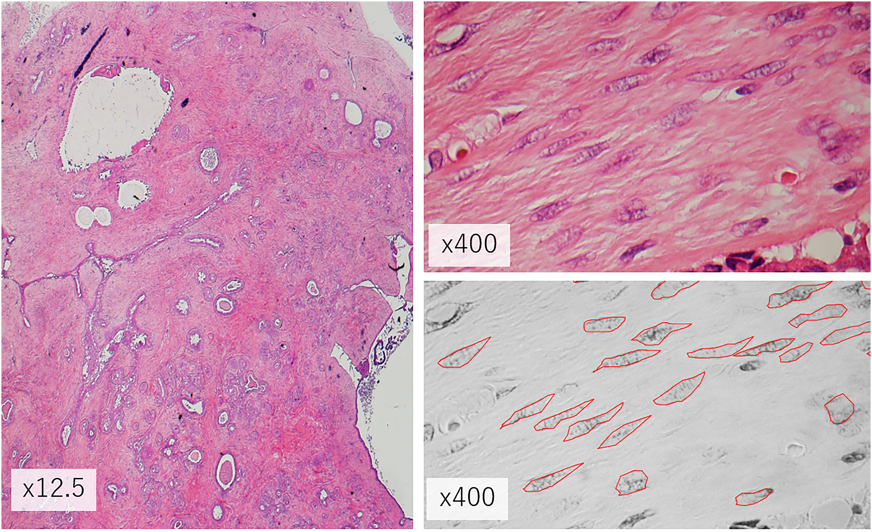
Measurement of length and width of tumor nuclei. Three random fields at high power were selected for morphological measurement. The length and width of tumor nuclei were measured along with the nuclear membrane (red enclosed lines). Lymphocytes and blood capillaries were excluded from the assessment.
Immunohistochemical Assessment
To determine targets for immunohistochemistry (IHC), we first conducted a preliminary analysis employing publicly available gene expression data in Gene Expression Omnibus (GES55224) obtained from a previous study by Vidal et al. 13 In a comparison of 34 FA and 20 benign PT, 10 genes (ERBB4, UBE2C, ANLN, MIA, CCNE1, CAV1, RAB25, CRYAB, MKI67, and EVI2A) were statistically differentially expressed between these 2 tumors (Supplemental Figure 1). In addition, CD10 and MED12 were also investigated. CD10 is a neutral endopeptidase on the cell surface and is significantly more highly expressed in PT than in FA.18-20 As mentioned before, MED12 is more highly expressed in PT than in FA. Additionally, MED12 expression is correlated with the estrogen receptor alpha (ERα) in the epithelium in fibroepithelial lesions.11,12 Therefore, the corresponding proteins of the above 10 genes, as well as CD10, MED12, and ERα, were selected as IHC targets (Supplemental Figure 2). Details of primary antibodies: ERBB4 (mouse monoclonal; Abcam), UBE2C (rabbit polyclonal; Abcam), ANLN (mouse monoclonal; Abcam), MIA (rabbit polyclonal; Funakoshi), CCNE1 (mouse monoclonal; Santa Cruz), CAV1 (rabbit monoclonal; Abcam), RAB25 (rabbit polyclonal; My Biosource), CRYAB (mouse monoclonal; Abcam), Ki67 (mouse monoclonal; DAKO), EVI2A (rabbit polyclonal; Funakoshi), CD10 (mouse monoclonal; Leica Biosystems), ERα (rabbit monoclonal; HISTOFINE), MED12 (rabbit polyclonal; Cosmo Bio Co.).
Immunohistochemical staining was assessed by YM and HS with the criteria of evaluation in 5% increments with random counts for 3 spots of each section for ERBB4, UBE2C, ANLN, MIA, CCNE1, CAV1, RAB25, CRYAB, EVI2A, CD10, MED12, and ERα. Ki67 is used in routine diagnosis, but optimal assessment as an average or at a hotspot is controversial. Therefore, we employed both methods by assessing 3 random spots and a hotspot separately. Samples in which ≥1% of tumor cells were stained were considered positive. Histological data are expressed as (%).
Statistical Analysis
Gene Expression Omnibus database variables and morphological appearance, as well as immunohistochemical assessment, were analyzed using a Mann–Whitney U test by R software version 4.4.0 (R studio, PBC). Comparisons of mean values were conducted with 2-sided t tests. To evaluate the independence of 2 categorical variables, Fisher's exact test was used. The optimal Ki67 cutoff value distinguishing FA from benign PT was determined using receiver operating characteristic curve analysis with SPSS software version 23.0 (IBM Corp.). The test level p < .050 and fold change >1.5 were considered statistically significant.
Results
Differences in Morphological Appearance
First, we evaluated morphological differences between FA and benign PT. Twenty surgical sections from the discovery set (10 FA and 10 benign PT) were employed. There were significant differences in the length of tumor cells’ nuclei (p < .001), area (p < .001), and ratio (p = .005) between FA and PT (Figure 2A). Tumor nuclei of benign PT were laterally more enlarged than those of FA (Figure 2B).

Differences in morphological appearance between fibroadenoma (FA) and benign PT. (A) Comparison of tumor cell nuclei in length, width, ratio, and area, assessed in a total of 1186 cells of FA and 1441 cells of benign PT. (B) Representative morphological features of FA and PT are shown (400×).
IHC in the Discovery set
Next, we immunohistochemically assessed these 20 surgical sections from the discovery set (10 FA and 10 benign PT) (Supplemental Table 2). Ki67 random (Stroma), Ki67 hotspot (Stroma), MED12 (Stroma), and EVI2A (epithelium and stroma) showed significant differences between FA and benign PT. Among them, only stromal Ki67 was consistent with the aforementioned gene expression analysis and previous reports because Ki67 expression was significantly higher in PT than in FA. Therefore, we assessed only stromal Ki67 (both random and hotspot) in further IHC on the remaining 40 surgical specimens. The final results of the total of 60 tumors are shown in Figure 3. Benign PT showed significantly higher stromal Ki67 expression both in random fields (p < .001) and at hotspots (p < .001).

Comparison of Ki67 expression between FA (n = 30) and benign PT (n = 30). Stromal Ki67 expression was counted (A) in random fields and (B) at a hotspot.
Determination of Cutoff Values of Ki67 for Distinguishing FA From PT
We next determined the optimal stromal Ki67 cutoff values to distinguish FA from benign PT using a receiver operating characteristic curve analysis. We employed stromal Ki67 values assessed both in random fields (Model 1) and a hotspot (Model 2) (Figure 4). Consequently, we identified 3.5% (specificity, 83.3%; sensitivity, 83.3%) and 8.5% (specificity, 86.7%; sensitivity, 70%) as the optimal cutoff values for distinguishing these 2 tumors in Models 1 and 2, respectively. The area under the curve of Model 1 was 0.87 and that of Model 2 was 0.86.

Receiver operating characteristic curves of stromal Ki67 values differentiating FA and benign PT. (A) Model 1: using data obtained from random fields. (B) Model 2: using data obtained from hot spots.
IHC in the Validation Cohort
Finally, we examined the validation cohort of needle biopsy specimens (10 FA and 10 benign PT), employing the cutoff values of stromal Ki67 established in the discovery set (Table 1). We confirmed that both Model 1 and Model 2 properly classified these 2 tumors (p = .043 and .029, respectively).
Application of Models to Stromal Ki67 Expression in Validation Sets.

Abbreviations; FA: fibroadenoma, PT: phyllodes tumor.
*Defined by the cutoff values of 3.5% and 8.5%.
Discussion
In the present study, we revealed that stromal Ki67 in benign PT was significantly higher than that in FA. We confirmed this finding in a validation cohort. Only a few studies have previously compared Ki67 expression between FA and benign PT. One report found no difference between these 2 tumors but their sample size was small and the evaluation method was imprecise. 21 Meanwhile, Jara-Lazaro et al 22 reported that benign PT (n = 17) frequently featured higher expression of Ki67 than FA (n = 21), which is consistent with our results. While they assessed Ki67 expression in 5% increments, we analyzed measured values with larger samples. Furthermore, this method enabled us to determine a cutoff value to distinguish the 2 tumors.
Assessment of Ki67 expression in random fields is theoretically more acceptable in practice, considering that biopsy specimens might not always contain the real hotspot of an individual tumor. However, our data indicate that both assessments, employing either random fields or a hotspot, are comparable. Meanwhile, Umekita and Yoshida 23 examined a large number of surgical specimens from fibroepithelial lesions and concluded that discriminating FA from benign PT with low Ki67 expression is difficult. In our cohort, we also found that both tumors had low Ki67 values but there was still a significant difference. We are aware that our cutoff values are not applicable in clinical diagnosis because of the need to count many cells. Further studies of technological developments employing automated scanning and image analysis are warranted to establish optimal Ki67 assessment methods.
As for MED12, many studies have reported that MED12 mutations are frequently observed in breast fibroepithelial lesions.4,11 However, the frequency of MED12 mutations is likely not different between FA and benign PT.12,14-16 Consistent with a study that examined MED12 protein levels, 12 we also did not observe a difference in MED12 protein levels between FA and benign PT. Taken together, MED12 seems inadequate to be utilized as a marker differentiating these 2 tumors.
We also examined the morphological appearance of tumor cell nuclei and confirmed that the nuclei of tumor cells were more elongated in FA, whereas those of benign PT were oval. Some previous studies have reported that FA and PT tumors are in the same lineage, supporting the difficulty of differentiating FA and benign PT.13,24 However, we revealed differences in morphological appearance and Ki67 expression. Morphological differences were a key interest, however, in daily practice, it is almost impossible to accurately measure these to distinguish between the 2 tumor types.
A major limitation of this study was its retrospective nature. Additionally, there might have been a selection bias, particularly in FA. FA is usually followed-up after a diagnostic biopsy and is removed surgically only when there is a reason, such as an increase in size. Thus, our surgical samples might not be representative of all FA. Another limitation was the small sample size, as this was a single-center study, and, as mentioned above, FA is rarely surgically removed. Recruiting more patients from multiple institutions would help validate our findings including a selection of the assessment field and an optimal cutoff value for Ki67. Technological advancements such as automated image analysis are also necessary to establish Ki67 assessment methods.
In conclusion, our data suggest that stromal Ki67 might be a potential marker for distinguishing between FA and benign PT.
Supplemental Material
sj-xlsx-1-ijs-10.1177_10668969231171132 - Supplemental material for Stromal Ki67 Expression Might be a Useful Marker for Distinguishing Fibroadenoma From Benign Phyllodes Tumor of the Breast
Supplemental material, sj-xlsx-1-ijs-10.1177_10668969231171132 for Stromal Ki67 Expression Might be a Useful Marker for Distinguishing Fibroadenoma From Benign Phyllodes Tumor of the Breast by Men Yuan, Harumi Saeki, Yoshiya Horimoto, Yumiko Ishizuka, Hiroko Onagi, Mitsue Saito, Takuo Hayashi, Atsushi Arakawa and Takashi Yao in International Journal of Surgical Pathology
Supplemental Material
sj-xlsx-2-ijs-10.1177_10668969231171132 - Supplemental material for Stromal Ki67 Expression Might be a Useful Marker for Distinguishing Fibroadenoma From Benign Phyllodes Tumor of the Breast
Supplemental material, sj-xlsx-2-ijs-10.1177_10668969231171132 for Stromal Ki67 Expression Might be a Useful Marker for Distinguishing Fibroadenoma From Benign Phyllodes Tumor of the Breast by Men Yuan, Harumi Saeki, Yoshiya Horimoto, Yumiko Ishizuka, Hiroko Onagi, Mitsue Saito, Takuo Hayashi, Atsushi Arakawa and Takashi Yao in International Journal of Surgical Pathology
Supplemental Material
sj-tif-3-ijs-10.1177_10668969231171132 - Supplemental material for Stromal Ki67 Expression Might be a Useful Marker for Distinguishing Fibroadenoma From Benign Phyllodes Tumor of the Breast
Supplemental material, sj-tif-3-ijs-10.1177_10668969231171132 for Stromal Ki67 Expression Might be a Useful Marker for Distinguishing Fibroadenoma From Benign Phyllodes Tumor of the Breast by Men Yuan, Harumi Saeki, Yoshiya Horimoto, Yumiko Ishizuka, Hiroko Onagi, Mitsue Saito, Takuo Hayashi, Atsushi Arakawa and Takashi Yao in International Journal of Surgical Pathology
Supplemental Material
sj-tif-4-ijs-10.1177_10668969231171132 - Supplemental material for Stromal Ki67 Expression Might be a Useful Marker for Distinguishing Fibroadenoma From Benign Phyllodes Tumor of the Breast
Supplemental material, sj-tif-4-ijs-10.1177_10668969231171132 for Stromal Ki67 Expression Might be a Useful Marker for Distinguishing Fibroadenoma From Benign Phyllodes Tumor of the Breast by Men Yuan, Harumi Saeki, Yoshiya Horimoto, Yumiko Ishizuka, Hiroko Onagi, Mitsue Saito, Takuo Hayashi, Atsushi Arakawa and Takashi Yao in International Journal of Surgical Pathology
Footnotes
Acknowledgements
We appreciate the efforts of the Laboratory of Molecular and Biochemical Research and the Laboratory of Morphology and Image Analysis, Research Support Centre, Juntendo University Graduate School of Medicine, for providing technical support. The authors also sincerely thank Clear Science Pty Ltd for language editing.
Declaration of Conflicting Interests
The author(s) declared no potential conflicts of interest with respect to the research, authorship, and/or publication of this article.
Ethical Approval
Ethical approval for this study was obtained from The Ethics Committee of Juntendo Hospital (No. E22-0008-H01).
Funding
The author(s) received no financial support for the research, authorship, and/or publication of this article.
Informed Consent
The Ethics Committee of Juntendo Hospital waived obtaining informed consent and instead, all participants were informed that the research policy was available on the homepage of the hospital and that they had the opportunity to opt out of the study at any time.
Trial Registration
Not applicable, because this article does not contain any clinical trials.

Supplemental Material
Supplemental material for this article is available online.
References
Supplementary Material
Please find the following supplemental material available below.
For Open Access articles published under a Creative Commons License, all supplemental material carries the same license as the article it is associated with.
For non-Open Access articles published, all supplemental material carries a non-exclusive license, and permission requests for re-use of supplemental material or any part of supplemental material shall be sent directly to the copyright owner as specified in the copyright notice associated with the article.
