






Abstract
Deficiency of fumarate hydratase (FH) protein expression in uterine corpus leiomyomas may be attributable to either germline or somatic mutations of the FH gene, the former being definitional for the hereditary leiomyomatosis and renal cell cancer syndrome. The authors assess whether, using previously reported FH-associated morphologic features, FH protein-deficient uterine corpus leiomyomas associated with a pathogenic germline mutations of the FH gene (group 1) are distinguishable from FH protein-deficient uterine corpus leiomyomas without such mutations (and whose FH protein loss is presumed to be attributable to somatic/epigenetic inactivation or other unknown phenomena: group 2). Groups 1 and 2 were compared regarding a variety of clinicopathologic features, including 7 core “FH-associated” tumoral morphologic features: staghorn vasculature; alveolar-type edema; bizarre nuclei; chain-like tumor nuclei; hyaline cytoplasmic globules; prominent nucleoli, intranuclear inclusions, and perinucleolar halos; and prominent eosinophilic/fibrillary cytoplasm. Among 2418 patients diagnosed with uterine corpus leiomyoma during the study period, FH-associated morphologic features were reported in 1.5% (37 patients), and FH immunohistochemistry was performed in 29 (1.19%). Fourteen (48.27%) of the 29 patients showed FH protein deficiency by immunohistochemistry. Twelve patients underwent germline testing, of which 8 (66.7%) were classified as group 1 and 4 (33.3%) as group 2. FH protein-deficient tumors were larger (10.44 vs 4.08 cm, P = 0.01) and associated with younger patients (42.05 vs 47.97, P = 0.004) than 370 randomly selected uterine leiomyoma controls. Groups 1 and 2 showed no significant differences in patient age and tumor size. In group 1 tumors, the FH-associated morphologic features were generally present diffusely; all group 1 tumors displayed ≥5 FH-associated features, whereas all group 2 tumors displayed <5 FH-associated features (means 6.5 ± 0.53 vs 3.5 ± 1.00, P < 0.001). Notably, eosinophilic/fibrillary cytoplasm and alveolar-type edema were each significantly more prevalent in group 1 tumors than group 2 tumors (P = 0.018 for both). No single morphologic feature was found to be completely sensitive and specific in making the distinction between group 1 and 2 tumors. Our findings suggest that groups 1 and 2 are unlikely to be morphologically distinguishable by individual morphologic features. Whether there is a combination of features that can reliably make this distinction is unclear and will require additional studies with larger cohorts.
Keywords
Introduction
Fumarate hydratase (FH) is a key enzyme of tricarboxylic acid cycle that serves as a catalyst in the conversion of fumarate to malate in the mitochondrial matrix.1,2 The earliest clinical reports of FH deficiency were of the severe autosomal recessive metabolic disorder that is caused by homozygous or compound heterozygous mutation in the FH gene.3,4 Heterozygous mutations in the FH gene, however, are associated with the syndrome of multiple cutaneous leiomyomas, uterine leiomyomas, and renal cell cancer that is known as hereditary leiomyomatosis and renal cell cancer (HLRCC) (OMIM # 150800).5–7 Although the FH gene, which is located at 1q42.3–q43, is thought to be a tumor suppressor gene,6,8,9 the precise mechanistic basis by which a FH mutation eventuates in tumorigenesis is presently unclear. 10 The Leiden Open Variation Database of FH gene variants reports numerous unique mutations that are pathogenic or “likely pathogenic.”11,12 Demonstration of pathogenic mutations, usually by Sanger sequencing or next-generation sequencing, 13 and typically in the setting of some clinical stigmata or familial history, establishes the diagnosis of HLRCC. 14 Clinical findings that may raise the possibility of HLRCC include single leiomyoma of the skin with a positive family history, early onset of renal tumors compatible with type 2 papillary morphology, and/or the presence of multiple uterine leiomyomas especially when occurring at an early age. 14 The potential for the latter to be an early harbinger of the syndrome has long been recognized. Compared to controls, patients with a germline FH mutation have a 5-fold elevated risk for uterine leiomyomas, more frequently present with multiple leiomyomata, are younger at initial diagnosis, and more frequently present with dysmenorrhea and pain.13,15-19 After surgical resection of the leiomyoma(s), the pathologist is called upon to segregate cases that may potentially be HLRCC-associated based on tumoral morphologic features, and a variety of such features have previously been associated with either loss of the FH protein or the presence of FH mutations.20-28 Low magnification features include thin-walled “staghorn”-shaped vessels (“hemangiopericytoma-like”), alveolar type of edema, chain-like arrangement of tumor cells, and leiomyomatosis-like architecture.21,23,26 Higher magnification features include eosinophilic/fibrillary cytoplasm, cytoplasmic globules, prominent nucleoli with perinucleolar halos, and occasionally bizarre nuclei.20,23,24,28 However, the sensitivity and specificity of each of the aforementioned features relative to an endpoint of FH protein loss and/or germline FH mutations have not been clearly determined.21,26,28-30 One screening approach for HLRCC is that tumors that display one or more of the aforementioned morphologic features are assessed by FH immunohistochemistry, with loss of staining for the protein being definitional for FH-deficient leiomyoma (FH-d), and FH-d leiomyoma triggering a referral for genetic counseling and, possibly, germline testing. 22 However, loss of FH protein expression is not completely sensitive for detecting an FH gene aberration, and retained staining has been observed in some tumors with pathogenic missense variant mutation.22,25 A second immunohistochemistry-based approach, which is reportedly more sensitive and specific, uses 2SC (S-[2-succinyl] cysteine), a covalent protein that becomes detectable in cells when the fumarate accumulates secondary to FH deficiency. 26 Only a subset of patients whose uterine leiomyomas show deficiency of the FH protein are found to have pathogenic germline FH gene mutations. For example, using targeted next-generation sequencing on tumoral tissue, Li et al 31 found germline mutations in 45% of tested FH-d uterine leiomyomas. FH deficiency in cases without a germline mutation is presumed to be attributable to somatic inactivation or other unknown factors. 29 Since only pathogenic germline mutations are definitional for HLRCC—along with its associated risk of renal cancers—it is essential to distinguish between patients with FH-d leiomyomas with a pathogenic germline mutation of the FH gene (group 1) and their counterparts whose FH protein deficiency is attributable to somatic mutations or other factors (group 2).32,33
The present study is a detailed comparative analysis with the aim of discerning whether there are differences in tumoral morphology between FH-d uterine leiomyoma with and without germline pathogenic mutations. We also describe the spectrum of morphologic features associated with these 2 groups in detail.
Materials and Methods
Study Cohort: Case Selection
A retrospective search of an institutional pathology electronic database was conducted for patients that were diagnosed with uterine leiomyoma between January 2017 and September 2022. From this cohort, we identified patients whose tumors were thought to display at least 1 morphologic feature suggestive of FH deficiency during their initial evaluation, primarily through a search using the word “fumarate” in the associated pathology reports. From the latter cohort, we identified patients on whose tumors’ FH immunohistochemistry had been performed during the routine clinical evaluation of the case. All slides associated with these tumors were retrieved and reviewed by 2 authors. Cases that displayed FH deficiency, inclusive of routine cases and additional that had been identified from 1 of the author's (OF) consultation files, constituted our study group. We also established a control group of 370 classic uterine leiomyoma cases devoid of any morphological feature suggestive of FH deficiency. The latter was selected from consecutive cases of uterine leiomyoma through a random selection mechanism that was ultimately designed to establish 10 control cases for each case of uterine leiomyoma with any FH-associated morphologic feature. Patient demographic data and other history were retrieved from the electronic database.
Immunohistochemical Studies
Immunohistochemical studies for FH were performed at a reference laboratory on sections from formalin-fixed paraffin-embedded tumor blocks, and stained slides were interpreted by the authors. A mouse monoclonal antibody (J-13 clone; Santa Cruz Biotechnology; dilution 1:1250) was used on Leica Bond autostainer with epitope retrieval. A case was classified as FH deficient if there was complete loss of cytoplasmic expression of the protein in tumor cells, with appropriately reactive internal controls (typically endothelial cells).
Germline Testing
Information on germline testing was obtained from each patient's medical record or from contributing pathologists. Following a diagnosis of FH-deficient leiomyoma, patients were generally referred for genetic counseling, and for the subset that elected to undergo germline testing for mutations of the FH gene, such tests were performed on blood or saliva samples that were obtained separately from the patient. For patients that were managed at the University of California San Diego, testing was performed at a reference laboratory, Ambry Genetics (Alisa Viejo, CA) using their multigene panel (CustomNext-Cancer® test), which evaluates for DNA sequence mutations by next-generation or Sanger sequencing of all coding domains as well as substantial portions of the flanking 5’ and 3’ ends of all the introns and untranslated regions. NCBI reference sequence was NM_000143.3. Results were reported as (1) negative (no clinically significant variants detected); (2) pathogenic mutations detected, ie, alterations for which sufficient evidence exists of their disease-causing capability; (3) variant, likely pathogenic detected [alterations with strong evidence in favor of pathogenicity]; and (4) variant(s) of unknown significance detected [ie, alterations with conflicting or limited evidence regarding their pathogenicity]. For consultation cases, testing was performed at Ambry as well as a variety of other laboratories within the United States; for the latter group, test methodology details were generally unavailable, and results or final reports were obtained from contributing pathologists.
Morphologic Review
Two pathologists independently reviewed the entire study cohort of patient specimens reported as FH-d, specifically reviewing for morphologic features that have previously been associated with FH protein deficiency and/or FH mutations [“FH-associated” features], as well as other potentially noteworthy morphologic findings. The authors assessed for the presence of alveolar type of edema, hemangiopericytoma-like vessels, necrosis [and subtype], hypercellularity, leiomyomatosis, mitotic index, nuclei details and arrangements, bizarre nuclei and pleomorphism, chain-like tumor cells, hyaline globules, prominent eosinophilic/fibrillary cytoplasm, schwannoma-like patterns, prominent nucleoli, intranuclear inclusions, and perinucleolar halos. Seven “primary FH-associated features” were identified as showing the most extensive evidentiary support in the literature. The presence or absence of these features was documented, and their frequencies were compared between group 1 and group 2 cases.
Statistics
Statistical analysis was conducted by Statistical Package for the Social Sciences 28.0 software (SPSS Inc., Chicago, IL). Categorical variables were compared by Fisher's exact test, and the numerical data analysis was performed by independent sample t-test. P < 0.05 is considered statistically significant.
Results
General Comparison
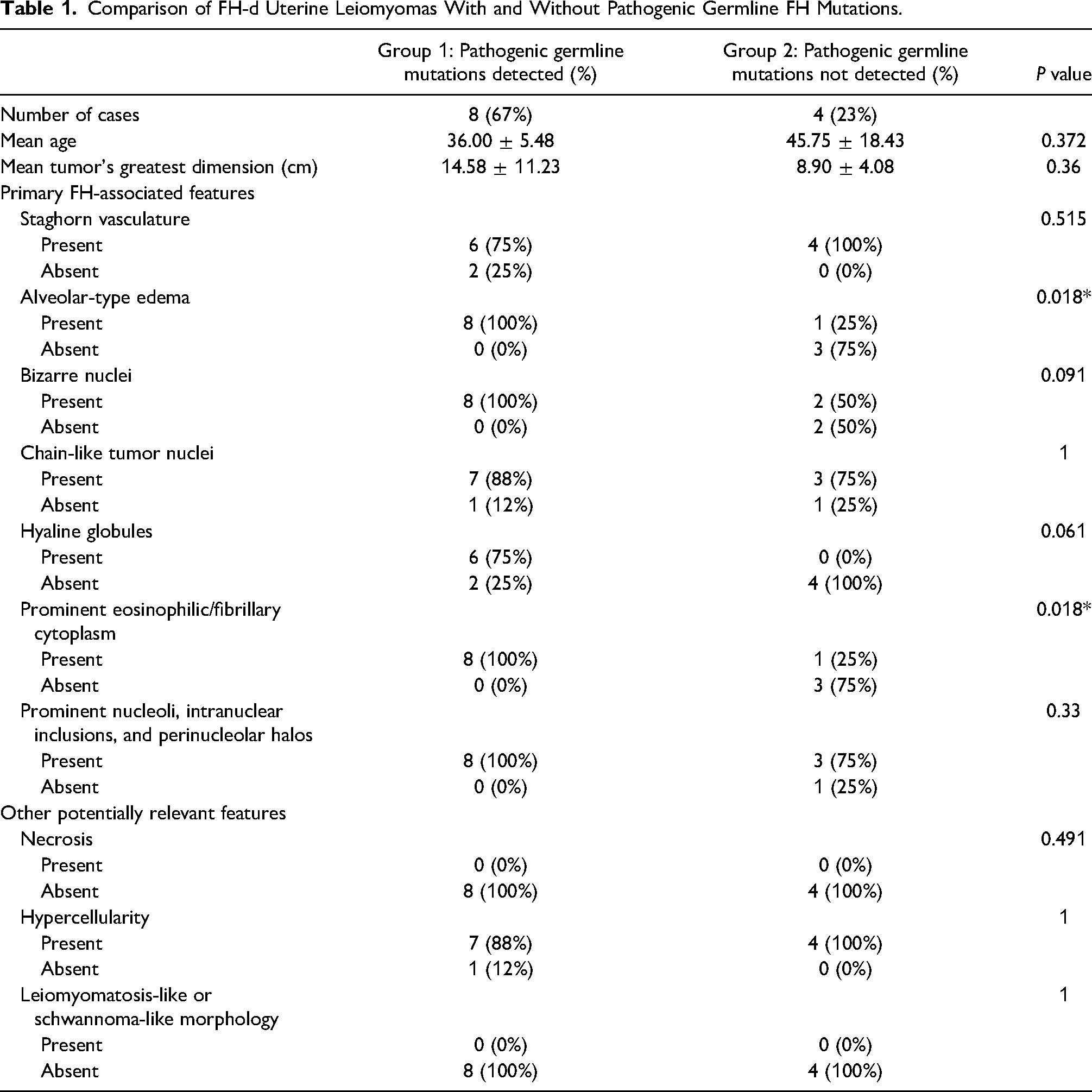
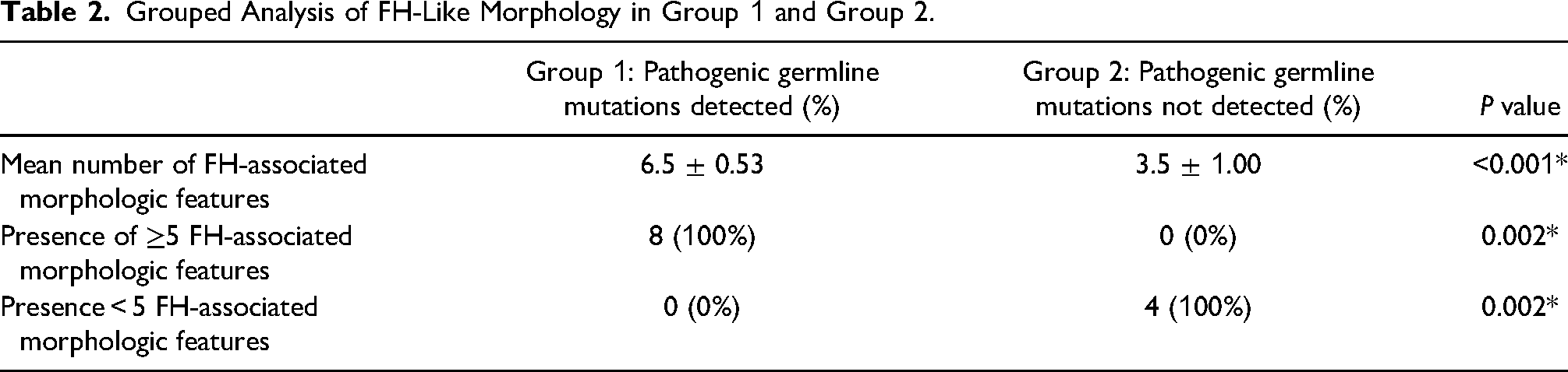
From 2418 uterine leiomyoma that were diagnosed during the 5-year study period, 37 (1.5%) patients had at least 1 tumor that had been classified by the reporting pathologist as displaying ≥1 FH-associated morphologic feature, and 29 of these had been assessed by FH immunohistochemistry. Fourteen (48.27%) of the 29 cases showed FH protein deficiency by immunohistochemistry (FH-d). FH-d tumors were larger (10.44 vs 4.08 cm, P < 0.01) and associated with younger patients (42.05 vs 47.97, P = 0.004) than a cohort of 370 randomly selected control patients with uterine leiomyoma. Twelve of the 14 patients with FH-d uterine leiomyoma elected to undergo germline testing; pathogenic FH mutations were present in 8 patients (66.67%) and were absent in 4 (33.33%). A comparison of morphologic features between the 2 tested groups is shown in Table 1, although all analyses were limited by small sample cohorts. Groups 1 and 2 showed no significant differences in patient age and tumor size. An analysis restricted to the 7 primary FH-associated features found that 100% of the group 1 cases showed ≥5 FH-associated features as compared to none of the group 2 cases (P = 0.002). However, 3 (75%) of the group 2 cases had 4 FH-associated features, and 1 (25%) had 3 features. The mean number of the FH-associated features was 6.5 ± 0.53 in group 1 cases, compared to 3.5 ± 1.00 in the group 2 cases (P < 0.001; Table 2). Two individual morphologic features, alveolar-type edema and prominent eosinophilic/fibrillary cytoplasm, were significantly more frequent in group 1, but none of the evaluated FH-associated morphologic features was individually found to be completely sensitive and specific in making the distinction between group 1 and 2 tumors. The combination of diffuse alveolar edema and either chain-like tumor cells (87.5% of group 1), scattered bizarre nuclei (100% of group 1), hyaline globules (75% of group 1), or prominent nucleoli/intranuclear inclusions/perinucleolar halos (100% of group 1) were not seen in any of the 4 group 2 counterparts. The morphological comparison of all 12 patients is presented in Figure 1.

Staghorn vessels with different configurations. Lumina ranged from dilated (A) to regular with smooth border (B) to slit like (C) with irregular intraluminal protrusions (D), clustered, and with perivascular edema.
Comparison of FH-d Uterine Leiomyomas With and Without Pathogenic Germline FH Mutations.
Grouped Analysis of FH-Like Morphology in Group 1 and Group 2.
Morphological Observations in FH-d Leiomyoma
A detailed description of morphologic features, including their spectrum in the context of group 1 versus group 2 status, is illustrated and presented in Figures 1–10.

All cases in group 1 showed diffuse alveolar type of edema (A and B). The single group 2 case with alveolar-type edema showed the changes focally (C and D). Other areas of the case displayed non-specific edema (E and F).

Bizarre cells with hyperchromatic nuclei and smudgy chromatin (A-F).
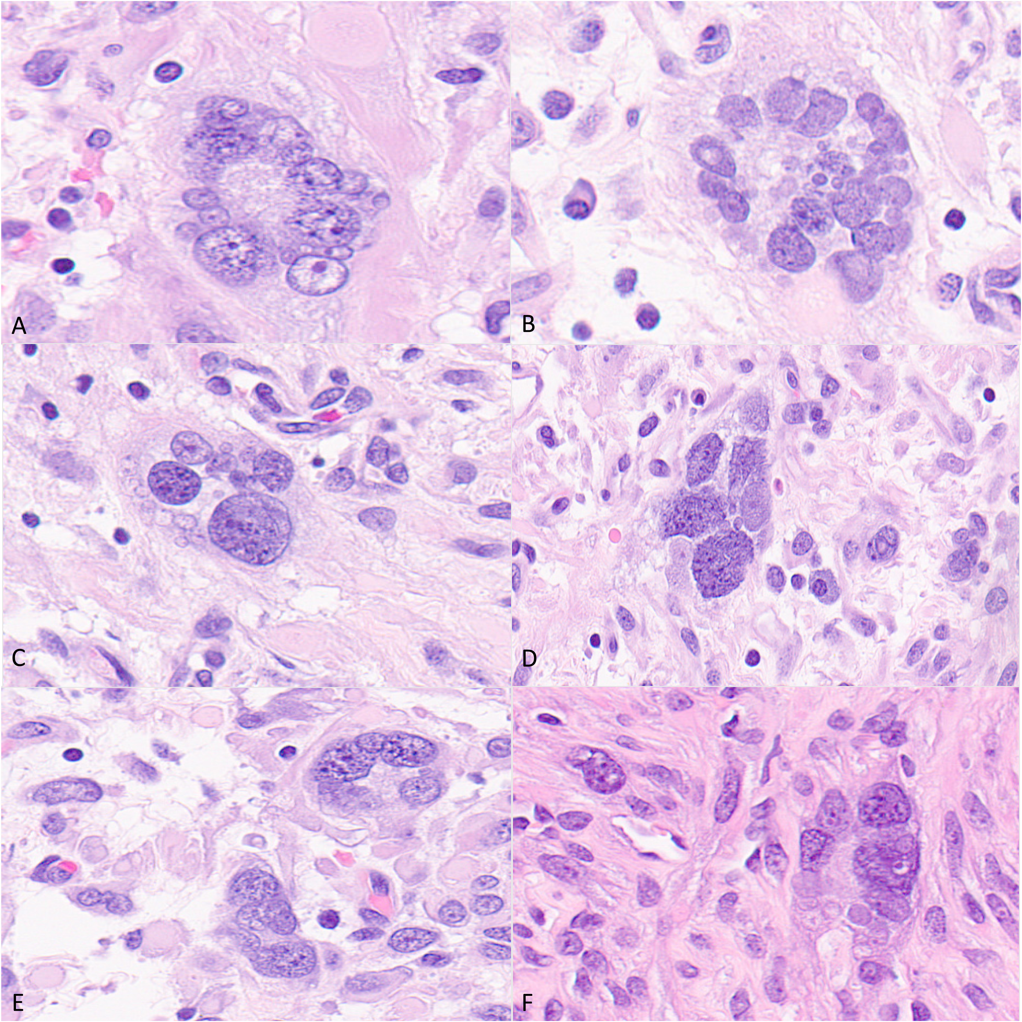
Bizarre large multinuclear giant cells with an variably open chromatin, forming rosette-like structures (A-F).
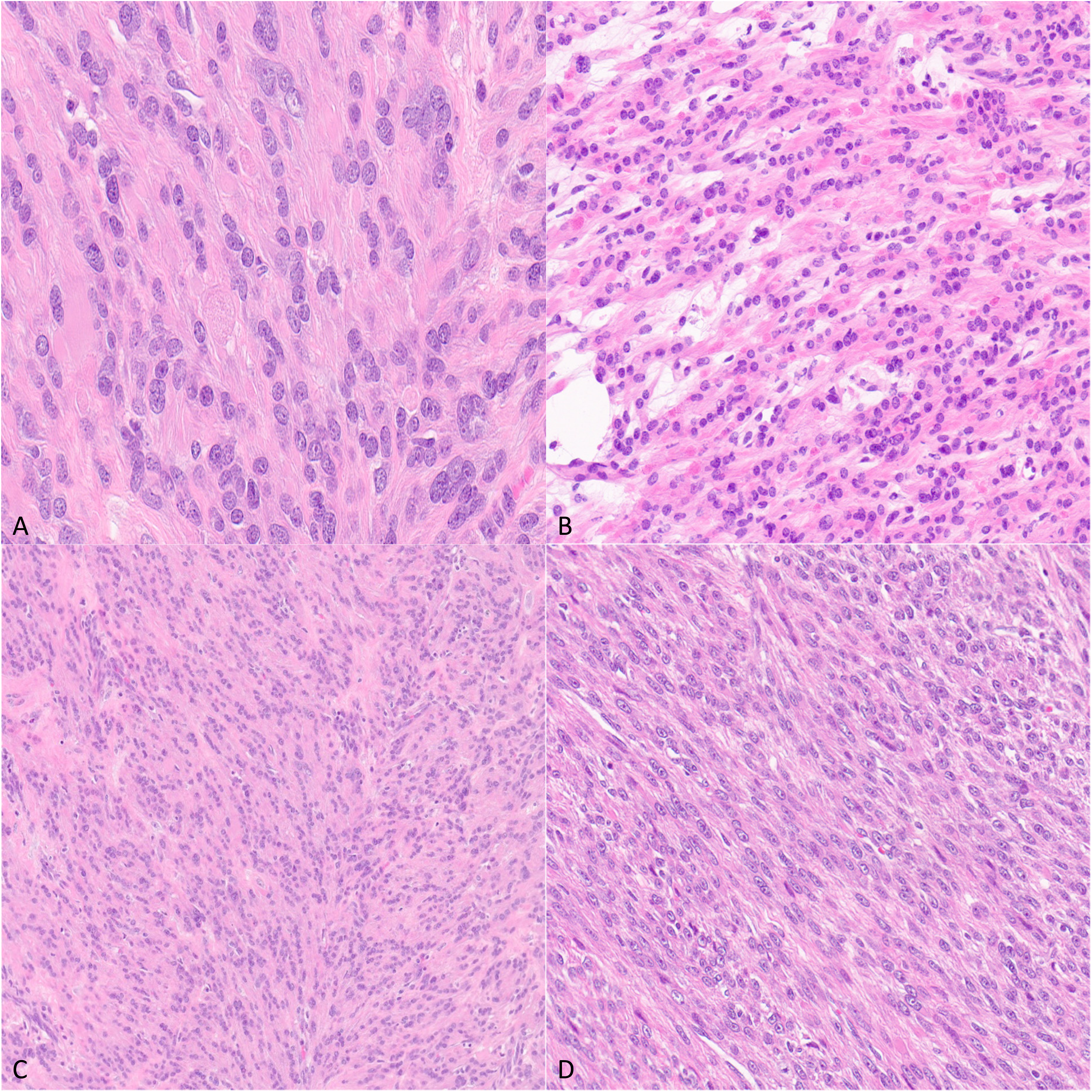
Chain-like tumor cells (A, B, and C). Foci with high cellularity resulting in a sheet-like pattern (D).

Eosinophilic hyaline globules (A-D; arrows). These were typically seen in a background of cells with notably eosinophilic cytoplasm.
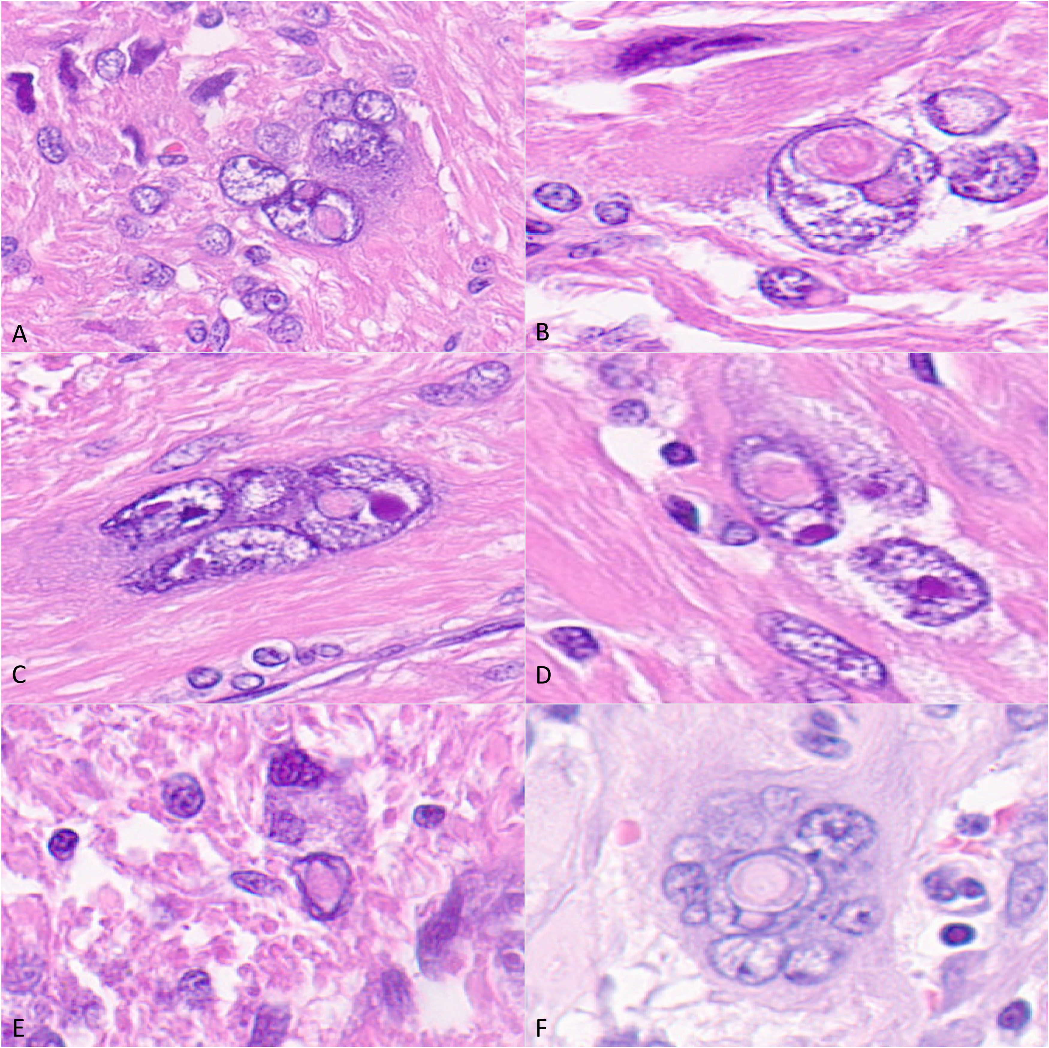
Intranuclear eosinophilic inclusions (A-F).
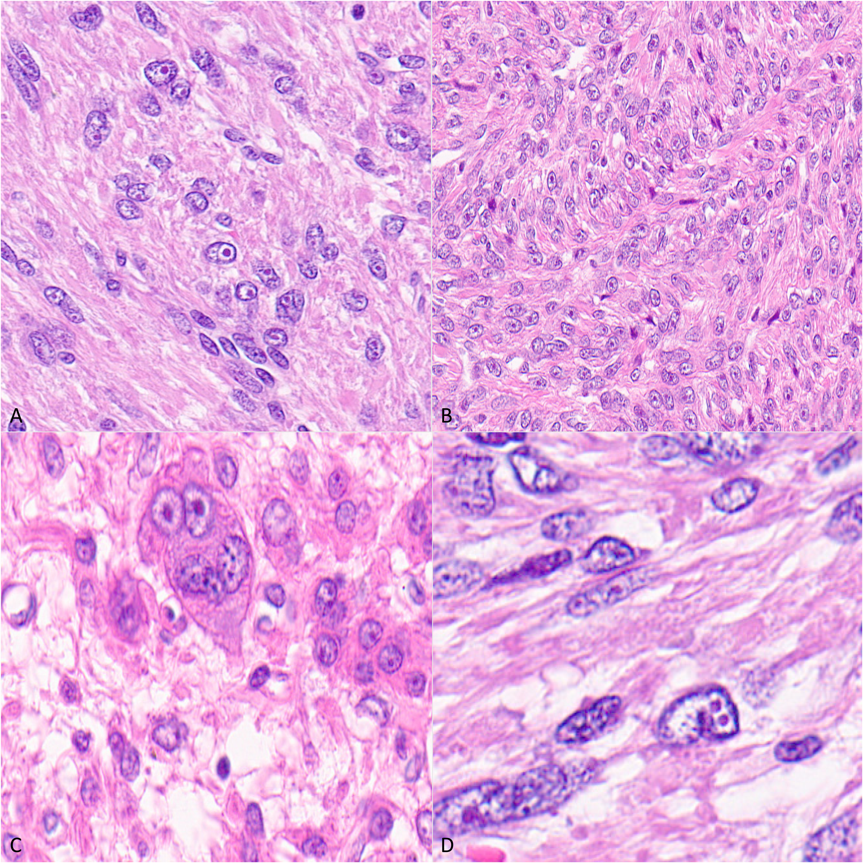
Prominent nucleoli with perinucleolar clearing.
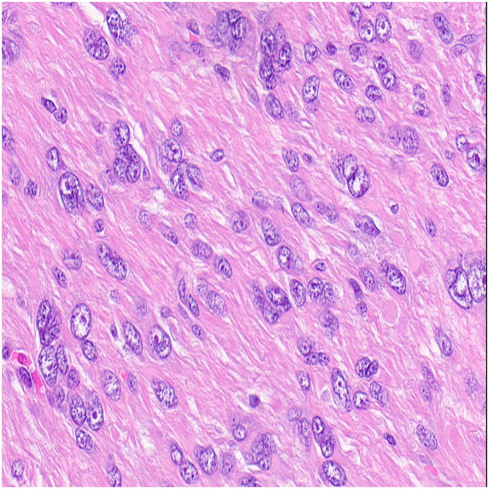
A group 1 case with prominent eosinophilic/fibrillary cytoplasm.
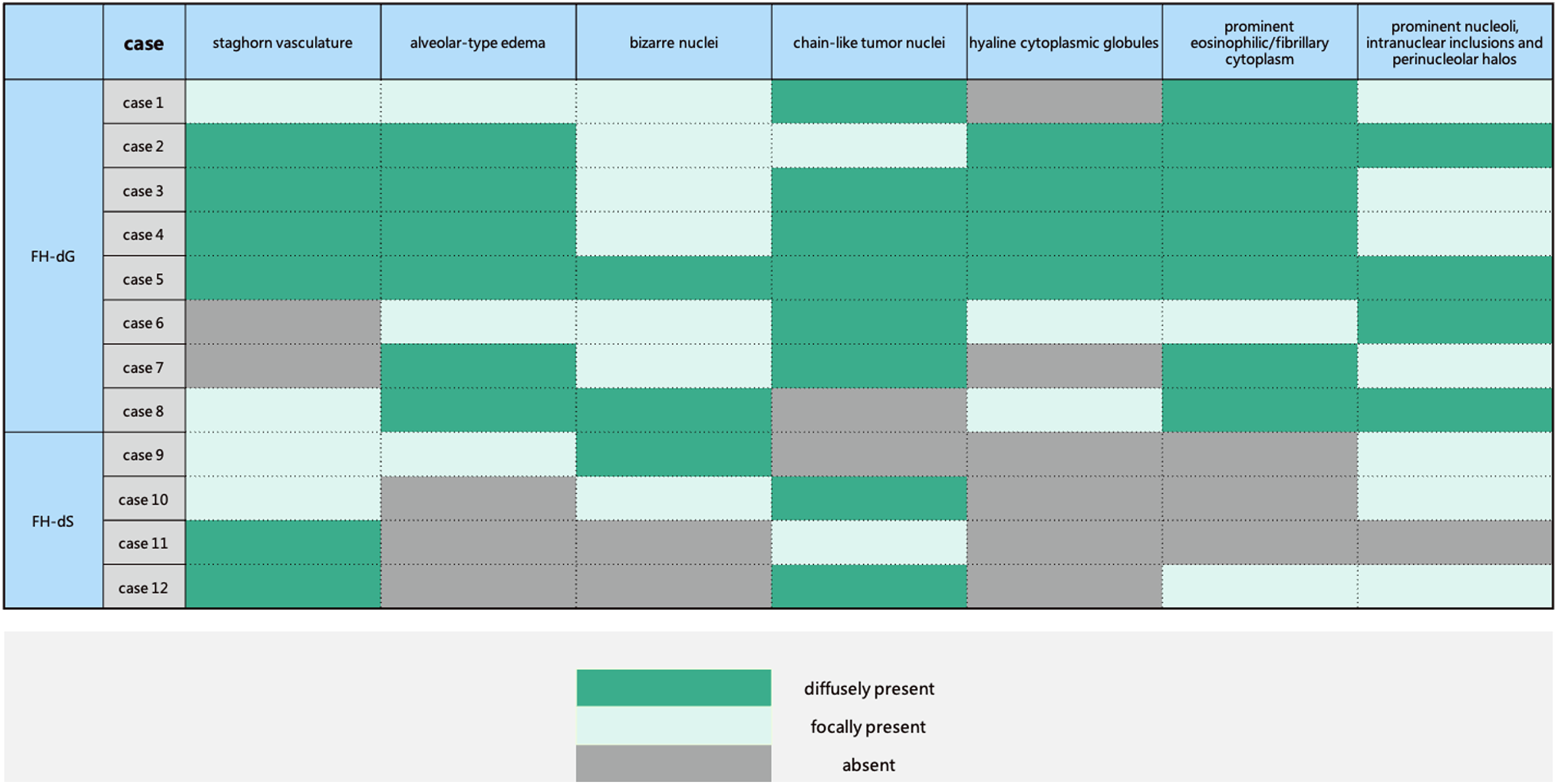
Summary of tumoral morphologic features in all 12 patients that underwent germline testing (FH-dG, fumarate hydratase protein-deficient leiomyoma of the uterine corpus with a pathogenic germline mutation of the FH gene; FH-dS, fumarate hydratase protein-deficient leiomyoma of the uterine corpus without a pathogenic germline mutation of the FH gene).
Hemangiopericytoma-Like Vessels
Staghorn vessels were present in 75% of the group 1 tumors as compared with 100% of group 2 tumors (P = 0.515). These vessels were easily discernable in most sections of the tumor in 83% of cases. The vessels were typically of small caliber and were mostly of venous or lymphatic type. Lumina ranged from dilated (Figure 1A) to regular with smooth borders (Figure 1B) to continuously slit-like (Figure 1C) or with irregular intraluminal protrusions (Figure 1D). Most cases showed admixtures of the above. The vessels were occasionally aggregated or clustered (Figure 1E). Some staghorn vessels were seen in a background of edema (Figure 1F) or showed perivascular edema (Figure 1F), whereas others showed neither of these features. Overall, the combination of alveolar edema and staghorn vessels was present in 75% of the group 1 cases, as compared with 25% of the group 2 cases.
Alveolar-Type Edema
Alveolar-type edema, wherein edematous zones displayed interspersed strands of residual smooth muscle, thereby mimicking alveolar tissue, was observed in 100% of the group 1 tumors as compared with 25% of the group 2 tumors (P = 0.018). The alveolar type of edema is generally present diffusely in group 1 (Figure 2A and B). The single case of group 2 uterine leiomyoma that had alveolar-type edema showed the changes only focally (Figure 2C and D). Other areas of the same tumor display non-specific (ie, not clearly alveolar-type) edema; the latter pattern appeared to be more common in group 2 (75%, Figure 2E and F). The combination of diffuse alveolar edema with either chain-like tumor cells, scattered bizarre nuclei, hyaline globules, or prominent nucleoli/intranuclear inclusions/perinucleolar halos was not seen in any of the group 2 cases. Thus, the diffuse presence of the typical alveolar type of edema is a potentially helpful pattern that is highly suggestive of group 1.
Bizarre Nuclei
Bizarre nuclei, wherein tumoral nuclei display irregular enlargement, “smudged” chromatin hyperchromasia, and irregular nuclear membranes (Figure 3A-F), were identified in 100% of the group 1 cases but also in 50% of the group 2 cases (P = 0.091). Another iteration of bizarre nuclei, characterized by large multinuclear giant cells with an open chromatin forming a rosette (Figure 4A-F), was also identified in multiple cases. Overall, bizarre nuclei were typically focal, and none of the cases in either group displayed bizarre nuclei diffusely.
Chain-Like Tumor Cells
Chain-like arrangement of tumoral nuclei was identified in 7 of 8 (88%) cases in the group 1 cases and 75% of the group 2 cases. In most cases, the change was diffuse (Figure 5A-C). No distinct morphological differences associated with chain-like nuclei were identified, although a spectrum of varying cellularity was observed, with some cases being notably more cellular than others (Figure 5D).
Hyaline Globules
Hyaline eosinophilic globules were identified in 6 (75%) of the 8 group 1 cases and in none of the group 2 cases (P = 0.061). The changes were diffuse in 4 of the 6 cases (Figure 6A-D).
Cells with hyaline globules were generally seen in a background of cells with eosinophilic cytoplasm, which were either granular or fibrillary. Other FH-associated features were typically present and in no case were hyaline globules, the only morphologic feature suggestive of FH deficiency.
Prominent Nucleoli, Intranuclear Inclusions, and Perinucleolar Halos
The spectrum of nuclear changes that have previously been associated with HLRCC-related tumors, including prominent nucleoli, intranuclear inclusions (Figure 7), and perinucleolar halos, was observed in 100% of the group 1 tumors but also in 75% of the group 2 tumors (P = 0.33). There was a wide spectrum of appearances within an individual case and from case to case. For some cases, the nucleoli were large with cherry-red color, whereas others were more subtle. Cells with perinucleolar halo were diffusely present in approximately one-third of cases in both groups (Figure 8). Overall, no differences were clearly discernible between the 2 groups regarding these features.
Prominent Eosinophilic/Fibrillary Cytoplasm
All group 1 cases demonstrated striking eosinophilic and fibrillary cytoplasm (Figure 9). Only 25% of the group 2 tumors showed this feature (P = 0.018). When present, the changes were most discernible at intermediate and high magnification. The changes ranged from granular (Figure 6) to fibrillary (Figures 5A and 7) to non-specifically eosinophilic (Figure 9).
Other Potentially Relevant Features
Leiomyomatosis-like and schwannoma-like morphology was not identified in either group. None of the other features that were assessed were plausibly discriminatory, including hypercellularity and necrosis.
Discussion
A variety of screening approaches may theoretically be used to identify patients with HLRCC, a rare syndrome that is associated with a 21% lifetime risk of developing renal cancers.34-38 Patients with HLRCC usually present with large uterine leiomyomas during the first 3 or 4 decades of life, which offers an initial potential opportunity to identify such patients. 39 Approaches that are centered on tumoral evaluation of resected uterine leiomyoma have been extensively studied, although the fact that conventional, FH-proficient uterine leiomyomas may also be encountered in the young, and that the latter, in general, are substantially more common than their FH-deficient counterparts, poses a significant challenge. Furthermore, morphologic features that have characteristically been associated with FH deficiency in uterine leiomyomas are not entirely specific, as some may also be seen in leiomyoma with bizarre nuclei, conventional uterine leiomyomas, and cellular uterine leiomyomas,21,23,24,28 and there may be problems with their diagnostic reproducibility among pathologists. 31 Immunohistochemistry for FH, even in the context of FH-associated morphology, may miss a significant subset of HLRCC patients whose uterine leiomyomas display intact FH protein expression. 22 Immunohistochemistry for 2SC, which is reportedly more diagnostically robust, nonetheless requires an additional study to assess its performance in identifying germline FH mutations. Finally, germline mutations of the FH gene, even in the context of abnormal morphology and abnormal immunohistochemistry, are only identified in a subset of patients. 39 In this study, we sought to determine whether there are any clearly discernible differences in tumoral morphology between FH-d uterine leiomyomas with and without germline pathogenic germline mutations. This may have implications for screening and confirmatory strategies for patients with possible HLRCC, including the necessity for expansive germline testing in limited resource settings.
In 12 patients with FH-d uterine leiomyomas that underwent germline testing, 8 (66.7%) were found to show pathogenic FH mutations. This figure is slightly higher than the up to 50% that has been reported in the literature, 39 which we attribute to the overrepresentation of consultation cases from multiple geographic locations in our cohort. Overall, the same spectrum of morphologic changes was seen in both group 1 and group 2. Regarding individual FH-associated morphologic features, no single feature was found to be completely sensitive and specific in making the distinction between the tumors in both groups. However, the combination of diffuse alveolar-type edema and either chain-like tumor cells, scattered bizarre nuclei, hyaline globules, or prominent nucleoli/intranuclear inclusions/perinucleolar halos was not seen in any of the 4 group 2 cases. Group 1 tumors showed, on average, a statistically significant higher number of FH-associated morphologic abnormalities than group 2 tumors.
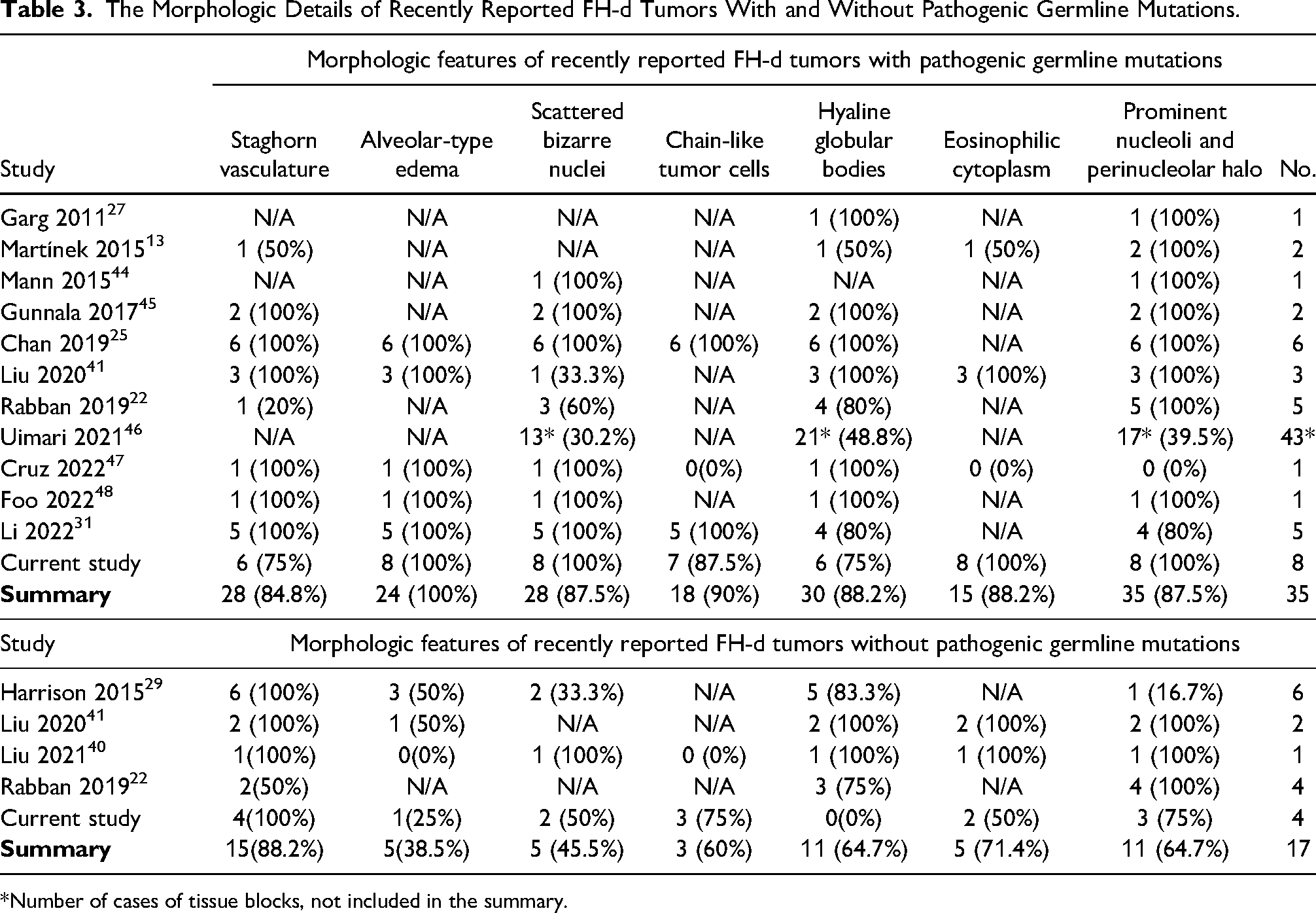
A brief literature review was conducted to identify the spectrum of morphologic features that have previously been reported to be differentially present in group 1 and 2 uterine leiomyomas. Two search phrases, “Fumarate hydratase deficient leiomyoma” and/or “HLRCC,” were applied in various combinations to the PubMed and Scopus databases for the period between 2007 and 2022. In total, 631 records were identified and were initially reviewed. Only studies with a description of histopathologic features that were correlated with FH germline testing results at the individual patient level were included. This yielded 35 group 1 cases and 17 group 2 cases. Many reports were devoid of full and detailed descriptions, which limited this analysis. Nonetheless, cases with extractable morphologic data are summarized in Table 3. This table shows that essentially every FH-associated morphologic feature has been reported in group 1 and 2 cases. Among the 17 group 2 cases, the 6 cases reported by Harrison et al 29 are the largest. In that series, at least 3 of those 6 cases combined alveolar edema with either hyaline globules, prominent nucleoli, or bizarre-type nuclear atypia. 29 Isolated cases by Liu et al40,41 also suggest that these features may be combined in the group 2 setting. Therefore, it is probable that the absence of these combinations in our group 2 can be attributed to the small size of the cohort. Overall, when our cases and previously reported cases are combined, no single morphologic feature was found is completely sensitive and specific in making the distinction between group 1 and 2 tumors. It is unclear, however, whether there is a specific combination of morphologic features that substantially increases the likelihood that a given FH-d uterine leiomyoma is associated with a germline mutation.
The Morphologic Details of Recently Reported FH-d Tumors With and Without Pathogenic Germline Mutations.
*Number of cases of tissue blocks, not included in the summary.
The finding that germline versus somatic mutations in the FH gene elicit a broadly comparable, but possibly not identical spectrum of tumoral morphologic alterations, is consistent with previous findings in the gynecologic tract. For example, in high-grade serous tubo-ovarian carcinomas, whether BRCA1 inactivation is due to germline or somatic mutations results in the same tumoral morphologic spectrum: overrepresentation of the so-called “SET” morphology. 42 For endometrial carcinomas, both sporadic MMRd (mismatch repair protein deficient) and Lynch syndrome-associated cancers are mostly of the endometrioid type, although sporadic MMRd tumors display fewer tumor-infiltrating lymphocytes and more likely show squamous and mucinous differentiation. 43 There are also plausibly gene-specific factors that ultimately affect tumoral morphology in a manner that is dependent or affected by whether its mutation is germline or somatic. These issues require additional analysis.
Some limitations of the current study are worthy of note, and the findings should be evaluated within that context. As is clear from Table 3, uterine leiomyomas associated with germline FH mutations are relatively rare. Our analysis only included 12 patients that underwent germline testing, which limited the analyses. Nonetheless, this study is one of the largest collections of cases to date with FH germline and somatic mutations that were directly correlated with tumoral morphologic features. Second, the retrospective nature of the current analysis meant that criteria for segregating cases with plausibly FH-associated features were necessarily different among different pathologists. In our group, cases were typically segregated for FH immunohistochemical analysis if they displayed at least one FH-associated morphologic feature. However, during the early part of the study period, some cases were classified as displaying FH-associated morphology, but did not undergo FH immunohistochemical testing. None of those patients underwent germline testing. As was previously noted, immunohistochemistry has some limitations in identifying uterine leiomyoma cases for which genetic counseling should be recommended or germline testing performed. Indeed, some authors believe “their role to be limited in the presence of well-developed FH-deficient morphology.” 39
In summary, our findings suggest that FH-d uterine leiomyomas with pathogenic germline FH mutations are unlikely to be morphologically distinguishable from FH-d uterine leiomyomas without such mutations. No single morphologic feature was found to be completely sensitive and specific in making this distinction. Whether there is a combination of morphologic features that can reliably make this distinction requires additional studies with larger cohorts.
Footnotes
Declaration of Conflicting Interests
The author(s) declared no potential conflicts of interest with respect to the research, authorship, and/or publication of this article.
Funding
The author(s) received no financial support for the research, authorship, and/or publication of this article.
Ethical Approval
The study was conducted in accordance with the Declaration of Helsinki and was classified as being exempt from the Institutional Review Board requirements under category 45 CFR 46.104(d) [USA] by the University of California San Diego IRB (IRB: #806223, approval date: January 24, 2023).
Informed Consent
Not applicable, because this article does not contain any clinical trials.
Trial Registration
Not applicable, because this article does not contain any clinical trials.
