
Abstract
Carcinomas of the head-and-neck region with squamous and glandular/mucinous features constitute a heterogeneous group, with a significant minority of tumors showing an human papillomavirus (HPV) association. The differential diagnosis is usually between mucoepidermoid carcinoma (MEC) and adenosquamous carcinoma. We present here two tumors that exemplify both the challenges of diagnostic classification, as well as the complex relationship to HPV: (a) a low risk HPV positive/p16 negative carcinoma that is most consistent with a relatively typical intermediate grade mucoepidermoid type carcinoma with complete MEC phenotype (three cell types), originating from intranasal sinonasal papillomas with exophytic and inverted patterns, and invading surrounding maxillary compartments, and (b) a p16 and keratin 7 (KRT7) positive carcinoma of the right tonsil, characterized by stratified squamous and mucinous cell (mucocyte) features. Whereas the first tumor represents a typical MEC ex-Schneiderian papilloma, the second is morphologically most consistent with the, novel for this anatomic location, diagnosis of “invasive stratified mucin producing carcinoma” (ISMC), pointing to an analogy to similar, high-risk HPV-driven malignancies recently described in the gynecologic (GYN) and genitourinary (GU) areas. Both tumors, despite their mucoepidermoid-like features had no connection to salivary glands and lacked the MAML2 translocation typical of salivary gland MEC, pointing to a mucosal/non-salivary gland origin. Using these two carcinomas as examples, we attempt to address questions related to: (a) the histological distinction between MEC, adenosquamous carcinoma, and ISMC, (b) similarities and differences between these histological entities in mucosal sites versus morphologically similar salivary gland tumors, and (c) the role of HPV in these tumors.
Keywords
Introduction
Human papillomavirus (HPV)-associated oropharyngeal squamous cell carcinoma is generally characterized by a distinctive, non-keratinizing/basaloid squamous morphology. The carcinomas with both squamous cells and mucocytes in the head and neck area are usually classified as either adenosquamous carcinoma or mucoepidermoid carcinoma (MEC). This nomenclature raises two issues: a. adenosquamous carcinoma of the head and neck have been often grouped together with high grade MEC in prior studies, even if adenosquamous carcinomas overall have a distinctly worse prognosis than MEC. 1 HPV-related adenosquamous carcinoma, on the other hand, appears to do significantly better than the typical adenosquamous carcinoma. Thus, both the correct classification, as well as the HPV status should be addressed in head and neck carcinomas with both squamous cells and mucocytes. Adenosquamous carcinoma can be separated from MEC by applying strict morphological criteria, such as, in adenosquamous carcinoma, the presence of a SCC-in-situ squamous component, definitive squamous differentiation (including keratinization), florid gland formation with “punched out” spaces with or without mucin. 1 MEC are characterized by the presence of three populations of cells including squamous cells, mucocytes, and intermediate cells, forming cystic and solid growth patterns. Most salivary gland MEC have translocation and gene fusion findings involving the MAML2 gene.
MEC can occur in the oropharynx and were presumed to share the generally better prognosis of carcinomas in this area, either due to the specific microenvironment of the oropharynx, or the association with HPV that has been shown to lead to a better clinical outcome for both SCC and adenosquamous carcinoma of the oropharynx. 1 Both possibilities have been disproven, with MEC of the oropharynx shown to be aggressive and alarmingly prone to metastasize irrespective of tumor grade or MAML2 status. 2
It was originally presumed that salivary gland MEC associated with HPV would have a better prognosis than HPV-negative tumors. However, this assumption has been brought into question by studies negating any HPV pathogenetic role in the salivary gland MEC.3,4
Some MEC arising outside of the salivary glands have been reported to originate from typical HPV-related tumors (eg, Schneiderian sinonasal papillomas),5,6 with variable clinical outcomes.
Unlike in the head and neck area where the differential diagnosis for tumors with both squamous cells and mucocytes is generally limited to adenosquamous carcinoma or MEC, in the gynecologic (GYN) and genitourinary (GU) areas a novel, HPV-associated, diagnostic entity has been introduced, termed invasive stratified mucin producing carcinoma (ISMC). 7 ISMC is characterized by stratified, immature epithelial cells displaying varying amounts of intracellular mucin throughout the majority of the lesional epithelium, with resulting spacing between adjacent tumor cell nuclei. These tumors are thought to arise from embryonic cells at the transformation zone of the uterine cervix as are most cervical squamous cell carcinomas and their precursor dysplastic lesions. 7
These tumors are strongly positive for p16, as a surrogate marker for HPV infection, and keratin 7 (KRT7), which is typically present in the cervical squamocolumnar junction/transformation zone. Recent studies, point to an analogy between the uterine cervical and the oropharyngeal “transformation zones” of the KRT7-positive tonsillar reticular crypt epithelium in the latter, corresponding to the transformation zone in the former. In both instances these histological areas would be providing cell targets for HPV mediated oncogenesis. 7 Accordingly, HPV-associated carcinomas of the oropharynx are positive for both p16 and KRT7. 8
The two examples of head and neck carcinomas we present here, both displaying squamous and mucinous features albeit in markedly different patterns, that we propose represent two interesting models of this complex pathogenetic and differential diagnostic spectrum: a classical type MEC related to low-risk HPV and an ISMC potentially related to high-risk HPV.
Case Presentation: Patient 1
Clinical and Radiologic Findings
A 49-year-old male, with a history of cigarette smoking and intranasal cocaine use, presented with a 2-year history of left nasal obstruction. His symptoms progressed over 4 to 6 months to include epistaxis, V2 numbness, and retroorbital pain.
Physical exam revealed an exophytic, left intranasal mass as well as a painful lesion of the hard palate. Imaging showed a unilateral sinonasal mass, extending posteriorly to the nasopharynx, inferiorly into the hard palate, laterally to involve the left pterygoid palatine fossa, and superiorly via the inferior orbital fissure into the inferior aspect of the left orbital apex and peri-orbital soft tissues. Perineural spread along the left trigeminal nerve (V2) was also noted within the foramen rotundum.
Pathologic Findings
Biopsies were taken from the left nasal cavity mass and the left hard palate defect. The left nasal cavity mass showed a typical sinonasal papilloma, exophytic and inverted type, whereas the hard palate biopsy was initially diagnosed as invasive squamous cell carcinoma.
The patient subsequently underwent a total maxillectomy and orbital exenteration. Grossly, the floor of the left nasal cavity showed a brown, friable, irregular surface with an underlying lesion extending into the nasal septum and eroding through the hard palate. This lesion was contiguous with a mucosal defect of the hard palate, measuring approximately 1 cm in diameter.
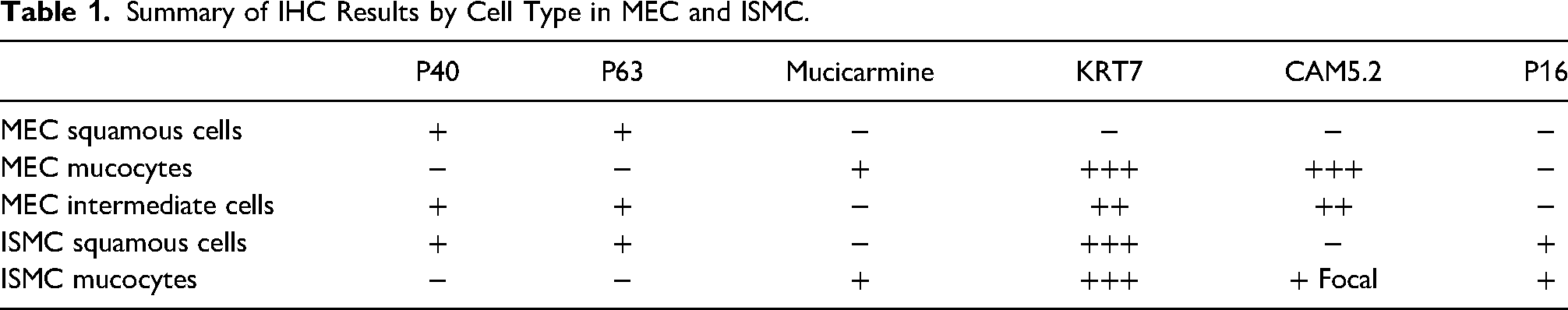
On microscopic examination, the floor of the left nasal cavity showed numerous sinonasal papillomas, with both exophytic and inverted growth patterns, containing scattered mucocytes and foci of high-grade squamous dysplasia (Figure 1c). Underlying and focally contiguous with the papillomas was an invasive carcinoma showing predominantly non-keratinizing squamous cells with interspersed mucocytes highlighted by both p40 and p63 immunohistochemistry (IHC) and by a mucicarmine special stain (SS), in the former and latter cells respectively (Figures 2a, c and 3c). There were also intermediate cells present, which stained weakly with p40 and p63. Prominent glandular lumina were present, lined predominantly by mucocytes. The glandular lining cells were strongly positive for both KRT7 and CAM5.2 (Figure 2e and g) whereas the intermediate cells were slightly less strongly positive for CAM5.2, and markedly less for KRT7. The squamous cells were negative for both KRT7 and CAM5.2 (Figures 2e and g). Overall, the lesion was composed of a significantly more prominent squamous and intermediate cell component, compared to the mucocytes. The lesional cells showed moderate atypia, some necrosis, and a relatively low mitotic rate. Ki67 proliferation rate was calculated at 44% (Figure 3g). The mucocytes displayed a prominent intracystic/glandular component, consistent with an intermediate grade MEC. p53 IHC showed a “wild-type” (ie, unmutated phenotype) staining pattern (see Table 1 for summary of IHC findings).

(a) Patient 1 low power view of hard palate and nasal septum showing squamous papillomas within the left nasal cavity and invasive carcinoma extending to and eroding the mucosal surface of the hard palate. (b) Patient 1 higher power view of invasive carcinoma eroding the mucosal surface of the hard palate. (c) Patient 1 sinonasal papilloma with benign mucocytes. (d) Patient 2 invasive carcinoma. (e) Patient 2 PAS special stain highlighting the mucocytes, as well as most of the squamous cells cytoplasm. (f) Patient 2 PAS-D special stain highlighting the mucocytes, isolated or in groups; the squamous cell cytoplasm appears predominantly clear. (g) Patient 2 nuclear palisading along the periphery of the tumor cell nests (arrows). (h) Patient 2 mucicarmine special stain highlighting both intracellular mucin (single arrow) and extravasated pools of mucin (double arrows).

(a) Patient 1 p40 IHC, positive in squamous and intermediate cell populations. (b) Patient 2 p40 IHC, diffusely positive. (c) Patient 1 p63 IHC, positive in squamous and intermediate cell populations. (d) Patient 2 p63 IHC, diffusely positive. (e) Patient 1 KRT7 IHC, 3+ positivity in mucocytes (single arrow), 2+ positivity in intermediate cells (double arrow), and negative in squamous cells (triple arrow). (f) Patient 2 KRT7 IHC, diffusely positive. (g) Patient 1 CAM5.2, 3+ positivity in mucocytes (single arrow), 2+ positivity in intermediate cells (double arrow), and negative in squamous cells (triple arrow). (h) Patient 2 CAM5.2, focally weakly positive, mostly in the pseudoglandular areas.

(a) Patient 1 perineural invasion. (b) Patient 1 mucicarmine SS, abundant positivity in areas of perineural invasion. (c) Patient 1 mucicarmine SS, positive in scattered cells. (d) Patient 2 mucicarmine SS, positive in scattered, isolated cells, or cell groups. (e) Patient 1 p16 IHC, negative. (f) Patient 2 p16 IHC, diffusely positive, except for the areas of mucus inclusions. (g) Patient 1 ki67 IHC, positive in 44% of lesional cells. (h) Patient 2 ki67 IHC, positive in 72% of lesional cells.
Summary of IHC Results by Cell Type in MEC and ISMC.
The carcinoma invaded into but not through the nasal septum, through the maxilla, and eroded through the mucosa of the underlying hard palate resulting in focal mucosal ulceration (Figure 1a and b). Additionally, perineural invasion of the inferior orbital nerve with associated extensive mucin production was present (Figure 3a and b). The carcinoma did involve the orbital apex and periorbital soft tissues, resulting in a pathologic tumor stage designation of T4b.
The sinonasal papillomas as well as the invasive carcinoma were negative for p16 by IHC (Figure 3e), negative for HR HPV by in-situ hybridization (ISH), but were positive for LR HPV by ISH. The invasive carcinoma was negative for the MAML2 (11q21) translocation by fluorescence in situ hybridization (FISH).
Given the presence of squamous cells, interspersed glandular elements, and intermediate cells, a final diagnosis of an intermediate grade MEC ex-inverted papilloma was made. Based on the morphological, immunohistochemical, and molecular studies it was concluded that the MEC clearly arose not from salivary gland tissue, but from the numerous adjacent sinonasal papillomas present within the left nasal cavity.
Case Presentation: Patient 2
Clinical and Radiologic Findings
The second patient is a 56-year-old woman with HIV and a history of oral cavity squamous cell carcinoma involving the mandibular bone, who underwent a segmental mandibulectomy and adjuvant radiation 4 years prior. She presented following mandibular trauma to address exposed hardware and concern for osteomyelitis of her reconstructive surgery. She was incidentally noted to have right-sided globus pharyngeus contralateral to her exposed hardware.
On physical examination, a new right oropharyngeal mass and cervical adenopathy were identified contralateral to her prior malignancy. She presented with trismus at that time. Imaging revealed a right-sided tonsil mass measuring 2.9 cm with extension into the parapharyngeal fat as well as a right-sided jugulodigasric lymph node conglomerate measuring 3.4 cm.
Pathologic Findings
A biopsy of the mucosal mass was performed and showed an invasive carcinoma with a dual cell population composed of stratified, non-keratinizing squamous cells and mucocytes, highlighted respectively by both p40 and p63 IHC in the former, and a mucicarmine and PAS +/- diastase special stains in the latter, (Figures 1e, f, 2b, d, and 3d). The squamous cell nests showed prominent peripheral cell palisading (Figure 1g). Although the mucocytes were often clustered in groups, no well-defined glandular lumina were present; instead isolated mucocytes and “pseudoglandular” structures/mucocyte aggregates, without true lumina were seen, in addition to areas of mucus extravasation (Figure 1h). In contrast to the previously described tumor above (patient 1), in this carcinoma all lesional cells were strongly and diffusely positive for KRT7. The squamous type cells showed strong and diffuse cytoplasmic KRT7 staining, whereas the mucocytes showed mostly peripheral subplasmalemmal staining, surrounding the intracellular mucus inclusions (Figure 2f). CAM5.2 showed only focal staining predominantly in the “pseudoglandular” structures (Figure 2h). Ki67 proliferation rate was calculated at 72% (Figure 3h). The tumor cells, including the mucocytes, were strongly and diffusely positive for p16 IHC (Figure 3f). Despite the frequent use of a p16 positivity as a proxy for HPV association, both high- and low-risk HPV ISH testing was negative, as was MAML2 translocation FISH testing (11q21). Unfortunately following these tests, tissue was no longer available for further analysis (eg, PCR-based testing). p53 IHC showed a “wild-type” (ie, unmutated phenotype) staining pattern. Please, see Table 1 for summary of IHC findings.
Discussion
Patient 1
Carcinomas displaying mixed squamous and glandular differentiation can be classified principally as either adenosquamous carcinoma or MEC, with other diagnoses being far less likely. The diagnosis of adenosquamous carcinoma requires the presence of both squamous cells and well-formed, “punched out” glandular lumina with mucin within the lumen and/or the lining cells. Additionally, the majority of adenosquamous carcinomas show mutated p53 type staining, which was not the case in our tumor. 1 Although the distinction between adenosquamous carcinoma and MEC can be controversial, 5 the diagnosis of intermediate grade MEC for tumor 1 appears most appropriate, based on the absence of overt keratinization, the intermingling of squamous and glandular/mucous cells, the the widespread glandular lumina formation with lobular arrangement, and the unequivocal presence of intermediate cells both histologically as well as immunohistochemically. Based on these findings this diagnostic classification is consistent with the previous literature on the topic of carcinomas ex-sinonasal papillomas in general, and distinction between adenosquamous carcinoma and MEC in particular. 5
Inverted papillomas are the most common subtype of sinonasal papillomas.8,9 Malignant transformation is seen in 7.6% of sinonasal papillomas with inverted papillomas showing the greatest likelihood of malignant transformation at 8.9% to 11%.5,8,10
When these papillomas do undergo malignant transformation, 83% of patients develop squamous cell carcinoma. 5 While MEC is predominantly considered a salivary gland neoplasm, they have been seen in many other locations including the lacrimal sac, upper aerodigestive tract, and conjunctiva, as well as the sinonasal region, as in this patient.11-14 In a review of the English language literature between 1962 and 2013, MEC was seen in approximately 10% of cases of carcinoma ex-sinonasal papilloma. 5 The nasal cavity is the second most common location to find MEC in the sinonasal region, with the most common location being the maxillary sinus. 11
MEC of salivary gland origin have been well described, including a characteristic MAML2 translocation (t(11,19) leading to a CRTC1(MECT)-MAML2 fusion). The overall frequency of MAML2 translocation in MEC-type tumors of the head and neck region has not been defined.
Inverted papillomas show HPV positivity in 38% of cases: most commonly low risk HPV types 6 and 11 are detected, with high-risk types also occasionally seen.8,15-17 p16 IHC staining in sinonasal papillomas has shown variable results. 12 Although p16 can be upregulated by mechanisms unrelated to HPV, within the oropharynx strong and diffuse p16 positivity is used as a surrogate diagnostic marker for high-risk HPV-related carcinomas, consistent with the negative result in this Low Risk HPV associated case.
Inverted papillomas have been reported to cause pseudoinvasion into the underlying soft tissue and even bone erosion. Additionally, as in our patient, they are known to occasionally harbor benign goblet cells. 18 Characteristics favoring carcinoma include overt bone and tissue invasion, significant cytologic atypia, and the presence of any characteristic translocations.
Our patient's tumor did show overt invasion, including erosion of the underlying hard palate mucosa. Involvement of a secondary mucosal surface has been described previously. 10 Congruent IHC and ISH results between the sinonasal papillomas and underlying carcinoma can help identify the tissue of origin, as it did in this example.
This tumor demonstrated abundant mucin production in overtly invasive areas, including the areas of perineural invasion. This finding supports the conclusion that the mucocytes were part of the neoplastic process as opposed to the native mucinous glands.
Hyrcza et al described a similar example of MEC arising from an inverted papilloma of the nasal cavity which was also MAML2 translocation negative and low risk HPV positive by linear array analysis. However, in their repoprt p16 IHC did show patchy strong positivity in 30% of tumor cells. 12
In summary, MEC ex-inverted papilloma is the most appropriate diagnosis for
Patient 2
The differential diagnosis for this tumor includes MEC, adenosquamous carcinoma, and the recently described entity of ISMC.
While MEC has been reported in the oropharynx, the most common malignancies of the tonsillar region are squamous cell carcinomas, very often high-risk HPV-related, and lymphoproliferative diseases.19,20 The lack of well-formed glandular lumina, intermediate cells, and MAML2 translocation argues against a diagnosis of MEC in this case. Similarly, regarding the differential diagnosis of adenosquamous carcinoma, the presented carcinoma lacks the two clearly distinct cell populations, the origination from a dysplastic epithelial surface component and the well-formed, “punched out” glandular lumina and abnormal/mutated p53 staining. 1
ISMC is a recently described entity in the GYN and genitourinary regions, described as a high-risk HPV-driven lesion with squamous cells and mucocytes, presumably originating from embryonic cells at the corresponding transformation zones of these areas. They show strong and diffuse positivity for p16 and KRT7 IHC. 21
Cytokeratin 7 has been strongly associated with HPV carcinogenesis in the uterine cervical epithelium. Specifically, KRT7 identifies the embryonic epithelial cells at the cervical squamocolumnar junction involved in cervical squamous cell HPV-mediated dysplasia and carcinoma. 7 The reticular crypt tonsillar epithelium, which is also positive for KRT7 IHC, is thought to represent a similar transition zone, prone to involvement by HPV-mediated carcinogenesis in that location. 7
Unlike the first tumor presented above, this second carcinoma showed strong and diffuse p16 and KRT7 positivity throughout, and a significantly higher Ki-67 proliferation rate, similar to previously reported ISMC. 22 While the second tumor was HPV negative by ISH, it is known that approximately 42% of squamous cell carcinomas in this region which are p16 IHC positive and HPV negative by ISH are in fact HPV positive using polymerase chain reaction (PCR) testing. Previously it was concluded that there is no significant difference in survival between p16 positive HPV positive lesions and p16-positive HPV-negative lesions. 23 On the other hand the presence of discordance between p16 and HPV findings is a real issue and is most likely related to factors such as lower viral copy number. 24 Patients with p16+/HPV− or p16−/HPV+ tumors have signifintly worse prognosis than patients with with p16+/HPV+ oropharyngeal cancer, but a significantly better prognosis than patients with p16−/HPV− oropharyngeal cancer. 24 In the absence of a history of cigarette smoking, p16+/HPV- tumors are considered as most likely HPV mediated, but possibly at lower viral copy numbers than the p16+/HPV+ tumors. 24 Therefore, given the general histological appearance (ie, basaloid type p16 and KRT7 positive carcinoma) and the anatomic location (oropharynx), this is most likely a high-risk HPV-driven lesion.
ISMC has not been previously described in the head and neck region. There are significant morphological and immunohistochemical similarities between this carcinoma and the ones previously reported in the GYN and genitourinary areas, including the stratified, immature epithelial cells, displaying varying amounts of intracellular mucin throughout the majority of the lesional epithelium, as well as strong p16 positivity (as a surrogate marker for HPV), strong KRT7 positivity, and wild-type p53. Given the general pathogenetic and histological parallels generally seen in the GYN/GU and head and neck regions regarding HPV-related carcinomas in general and the morphologic and immunohistochemical similarities described above, we postulate that this carcinoma is consistent with the diagnosis of head and neck ISMC.
Footnotes
Declaration of Conflicting Interests
The author(s) declared no potential conflicts of interest with respect to the research, authorship, and/or publication of this article.
Funding
The author(s) received no financial support for the research, authorship, and/or publication of this article.
Ethical Approval
Ethical approval for this study was waived because based on the institutional review board (UMAB IRB) rules, specifically, retrospective studies of 3 cases or less do not require approval.
Informed Consent
Not applicable, because this article does not contain any studies with human or animal subjects.
Trial Registration
Not applicable, because this article does not contain any clinical trials.